
Glyphocrangon juxtaculeata Chace, 1984
|
publication ID |
https://doi.org/ 10.26107/RBZ-2020-0079 |
|
publication LSID |
lsid:zoobank.org:pub:4A6F3831-7B0E-4A3F-B7A7-A0663846372F |
|
persistent identifier |
https://treatment.plazi.org/id/03997B5A-8D29-FFC0-FF70-9AE4FA59FB91 |
|
treatment provided by |
Diego |
|
scientific name |
Glyphocrangon juxtaculeata Chace, 1984 |
| status |
|
Glyphocrangon juxtaculeata Chace, 1984 View in CoL
( Figs. 2–5 View Fig View Fig View Fig View Fig )
Glyphocrangon juxtaculeata Chace, 1984: 15 View in CoL (in part), fig. 3 (type locality: southern Buru , Indonesia). –– Brand & Takeda, 1996:
279 (list). Glyphocrangon regalis, Komai, 2004b: 542 View in CoL (in part).? Glyphocrangon juxtaculeata, Han & Li, 2007: 548 View in CoL , fig. 3. Not Glyphocrangon juxtaculeata, Hayashi & Araki, 1999: 626 View in CoL
(list). [= Glyphocrangon perplexa Komai, 2004b ]
Material examined. Holotype: “Albatross”, stn 5638, off S Buru, Banda Sea, Indonesia, 03°47.15′S, 126°23.40′E, 946 m, 10.12.1909, juvenile (cl 9.5 mm), USNM 205090 View Materials . GoogleMaps
Other material: SJADES 2018, stn CP 35, S of Tg. Boyongkareuceng , 07°47.68′S, 107°41.90′E to 07°47.68′S, 107°42.48′E, 603–686 m, rocks, mud and clay, 29 March 2018, beam trawl, 1 female (cl 17.2 mm), ZRC 2020.0296 View Materials GoogleMaps .
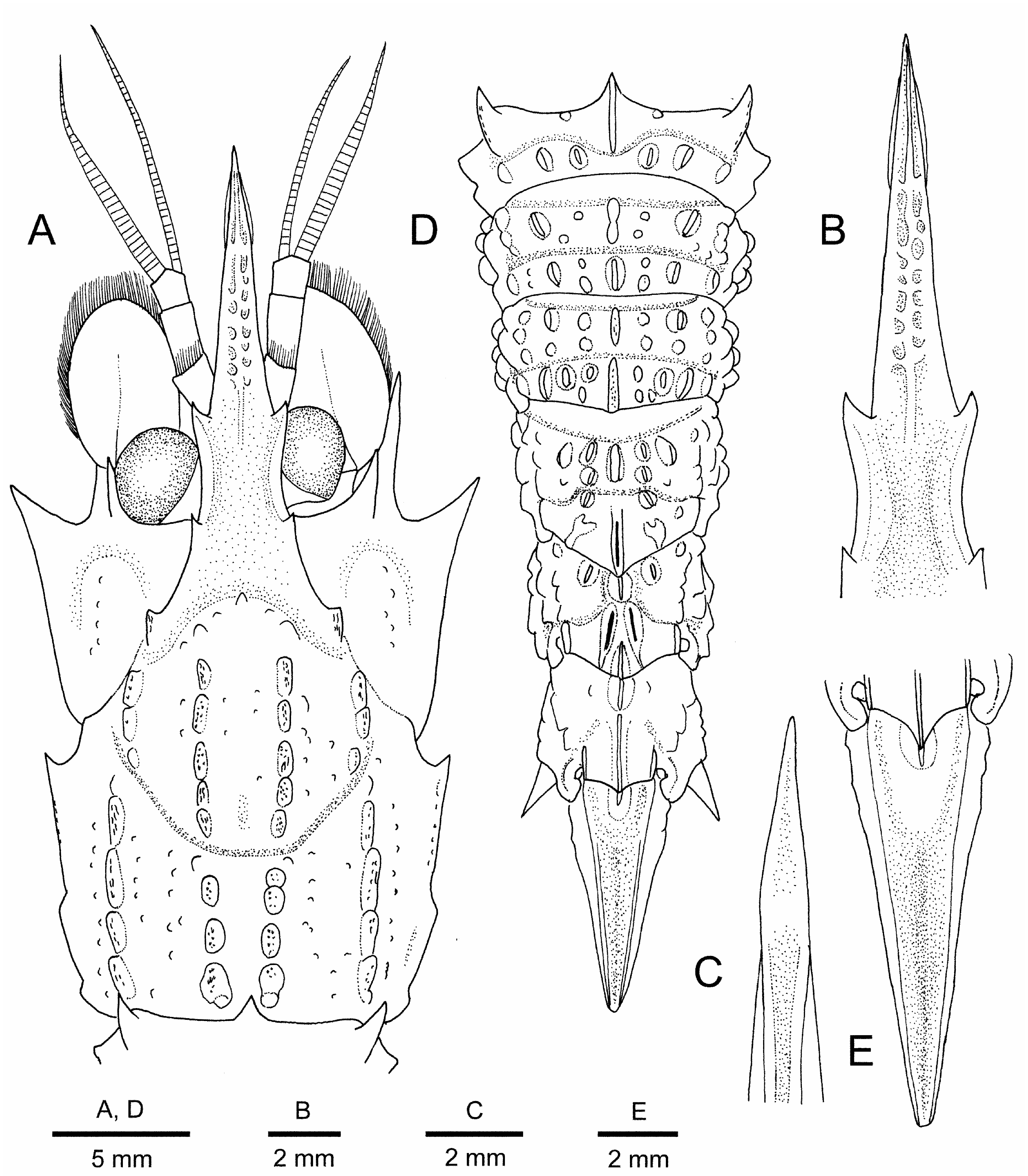
Description of newly collected specimen. Body ( Figs. 2 View Fig , 3A, D View Fig ) moderately robust for genus. Integument glabrous, without dense pubescence or scattered short setae.
Rostrum ( Figs. 2 View Fig , 3A–C View Fig ) slightly descending proximally and upturned in distal one-third, gradually tapering to acute tip, 0.75 times as long as carapace, armed with 2 pairs of spines on dorsolateral ridges; anterior pair located at 0.3 of rostral length, proximal pair just above orbital margin; dorsal margin of proximal spine faintly sinuous; dorsal surface distinctly faveolate anterior to anterior pair of lateral spines (middorsal ridge obsolescent in proximal 0.3 of rostrum); dorsolateral ridges between 2 lateral spines raised, bluntly carinate, those anterior to lateral spines weakly elevated, also bluntly carinate; ventral surface medially sulcate (sulcus becoming wider and shallower toward distal), ventrolateral margins sharply carinate.
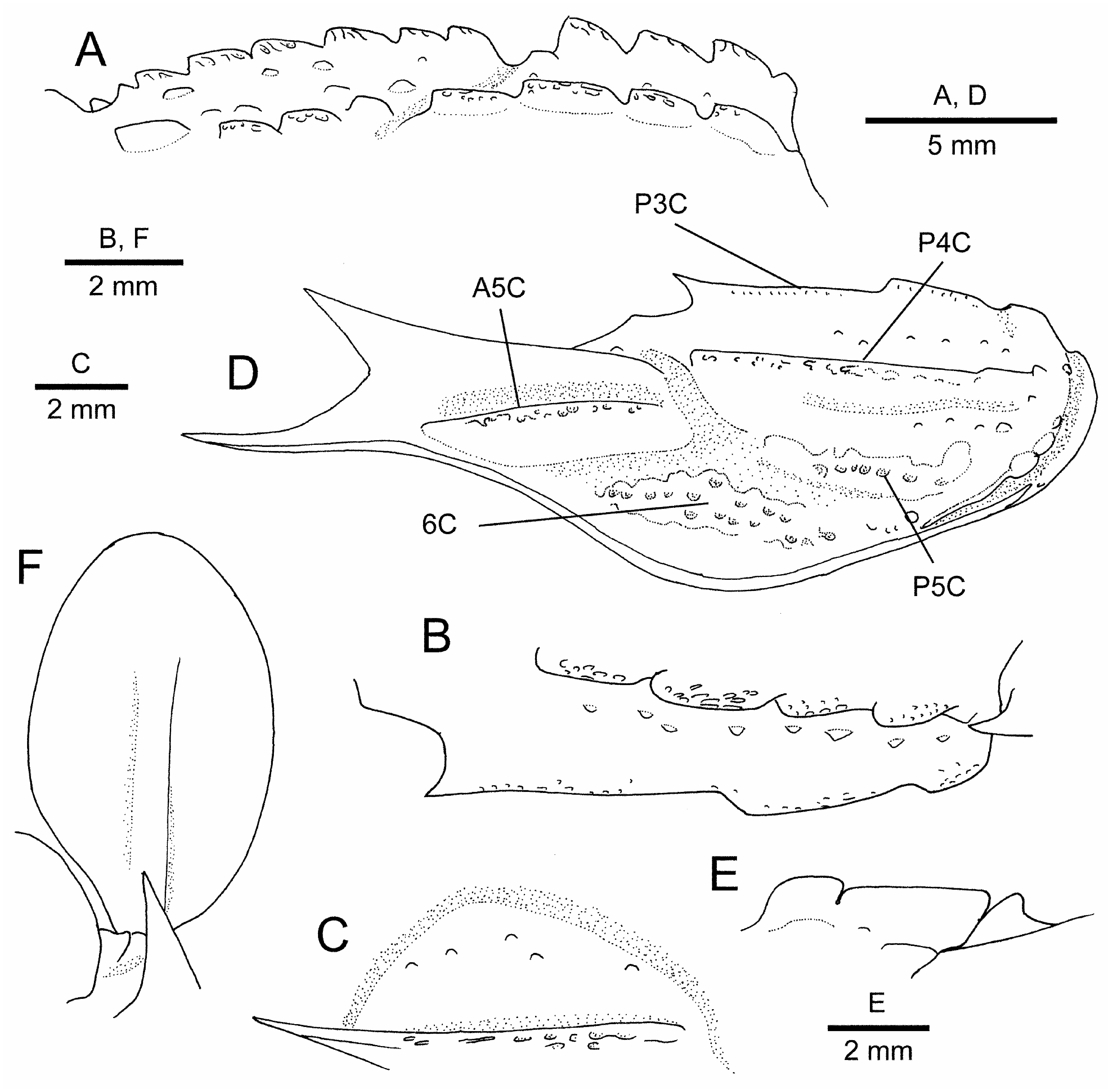
Carapace ( Figs. 2 View Fig , 3A View Fig ) 1.25 times as long as width between anterior ends of posterior third carinae; surfaces of carinae and tubercles etched with reticulate pattern of minute pits; major carinae moderately high; anterior and cervical grooves well demarcated. Anterior first (submedian) carina composed of 6 tubercles, these tubercles forwardly directed with blunt tips, compressed laterally ( Fig. 4A View Fig ); posterior first carina composed of 4 forwardly directed, unequal tubercles, first tubercle strongest, subacute, others bluntly pointed ( Fig. 4A View Fig ). Anterior second (intermediate) carina composed of 4 tubercles, anteriormost one larger than others, subacute, others blunt; posterior second carina slightly arched in lateral view, divided into 4 lobes (none sharply pointed) and 1 small blunt tubercle ( Fig. 4A View Fig ). Anterior third (antennal) carina confined to antennal spine; posterior third carina divided into 3 unequal lobes, anteriormost lobe terminating into small spine directed anteriorly, others bluntly pointed ( Fig. 4B, D View Fig ). Anterior fourth (lateral) carina expanded into relatively slender, vertically compressed, acute lamina slightly exceeding beyond sinus between antennal and branchiostegal spines, distance between tips of laminae 0.9 times of carapace length; posterior fourth carina faintly divided into 2 lobes, followed by 2 blunt tubercles posteriorly, anterior end bluntly pointed ( Fig. 4D View Fig ). Anterior fifth (sublateral) carina well demarcated ( Fig. 4D View Fig ); posterior fifth carina low ( Fig. 4D View Fig ). Sixth (submarginal) carina represented by low, broad elevation with roughly eroded surface ( Fig. 4D View Fig ). Orbital margin slightly elevated; postorbital region slightly depressed, unarmed. Intercarinal tubercles tiny, all blunt. Median part of gastric region with only 3 tubercles along anterior groove and few tubercles on either side of midline; posteromedian part without additional tubercles. Lateral part of gastric region with space between first and second carinae having about 6 low tubercles; space between second carina and lateral groove devoid of conspicuous tubercles. Posterior dorsolateral region with several low tubercles arranged in 2 irregular longitudinal rows. Upper part of hepatic region slightly convex, with some obsolescent tubercles ( Fig. 4C View Fig ); lower part of hepatic region shallowly concave, unarmed ( Fig. 4D View Fig ). Upper part of branchial region bearing 8 (left) or 10 (right) tiny tubercles ( Fig. 4B View Fig ); middle part with 7 (right) or 8 (left) tiny tubercles arranged in single longitudinal row; lower part posteriorly with 5 (left) or 7 (right) obsolescent tubercles. Antennal spine strong, ascending in lateral view (angle about 30° against horizontal plane of carapace), directed forward in dorsal view, falling short of distal corneal margin. Branchiostegal spine very strong, directed anteriorly and slightly curving dorsally in lateral view, slightly curving laterally in ventral view, reaching nearly to midlength of antennal scale, hardly visible in dorsal view; lateral face without conspicuous ridges, but sharply carinate ventrally. Marginal posterolateral corner slightly produced, forming light angle.
Pleon ( Figs. 2 View Fig , 3D View Fig ) covered with blunt tubercles. Pleomere 1 with middorsal elevation defined by shallow transverse groove; middorsal carina forming large, laterally compressed tooth with forwardly directed, acute apex; posterior part of tergum with 2 laterally compressed tubercles on either side of midline. Dorsolateral carina developed into strong triangular tooth with forwardly directed, acute apex. Lateral carina composed of 2 blunt tubercles. Pleuron ornamented anteriorly with 3 tubercles; posterior depression abruptly delimited; anteroventral corner produced anteriorly, subacutely pointed. Pleomeres 2–4 with crest-like median carina, each deeply divided into 2 parts. Tubercles or prominences composing submedian or dorsolateral carinae strongly compressed laterally; surfaces of carinae and tubercles weakly etched with minute pits. Posterior transverse grooves all moderately deep. Vertical ridges on pleura 3 and 4 terminating ventrally in small blunt tubercle. Pleural teeth moderately strong, acute, slightly unequal. Pleomere 2 with anterior part of median carina somewhat produced anteriorly, posterior part semicircular in lateral view. Pleomere 3 with anterior part of median carina slightly produced anteriorly, posterior part subrectangular, produced posteriorly. Pleomere 4 with anterior part of median carina much shorter than posterior part, subrectangular; posterior part bearing median groove, produced posteriorly in blunt, triangular tooth.
Pleomere 5 with anterior median carina posteriorly directed, non-spiniform; posterior median carina produced posteriorly in rounded lobe. Tergum with shallow oblique groove; anterior submedian carinae represented by small, laterally compressed tubercle with posteriorly directed apex; posterior submedian carinae diverging posteriorly, abruptly narrowed posteriorly, each with median groove. Pleuron with 2 strong, slightly unequal ventral teeth.
Pleomere 6 with crested median carina, divided into 2 parts by narrow notch ( Fig. 4E View Fig ), posterior part weakly produced posteriorly into blunt tooth. Tergum with 1 minute tubercle anteriorly. Lateral carina composed of 4 unequal, somewhat compressed tubercles, none spiniform. Pleuron with blunt carina laterally; posteroventral tooth strong, flared laterally, without supporting carina on lateral face.
Telson ( Fig. 3D, E View Fig ) 0.6 times as long as carapace; tapering to acute apex; anterodorsal projection prominent, laterally compressed, with posteriorly directed, subacute apex, without accessory tubercle. Dorsolateral and ventrolateral carinae sharply defined, former smooth, latter with trace of few tubercles proximally.
Cornea ( Figs. 2 View Fig , 3A View Fig ) large (width 0.2 of carapace length), darkly pigmented in preservative. Ocular peduncle with minute papilla-like process on anteromesial face (not illustrated).
Antennular peduncle ( Figs. 2 View Fig , 3A View Fig ) slightly overreaching anterior margin of antennal scale; article 2 subcylindrical, about 2.5 times longer than wide; aesthetasc-bearing portion of outer flagellum about 0.3 times as long as carapace.
Antennal scaphocerite ( Figs. 2 View Fig , 3A View Fig , 4F View Fig ) oval, 0.4 times as long as wide, without trace of lateral tooth; lateral margin entirely setose. Carpocerite falling short of anterior margin of scaphocerite.
Maxilliped 3 endopod ( Fig. 5A, B View Fig ) moderately stout, just reaching anterior margin of antennal scale. Ultimate article tapering to acute unguis demarcated basally, with 4 and 3 strong spiniform setae on inner and outer margins respectively, and 7 strong spiniform setae on inner face. Penultimate article with 1 spiniform seta on inner surface distally and 3 spiniform setae on outer margin. Antepenultimate article with dorsolateral margin sharply carinate. Exopod overreaching midlength of antepenultimate article; flagellum not reaching distal end of antepenultimate article.
Pereopod 1 ( Fig. 5C View Fig ) not reaching midlength of antennal scale. Flexed dactylus not reaching midlength of palm; palm glabrous on lateral surface (without short pubescence), with longitudinal row of tufts of stiff setae on extensor to mesial surface. Carpus short, cup-shaped. Merus with 2 inconspicuous longitudinal ridges dorsally and ventrally on lateral surface. Ventral lamina of ischium bluntly pointed distally.
Pereopod 2 unequal. Left ( Fig. 5D View Fig ) shorter and stouter than left, overreaching midlength of antennal scale by length of chela; chela small, as wide as distalmost carpal article; carpus subequal in length to merus, ischium, and basis combined, divided into 19 articles; merus and ischium similar in structure to that of right. Right ( Fig. 5E View Fig ) overreaching antennal scale by length of chela and 0.2 of carpus; chela small, as wide as distalmost article of carpus; carpus subequal in length to merus, ischium, and basis combined, divided into 30 articles; merus subequal in length to ischium; ventral margin of ischium slightly expanded proximally to accommodate chela when flexed.
Pereopod 3 ( Fig. 5F View Fig ) reaching anterior margin of antennal scale by tip of dactylus; dactylus compressed laterally, 0.3 times as long as propodus; propodus slightly narrowing distally; carpus 0.7 times as long as propodus.
Pereopod 4 ( Fig. 5G View Fig ) moderately slender, reaching anterior margin of antennal scale by tip of dactylus. Dactylus ( Fig. 5H View Fig ) subspatulate, 0.4 times as long as propodus, terminating in acute tip; extensor surface with trace of longitudinal sulcus along midline, and with submarginal row of minute setae adjacent to lateral margin in distal one-fourth. Propodus slightly narrowing distally, with tuft of stiff bristle-like setae on distal margin partially covering base of dactylus. Carpus 0.7 times as long as propodus.
Pereopod 5 ( Fig. 5I View Fig ) similar to fourth pereopod, falling slightly short of anterior margin of antennal scale. Dactylus
( Fig. 5J View Fig ) 0.3 times as long as propodus, shorter than that of fourth pereopod; extensor surface also with trace of median sulcus. Carpus 0.6 times as long as propodus.
Pleopods and uropod ( Fig. 2 View Fig ) typical of genus, without distinguishing features.
Colouration in fresh condition. Body and appendages generally orange; rostrum, carapace antennal and branchiostegal spines, antennal scale, antennal flagella and maxilliped 3 ultimate article darker, reddish; cornea pigmented, reflective ( Fig. 2 View Fig ).
Remarks. Chace (1984) described Glyphocrangon juxtaculeata on the basis of a female or juvenile holotype from the Banda Sea, off the south coast of Buru, Indonesia; he tentatively identified a young male specimen from the Sulu Sea, the Philippines, with G. juxtaculeata , noting several differences from the holotype. Komai (2004b) remarked that it was not easy to determine the specific identify of the two type specimens of G. juxtaculeata , because juvenile specimens of Glyphocrangon do not fully develop diagnostic features of the species. Nevertheless, he indicated that the holotype and non-type specimen were specifically distinct because of some differences already noted by Chace (1984), and suspected that the holotype of G. juxtaculeata might represent a juvenile stage of G. regalis Spence Bate, 1888 , based on the geographical proximity and general morphological similarity between the holotypes of the two nominal taxa. The status of the non-type male specimen remained to be determined. Later, Han & Li (2007) referred a juvenile specimen from the South China Sea to G. juxtaculeata but did not give clear evidence why they believed that G. juxtaculeata was a valid species.
Our specimen from off Java could be referred to a species of the G. regalis species complex as diagnosed by Komai (2004b), and is morphologically similar to G. smithii Wood- Mason in Wood-Mason & Alcock, 1891, known from the Andaman Sea, and G. amblytes Komai, 2004b from the western Indian Ocean, in the relatively weak intercarinal tubercles on the carapace and pleon, and the small anterior spine of the carapace posterior third carina ( Komai, 2004b; Komai & Chan, 2017). As outlined below, however, the present specimen is specifically distinct from G. smithii and G. amblytes . The supposed synonymy of G. juxtaculeata with G. regalis by Komai (2004b) can now be rejected because the presence or absence of the anterior spine of the carapace posterior third carina in the female has been clarified to be stable in certain species of Glyphocrangon ( Komai, 2004b, 2011; Komai & Chan, 2013), although varying between male and female in some species: the holotype of G. juxtaculeata has this spine, whereas the holotype of G. regalis does not have such a spine ( Komai, 2004b). Considering the geographical point of view and the possession of a relatively small anterior spine of the carapace posterior third carina, the present specimen could be best assigned to G. juxtaculeata .
The newly collected specimen now enables us to reassess diagnostic features of G. juxtaculeata . As noted above, G. juxtaculeata closely resembles G. smithii and G. amblytes . Possible differentiating characters between G. juxtaculeata and G. smithii include: (1) the dorsal sculpture of the rostrum is more pronounced in G. juxtaculeata than in G. smithii (cf. Fig. 2B View Fig versus Komai & Chan, 2017 : fig. 7A); (2) tubercles consisting of the carapace first (submedian) carina are more blunt in G. juxtaculeata than in G. smithii ( Fig. 3 A View Fig versus Komai & Chan, 2017: fig. 7C); (3) tubercles on the pleon are fewer in G. juxtaculeata than in G. smithii ( Figs. 1 View Fig , 2D View Fig versus Komai & Chan, 2017 : fig. 6A, B); (4) the dactyli of the pereopods 4 and 5 have only a shallow median sulcus on the extensor surface in G. juxtaculeata ( Fig. 4H, J View Fig ), whereas in G. smithii , there is a deep median groove on each dactylus ( Komai & Chan, 2017: fig. 8I, K). From G. amblytes , G. juxtaculeata is distinguished by the following minor particulars: (1) the carapace first carina is higher in G. juxtaculeata than in G. amblytes (cf. Fig. 3 A View Fig versus Komai, 2004b: fig. 89); (2) the anteriormost tubercle of the carapace anterior second carina is subacutely pointed in G. juxtaculeata ( Fig. 2A View Fig ), rather than obtuse in G. amblytes (cf. Komai, 2004b: fig. 89); (3) the carapace posterior third carina is divided into three unequal lobes in G. juxtaculeata ( Figs. 2A View Fig , 3B View Fig ), while entire in G. amblytes (cf. Komai, 2004b: fig. 89); (4) the acute lamina formed by the carapace anterior fourth carina is less expanded in G. juxtaculeata than in G. amblytes (distance between tips 0.9 times of carapace length in G. juxtaculeata versus 1.0–1.1 times in G. amblytes ).
Considering species occurring outside the Indo-West Pacific, G. aculeata A. Milne-Edwards, 1881 from the Atlantic is similar to G. juxtaculeata . Indeed, Chace (1984) compared G. juxtaculeata only with G. aculeata . As Chace (1984) discussed, G. aculeata differs from G. juxtaculeata in the lack of transverse septa on the dorsal surface of the rostrum, the absence of tubercles on the upper hepatic region of the carapace, and the entire posterior third carina of the carapace. Furthermore, the pleural ventral teeth are longer and more acuminate in G. aculeata than in G. juxtaculeata ( Holthuis, 1971: fig. 10; Komai, 2004a: fig. 1 A versus Fig. 1 View Fig ); the posterior middorsal carinae of the pleomeres 5 and 6 are acuminate in G. aculeata (cf. Holthuis, 1971: fig. 10; Komai, 2004a: fig. 1A), rather than bluntly pointed in G. juxtaculeata ( Figs. 1 View Fig , 3E View Fig ).
Although the present molecular analysis shows that G. juxtaculeata has rather low COI sequence divergence (2.2–2.5%) from G. lowryi (Table 2), it is clearly different from the latter in the following morphological features: (1) the rostrum proximal part is devoid of submedian tubercles and lateral tubercle inferior to the first lateral spine in G. juxtaculeata ( Fig. 3A, B View Fig ), which are present in G. lowryi ( Komai, 2004b: fig. 100A); (2) intercarinal tubercles on the carapace and pleon are much fewer and less conspicuous in G. juxtaculeata than in G. lowryi ( Figs. 2 View Fig , 3A, B View Fig and Komai, 2004b: figs. 99, 100C, D); (3) the posterior tips of the two divisions of the pleomere 6 middorsal carina are rounded in G. juxtaculeata ( Fig. 3E View Fig ), while they are sharply pointed in G. lowryi ( Komai, 2004b: fig. 99); (4) the telson anterodorsal projection is simple and blunt in G. juxtaculeata .
outgroup 9 – 0.283 an is
)
bp
614 0.108 0.298 (8 – – lacazei 0.106 0.293 Aegaeon . 7 0.110 0.275
Glyphocrangon 6 –0.100 –0.294 of 0.099 0.291 species studied 5 0.107 0.287 amongst
bp
). 4 0.123 – 0.295 –
–
657
619 individuals 0.120 0.293
(of 0.115 0.278 segment number 3 – 0.108 0.276 – gene
COI
indicate 0.175 0.157 0.147
mitochondrial
in parentheses
2 – 0.167 – – 0.153 0.144 0.142 – 0.149 0.149 0.149 0.271
distance of
. Numbers 1 – 0.136 0.094
0.103
– –
0.083
0.081 0.057 –
0.062
0.058 0.097 0.067 0.097 0.290 pairwise divergence
))
model
Nei
–
intraspecific
3 (species
Tamura indicate (
2) () 2, new
(Corrected
.
shade in.
G
armata .
G
G
.. investigatoris
G
.
G
G
. () proxima .
G
.
G
G
Table
2
Numbers 1 2 3 4 5 6 7 8 9 10
( Fig. 4E View Fig ), while bifid with sharp apices in G. lowryi ( Komai, 2004b: fig. 99); (5) the telson dorsolateral and ventrolateral margins are nearly smooth or only faintly tuberculate in G. juxtaculeata ( Fig. 3E View Fig ), rather than strongly tuberculate in G. lowryi ( Komai, 2004b: fig. 99). Furthermore, the colouration in the fresh condition is substantially different between the two. In G. juxtaculeata , the overall body and appendages are generally orange ( Fig. 2 View Fig ). On the other hand, in G. lowryi , the anterior part of the carapace, including the rostrum, antennae, and pereopods are scarlet; the carapace has a tinge of red on the dorsum, and whitish on the lateral sides; the pleon is entirely reddish except for the whitish pleomere 6 and telson ( Komai & Chan, 2013: fig. 3B).
Komai (2004b) referred the record of G. juxtaculeata from the East China Sea by Hayashi & Araki (1999) to G. perplexa Komai, 2004b .
The male specimen from the Sulu Sea, provisionally referred to G. juxtaculeata by Chace (1984), is characterised by the sharply pointed, spiniform tubercles comprising the carapace first carina and the middorsal carinae on the pleomeres 2 and 3, which are strongly produced into acute teeth ( Chace, 1984: fig. 4). In these regards, it agrees with G. spinosissima Brand & Takeda, 1996 , originally described from the Sulu Sea ( Brand & Takeda, 1996; Komai, 2004b), and it is likely that the specimen might represent a juvenile of G. spinosissima .
The identity of the juvenile specimen referred to as G. juxtaculeata by Han & Li (2007) remains to be determined.


No known copyright restrictions apply. See Agosti, D., Egloff, W., 2009. Taxonomic information exchange and copyright: the Plazi approach. BMC Research Notes 2009, 2:53 for further explanation.
|
Kingdom |
|
|
Phylum |
|
|
Class |
|
|
Order |
|
|
Family |
|
|
Genus |
Glyphocrangon juxtaculeata Chace, 1984
| Komai, Tomoyuki, Yang, Chien-Hui & Chan, Tin-Yam 2020 |
Glyphocrangon juxtaculeata
| Chace FA Jr 1984: 15 |