
Glyphocrangon smithii Wood-Mason & Alcock, 1891
|
publication ID |
https://doi.org/ 10.11646/zootaxa.4303.1.4 |
|
publication LSID |
lsid:zoobank.org:pub:9062A5B8-B5A1-465F-81E3-556763103195 |
|
DOI |
https://doi.org/10.5281/zenodo.6015725 |
|
persistent identifier |
https://treatment.plazi.org/id/96367D76-FF82-FFE0-F6D8-2806FE3FFA8A |
|
treatment provided by |
Plazi |
|
scientific name |
Glyphocrangon smithii Wood-Mason & Alcock, 1891 |
| status |
|
Glyphocrangon smithii Wood-Mason & Alcock, 1891 View in CoL
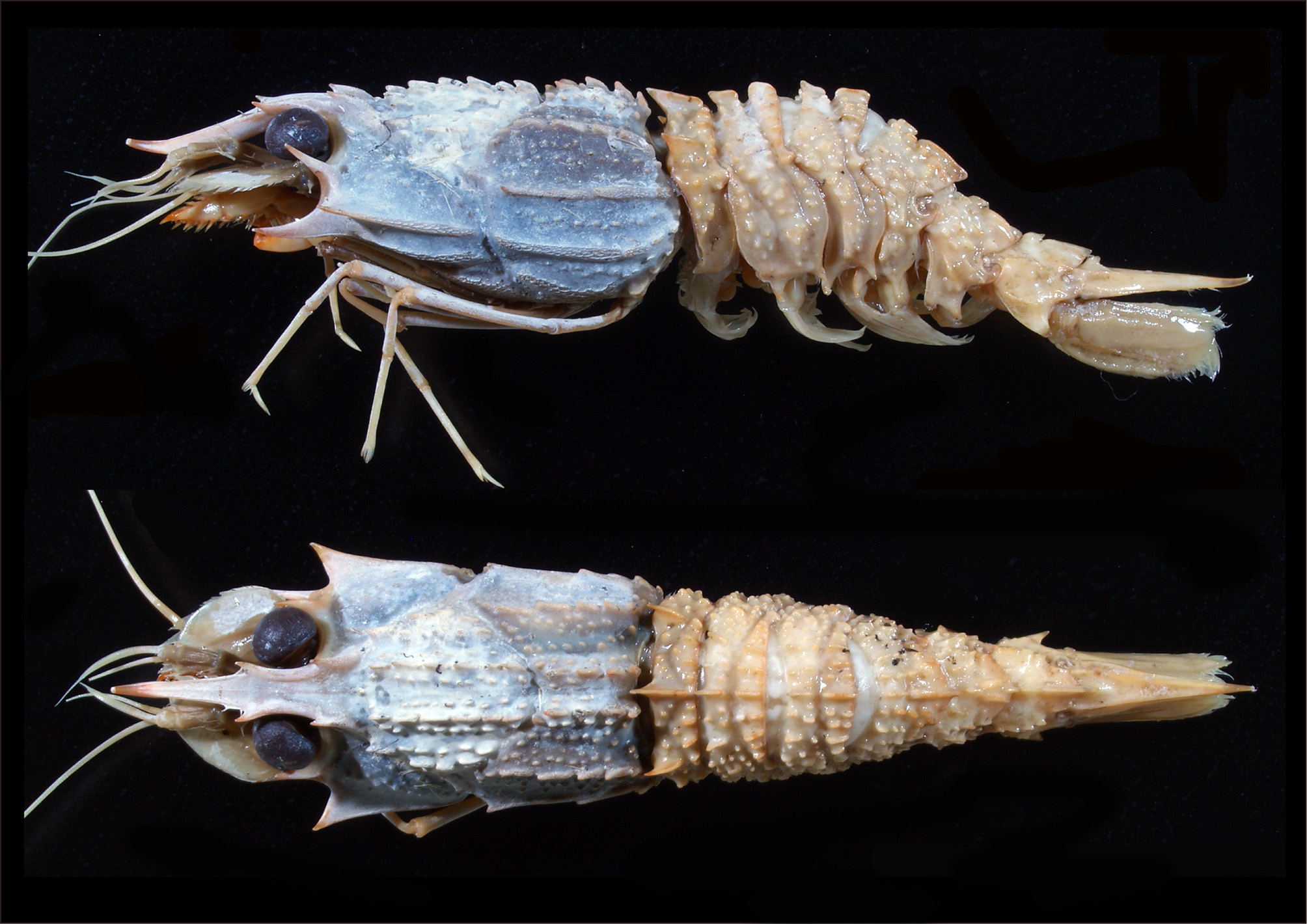
Figs 3–8 View FIGURE 3 View FIGURE 4 View FIGURE 5 View FIGURE 6 View FIGURE 7 View FIGURE 8
Glyphocrangon smithii wood-Mason & Alcock, 1891 View in CoL b: 357 (type locality: Bay of Bengal, near Andaman Islands, 561 fathoms).—wood-Mason 1894: pl. 7, fig. 3, 3a.— Alcock 1901: 129.— Chace 1984: 6 (key, in part).— Komai 2004: 583.
Not Glyphocrangon smithii: Calman 1939: 216 View in CoL [= Glyphocrangon boletifera Komai 2004 View in CoL (material from the Gulf of Aden ); Glyphocrangon amblytes Komai, 2004 View in CoL (material from the Maldives)].
Not Glyphocrangon smithii: Monod 1973: 124 View in CoL , fig. 34. (= G. armata Komai, 2004 View in CoL ).
Not Glyphocrangon smithii: Hayashi 1986: 149 View in CoL , 279, fig. 99.— Miya 1995: 194 (in part).— Takeda 1997: 250, pl. 3, fig. G.— Sakaji 2001: 211, pl. 2, fig. E. (= G. perplexa Komai, 2004 View in CoL ).
Material examined. Andaman Sea, “Dr. Fridtjof Nansen”, stn 135, 12°36.63’N, 96°62.18’E, 523 m, 23 May 2015, beam trawl, 1 ovigerous female (cl 31.5 mm), NTOU M02041 View Materials .
Description. Body ( Fig. 3 View FIGURE 3 ) moderately robust; integument firm, entirely glabrous.
Rostrum ( Figs 4 View FIGURE 4 A, B; 7A, B) descending at base and upturned distally, gradually tapering in dorsal view, 0.6 times as long as carapace, armed with 2 pairs of spines on dorsolateral ridges; anterior pair located at 0.7 of rostral length, proximal pair just above orbital margin; dorsal margin of proximal spine evenly convex; dorsal surface faintly rugose in distal half (middorsal ridge hardly discernible in proximal 0.3 of rostrum); dorsolateral ridges between 2 lateral spines strongly raised, bluntly carinate, those anterior to lateral spines weakly elevated, sharply carinate; ventral surface sulcate medially in proximal two-thirds, distal one-third nearly flat, ventrolateral margins bluntly carinate.
Carapace ( Fig. 4 View FIGURE 4 A, B) 1.3 times as long as width between tips of anterior spines of posterior third carinae; surfaces of carinae and tubercles etched with reticulate pattern of minute pits; major carinae moderately high; anterior and cervical grooves deep. Anterior first (submedian) carina composed of 6 tubercles, these tubercles forwardly directed with blunt tips, compressed laterally ( Fig. 7 View FIGURE 7 C); posterior first carina composed of 4 forwardly directed tubercles, first tubercle strongest, subacute, others bluntly pointed ( Fig. 7 View FIGURE 7 C). Anterior second (intermediate) carina composed of 4 tubercles, anteriormost one larger than others, subacute, others blunt ( Fig. 7 View FIGURE 7 C); posterior second carina slightly arched in lateral view, divided into 4 lobes and 1 small tubercle (none sharply pointed) ( Fig. 7 View FIGURE 7 C). Anterior third (antennal) carina confined to antennal spine; posterior third carina with 2 blunt tubercles on posterior part, terminating anteriorly in small spine directed anteriorly ( Fig. 7 View FIGURE 7 D); distance between tips of anterior teeth 0.9 of distance between tips of acute laminae of anterior fourth carinae. Anterior fourth (lateral) carina expanded into single moderately large, vertically compressed, acute lamina just reaching sinus between antennal and branchiostegal spines, distance between tips of laminae 0.9 times of carapace length; posterior fourth carina almost entire, followed by 1 blunt tubercles posteriorly, anterior end bluntly angled. Anterior fifth (sublateral) carina well defined; posterior fifth carina low, broad, surface roughly eroded, posteriorly followed by small tubercles. Sixth (submarginal) carina represented by low, broad elevation with roughly eroded surface. Orbital margin slightly elevated; postorbital region shallowly depressed, unarmed. Intercarinal tubercles tiny, conspicuous, none spiniform. Median part of gastric region with 2 longitudinal rows of tubercles and anteromedian tubercle with forwardly directed tip; posteromedian part also with 2 rows of tubercles. Lateral part of gastric region with space between first and second carinae having tubercles arranged in 2 longitudinal rows; space between second carina and lateral groove with 4 tiny tubercles. Posterior dorsolateral region with many tubercles arranged in 3 irregular longitudinal rows ( Fig. 7 View FIGURE 7 C). Upper part of hepatic region weakly convex, with scattered tubercles ( Fig. 7 View FIGURE 7 E); lower part of hepatic region shallowly concave, unarmed. Upper part of branchial region bearing about 30 tiny tubercles ( Fig. 7 View FIGURE 7 D); middle part with about 10 tubercles arranged in single longitudinal row; lower part with about 10 tubercles. Antennal spine strong, ascending in lateral view (angle about 45° against horizontal plane of carapace), slightly curved inward in dorsal view, falling far short of distal corneal margin. Branchiostegal spine very strong, directed anteriorly and slightly curving dorsally in lateral view, slightly curving laterally in ventral view, not reaching midlength of antennal scale, hardly visible in dorsal view; lateral face without ridges or carinae, but sharply carinate ventrally. Marginal posterolateral corner slightly produced, forming light angle.
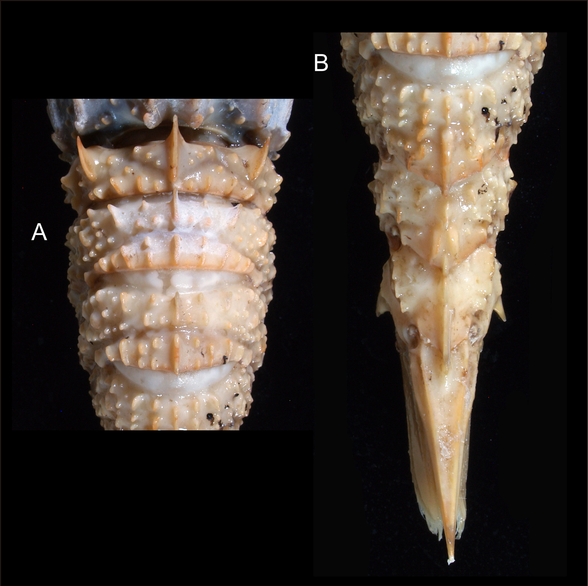
Pleon ( Figs 5 View FIGURE 5 A, B; 6A, B) covered with blunt or distally rounded, conspicuous tubercles. Somite 1 with middorsal elevation defined by deep transverse groove, with 3 tubercles arranged in obliquely transverse row on either side of middorsal carina; middorsal carina forming large, strongly compressed tooth with forwardly directed, acute apex; posterior part of tergum with 4 or 5 laterally compressed tubercles on either side of midline. Dorsolateral carina developed into strong tooth with forwardly directed, acute apex. Lateral carina composed of 2 blunt tubercles. Pleuron ornamented anteriorly with tubercles; posterior depression abruptly delimited; anteroventral corner produced anteriorly, subacutely pointed.
Pleomeres 2–4 with median carina strongly compressed laterally, each deeply divided into 2 parts. Tubercles or prominences composing submedian or dorsolateral carinae strongly compressed laterally; surfaces of carinae and tubercles weakly etched with minute pits. Posterior transverse grooves all deep. Vertical ridges on pleura 3 and 4 terminating ventrally in small blunt tubercle. Pleural teeth moderately strong, acute, slightly unequal. Pleomere 2 with anterior part of median carina somewhat produced anteriorly, posterior part entirely rounded. Pleomere 3 with anterior part of median carina slightly produced anteriorly, posterior part subrectangular. Pleomere 4 with anterior part of median carina much shorter than posterior part, subrectangular; posterior part bearing median groove, produced posteriorly in blunt, triangular tooth.
Pleomere 5 with anterior median carina posteriorly directed, non-spiniform; posterior median carina produced posteriorly in rounded lobe. Tergum with shallow oblique groove; anterior submedian carinae represented by small, laterally compressed tubercle with posteriorly directed apex; posterior submedian carinae diverging posteriorly, abruptly narrowed posteriorly, each with median groove. Pleuron with 2 strong, slightly unequal ventral teeth.
Pleomere 6 with crested median carina, divided into 2 parts by narrow notch, posterior part produced posteriorly into blunt tooth. Tergum with 1 minute tubercle anteriorly. Lateral carina composed of 4 unequal, somewhat compressed tubercles, none spiniform. Pleuron with blunt carina laterally; posteroventral tooth strong, flared laterally, without supporting carina on lateral face.
Telson ( Figs 5 View FIGURE 5 ; 6; 7F) 0.7 times as long as carapace; tapering to acute apex; anterodorsal projection prominent, laterally compressed, with posteriorly directed, blunt apex, without accessory tubercle. Dorsolateral and ventrolateral carinae sharply defined, almost smooth.
Cornea ( Fig. 4 View FIGURE 4 A, B) large (width 0.22 of carapace length), darkly pigmented even in preservative. Ocular peduncle with minute papilla-like process on anteromesial face (not illustrated).
Antennular peduncle ( Fig. 4 View FIGURE 4 A, B) slightly overreaching anterior margin of antennal scale; article 2 subcylindrical, about 2 times longer than wide; aesthetasc-bearing portion of outer flagellum 0.25 times as long as carapace.
Antennal scale ( Figs 4 View FIGURE 4 A, B; 7G) oval, 1.6 times as long as wide, without even trace of lateral tooth; lateral margin entirely setose; lateral part of dorsal surface slightly rugose. Carpocerite falling short of anterior margin of antennal scale.
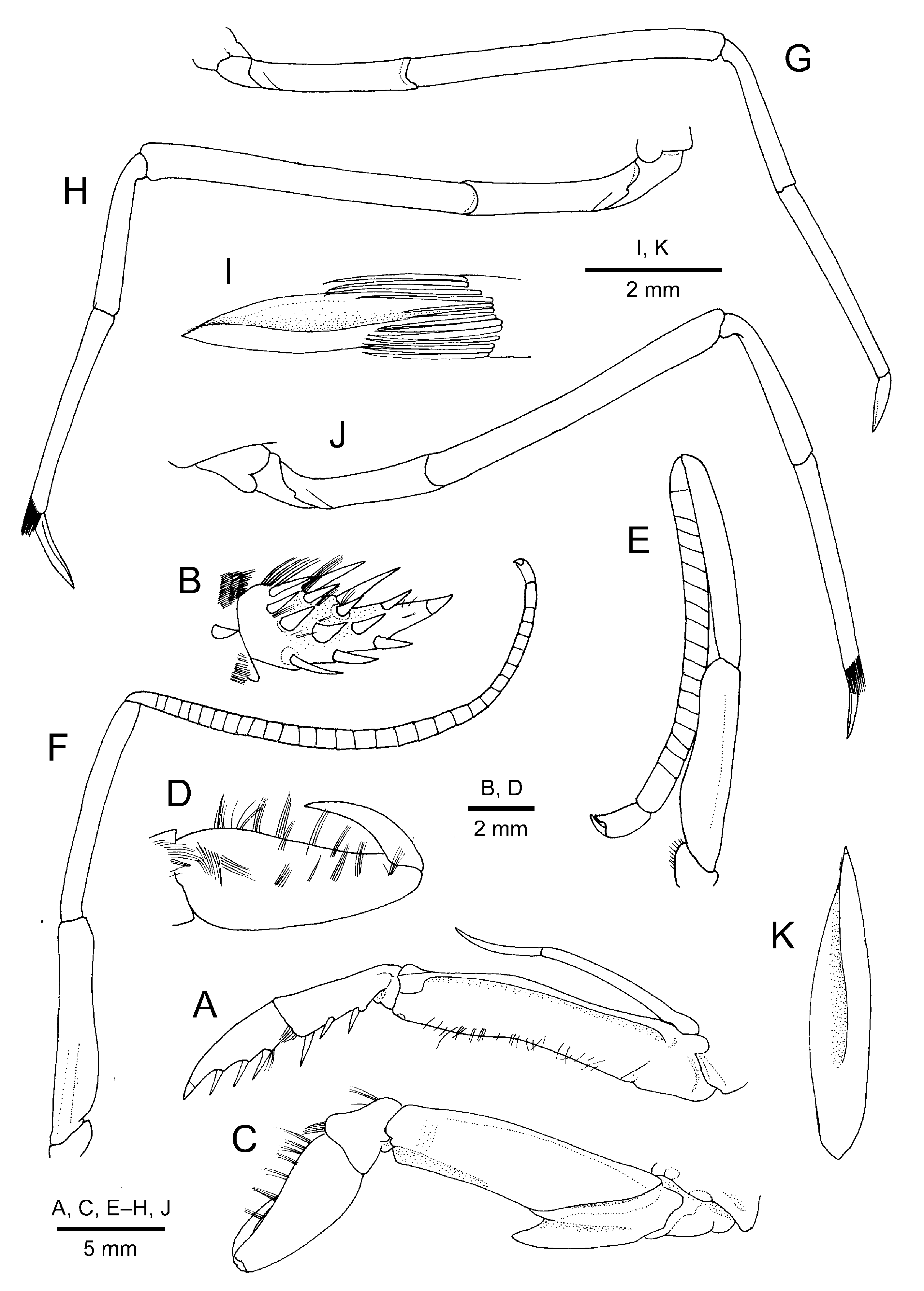
Maxilliped 3 endopod ( Fig. 8 View FIGURE 8 A) moderately stout, slightly overreaching anterior margin of antennal scale. Ultimate article tapering in acute unguis demarcated basally, with several strong spiniform setae on margins and mesial face ( Fig. 8 View FIGURE 8 B). Penultimate article with 1 spiniform seta on inner surface distally and 3 spiniform setae on ventrolateral margin. Antepenultimate article with dorsolateral margin sharply carinate. Exopod not reaching midlength of antepenultimate article; flagellum not reaching distal end of antepenultimate article.
Pereopod 1 ( Fig. 8 View FIGURE 8 C) slightly overreaching midlength of antennal scale. Flexed dactylus reaching midlength of palm; palm glabrous (without short pubescence), with longitudinal row of tufts of stiff setae on upper surface ( Fig. 8 View FIGURE 8 D). Carpus short, cup-shaped, Merus with 2 blunt longitudinal ridges dorsally and ventrally on lateral surface. Ventral lamina of ischium subacutely pointed distally.
Pereopods 2 ( Fig. 8 View FIGURE 8 E, F) unequal. Left ( Fig. 8 View FIGURE 8 E) overreaching antennal scale by length of chela; chela as wide as distalmost article of carpus, fingers with distinct hiatus proximally; carpus shorter than merus and ischium combined, divided into 18 articles. Right ( Fig. 5 View FIGURE 5 F) longer and slenderer than left, overreaching antennal scale by half length of carpus; chela small, narrower than distalmost carpal article; carpus subequal in length to merusischium combined, divided into 30 articles.
Pereopod 3 ( Fig. 8 View FIGURE 8 G) overreaching anterior margin of antennal scale by length of dactylus and 0.2 of propodus; dactylus compressed laterally, 0.3 times as long as propodus; propodus slightly narrowing distally; carpus 0.7 times as long as propodus.
Pereopod 4 ( Fig. 8 View FIGURE 8 H, I) moderately slender, overreaching anterior margin of antennal scale by length of dactylus. Dactylus ( Fig. 8 View FIGURE 8 I) subspatulate, 0.4 times as long as propodus, terminating in acute tip; extensor surface with deep longitudinal groove along midline, and with submarginal row of minute setae adjacent to lateral margin in distal one-fourth. Propodus slightly narrowing distally, with tuft of stiff bristle-like setae on distal margin partially covering base of dactylus. Carpus 0.8 times as long as propodus.
Pereopod 5 ( Fig. 8 View FIGURE 8 J, K) similar to fourth pereopod, falling slightly short of anterior margin of antennal scale. Dactylus ( Fig. 8 View FIGURE 8 K) 0.35 times as long as propodus, shorter than that of fourth pereopod; extensor surface also with deep median groove. Carpus 0.8 times as long as propodus.
Pleopods and uropod ( Fig. 5 View FIGURE 5 A, B) typical of genus, without distinguishing features.
Eggs 3.0 x 2.7 mm.
Colour in life. Described as bright crimson by Wood-Mason & Alcock (1891b) and Alcock (1901).
Remarks. Glyphocrangon smithii was originally described on the basis of two male specimens (syntypes) collected from the Bay of Bengal near the Andaman Islands , at a depth of 561 fathoms (about 1010 m) (Wood- Mason & Alcock 1891b). Illustration of a habitus in the dorsal and lateral views of G. smithii was subsequently published in 1894 ( Wood-Mason 1894: plate 7, fig. 3, 3a). Alcock (1901) briefly described the species with additional specimens from the Andaman Sea at depths of 188–220 fathoms (= 338–396 m) and the Arabian Sea , off the Maldives, at a depth of 459 fathoms (= 826 m). Since then, there have been several records under this name from various Indo-West Pacific localities, viz., the Gulf of Aden and the Maldives ( Calman 1939), New Caledonia ( Monod 1973) and Japan ( Hayashi 1986; Miya 1995; Takeda 1997; Sakaji 2001). In a taxonomic review of Glyphocrangon, Komai (2004) argued that none of the records outside the eastern Indian Ocean does represent G. smithii , although the type or topotypic materials were not available at the time. He tried to diagnose G. smithii on the basis of the type description by Wood-Mason & Alcock (1891b) and the published illustration by Wood-Mason (1894), and referred the species to the G. regalis Spence Bate, 1888 species complex. One of the two specimens from Maldives, studied by Calman (1939), was provisionally referred to G. amblytes ; Calman’s (1939) specimens from the Gulf of Aden were described as a new species, G. boletifera Komai, 2004 ; Monod’s (1973) specimens were re-identified with G. armata Komai, 2004 ; Japanese records of G. smithii (cf. Hayashi 1986; Miya 1995; Takeda 1997; Sakaji 2000) were all referred to G. perplexa Komai, 2004 ( Komai 2004) .
The present specimen, collected from the Andaman Sea, agrees well with the rather brief descriptions of G. smithii by Wood-Mason & Alcock (1891b) and Alcock (1901), and is identified with little hesitation with the species. Examination of the present topotypic specimen clarified the diagnostic features of G. smithii , which have remained to be reassessed. In the presence of a small anterior spine on the posterior third carina on the carapace and the less developed, tiny tubercles on the intercarinal spaces of the carapace, G. smithii closely resembles G. amblytes Komai, 2004 (western Indian Ocean) and G. wagini Burukovsky, 1990 (Sala y Gomez Ridge, eastern Pacific), as Komai (2004) discussed. Glyphocrangon smithii , however, appears characteristic in having a very deep longitudinal groove on the dorsal surface of the pereopod 4 and 5 dactyli. In the other species of the G. regalis species complex, not only in G. amblytes and G. wagini , the grooves or depressions of the pereopod 4 and 5 dactyli are not so deep as in G. smithii (cf. Komai 2004; 2011 l; Komai & Chan 2013). Komai (2004) interpreted from the descriptions by Wood-Mason & Alcock (1891) and Alcock (1901) that the dorsal surface of the rostrum is smooth in G. smithii , but it is not the case. In the present specimen, the dorsal surface of the rostrum is faintly rugose in the distal half, though indeed the development is much weaker than in the other species in the G. regalis species complex ( Komai 2004) except for G. wagini .
Glyphocrangon amblytes further differs from G. smithii in the following particulars (see also Komai 2004): (1) the dorsal surface of the rostrum has more clearly defined transverse septa in G. amblytes ; (2) intercarinal tubercles on the carapace are weaker in G. amblytes than in G. smithii ; (3) the posterior third carina on the carapace is smooth in G. amblytes , while there are two blunt tubercles in the posterior part in G. smithii ; (4) the acute laminae of the anterior fourth carinae is better developed in G. amblytes than in G. smithii , of which the distance between tips being equal or slightly greater than the carapace length in G. amblytes , rather than smaller in G. smithii (0.9 times of the carapace length); (5) the middorsal carina on the pleomeres 1–4 is lower in G. amblytes than in G. smithii ; (6) tubercles on the pleon are also smaller and lower in G. amblytes than in G. smithii .
Glyphocrangon wagini View in CoL differs from G. smithii View in CoL in the following features ( Burukovsky 1990; Komai 2004): (1) the dorsal margin of the posterior pair of the lateral rostral spines is distinctly sinuous in G. wagini View in CoL , rather than evenly convex in G. smithii View in CoL ; (2) the anterior terminal tooth of the posterior third carina on the carapace is directed laterally in G. wagini View in CoL , but anteriorly in G. smithii View in CoL ; (3) acute laminae of the anterior fourth carina is more strongly expanded in G. wagini View in CoL , of which the distance of tips is greater than the carapace length (1.13 times of the carapace length); (4) the cornea is comparatively small in G. wagini View in CoL than in G. smithii View in CoL (corneal width 0.17 times of the carapace length versus 0.22 times);
Amongst the species described from the northeastern part of the Indian Ocean, G. caeca Wood-Mason in Anonymous, 1891 View in CoL , G. cerea Alcock & Anderson, 1894 View in CoL , and G. unguiculata Wood-Mason, in Wood-Mason & Alcock, 1891b View in CoL remain to be reassessed based on examination of the type or topotypic material (see Komai 2004, 2006).
Distribution. Known with certainty from the eastern Indian Ocean: Bay of Bengal and Andaman Sea; at depths of 338–1010 m.


No known copyright restrictions apply. See Agosti, D., Egloff, W., 2009. Taxonomic information exchange and copyright: the Plazi approach. BMC Research Notes 2009, 2:53 for further explanation.
|
Kingdom |
|
|
Phylum |
|
|
Class |
|
|
Order |
|
|
Family |
|
|
Genus |
Glyphocrangon smithii Wood-Mason & Alcock, 1891
| Komai, Tomoyuki & Chan, Tin-Yam 2017 |
Glyphocrangon smithii:
| Sakaji 2001: 211 |
| Takeda 1997: 250 |
| Miya 1995: 194 |
| Hayashi 1986: 149 |
Glyphocrangon smithii:
| Monod 1973: 124 |
Glyphocrangon smithii wood-Mason & Alcock, 1891
| Komai 2004: 583 |
| Chace 1984: 6 |
| Alcock 1901: 129 |