
Reinhardorhynchus riae Diez, Reygel & Artois, 2021
|
publication ID |
https://doi.org/ 10.11646/zootaxa.4948.4.1 |
|
publication LSID |
lsid:zoobank.org:pub:44061E80-81B7-46AF-AD51-9B461C2E2B67 |
|
DOI |
https://doi.org/10.5281/zenodo.4669986 |
|
persistent identifier |
https://treatment.plazi.org/id/65D70F61-74DB-458B-B5EA-18AB0F972270 |
|
taxon LSID |
lsid:zoobank.org:act:65D70F61-74DB-458B-B5EA-18AB0F972270 |
|
treatment provided by |
Plazi |
|
scientific name |
Reinhardorhynchus riae Diez, Reygel & Artois |
| status |
sp. nov. |
Reinhardorhynchus riae Diez, Reygel & Artois sp. n.
( Fig. 7–8 View FIGURE 7 View FIGURE 8 )
urn:lsid:zoobank.org:act:65D70F61-74DB-458B-B5EA-18AB0F972270
Material and distribution. Observations on live specimens. Three whole mounts, one designated holotype ( FMNH https://id.luomus.fi/ KV.651), the others reference material (HU XIII.3.33– XIII.3.34), and one serially-sectioned specimen (HU XIII.3.35) collected in Mala (Lanzarote, Canary Islands ) (29°05’01”N; 13°26’59”W), in front of “Cuevita de Mala” (October 10, 2011), sand patch under loose macroalgae, coarse shell gravel, very clean, 12 m deep (Type Locality). One whole mount from the same locality (HU XIII.3.36) (October 8, 2011), medium-fine, calcareous sand from a large parch among rocks, poorly-oxygenated redox layer just below surface, 20 m deep. Four whole mounts and seven serially-sectioned specimens (HU XIII.3.37– XIII.3.47) collected in a sheltered beach, a bit south of Orzola (Lanzarote, Canary Islands ) (29°13’23”N; 13°27’05”W) (October 6, 2011), medium-coarse sand, with holes from burrowing animals, taken at low tide, just below the water line. One whole mount (HU XIII.3.48) collected at the same locality (October 7, 2011), sample taken 0.4–0.5 m deep, coarse sand with lava rocks scattered around. Salinity 35 ‰ in all the localities. One whole mount (HU XIII.3.49) from Punta Negra (40°57’12”N, 08°13’43”E), Stintino, Sardinia, Italia (September 2018), on silty algae, 0.5 m deep, salinity 40 GoogleMaps ‰.
Etymology. Species dedicated to Ria Vanderspikken (Hasselt University), in acknowledgement of all her help in organising the sampling campaigns, archiving of literature and taking care of the HU specimen collection.
Diagnosis. Species of Reinhardorhynchus gen. n. with a copulatory organ armed with two transverse spiny belts, a penis papilla, and two distal hooks. Prostate vesicle enclosed in a muscular bulb. This bulb ends in a pseudocuticular plate and is armed with two spiny rows being ±72 μm and ±55 μm long, respectively. Spines ±3 μm long at the sides and ±9 μm long in the middle of the rows. Penis papilla covered by a pseudocuticula, which carries the distal hooks. Hooks flattened, with a broad and rounded distal end, ±14 μm long and ±32 μm wide at their base. Female duct bipartite: proximal compartment lined by a nucleated epithelium and without muscles, distal compartment surrounded by a thick layer of circular muscles.
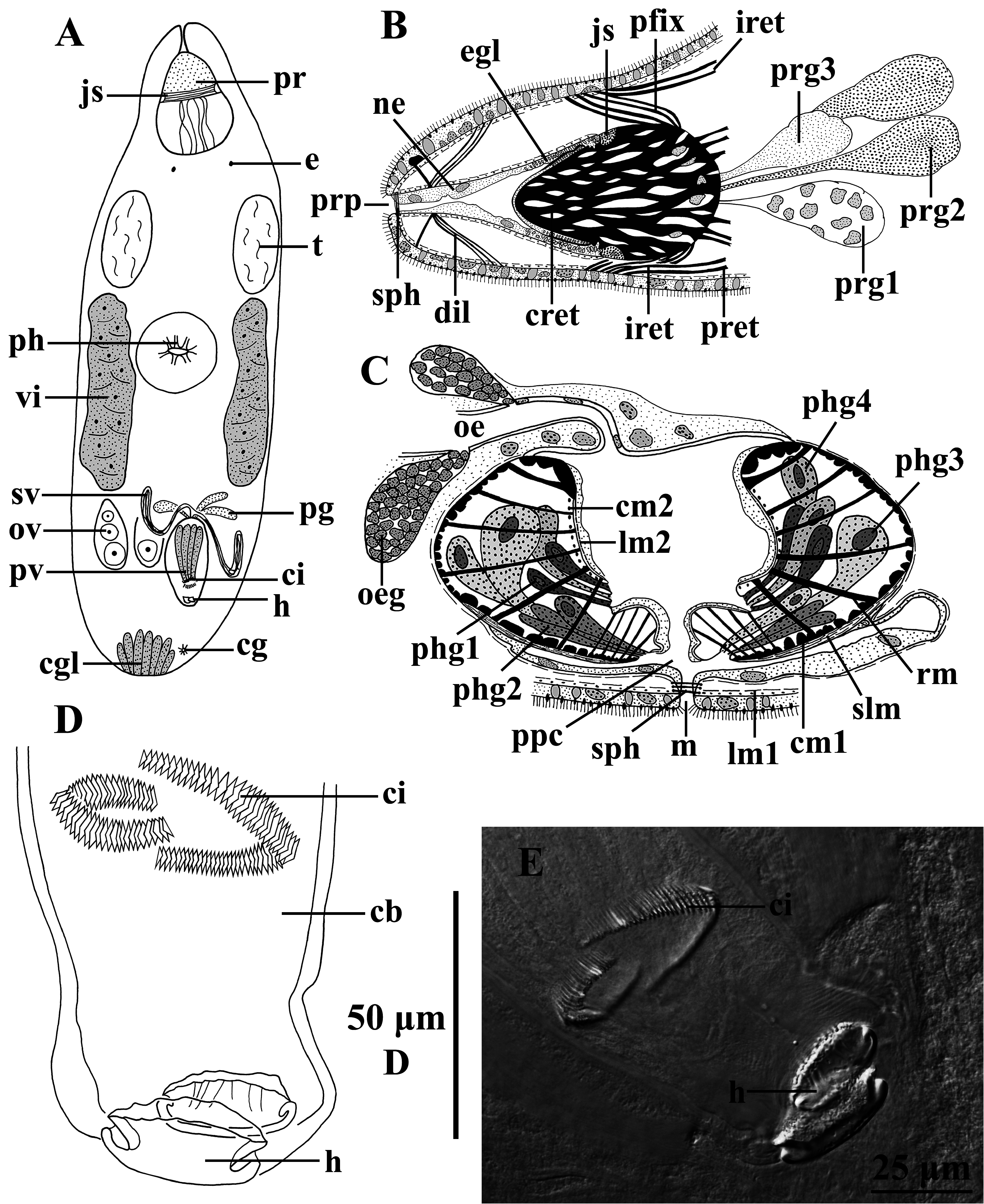
Description. The specimens are 1.8–3 mm long (x̄ = 2.2 mm; n = 8), translucent, with a pair of eyes ( Fig. 7A View FIGURE 7 : e). The coloration of the specimens is due to pinkish glands within the parenchyma. The syncytial epidermis is 7–8 µm thick and completely ciliated; cilia ± 4 µm long. The epidermis contains many vacuoles, which are either empty or filled with a dark secretion (coarse- or fine-grained). The 1–2 µm-long rhabdites are located near the apical surface of the epidermis. Caudally in the body some eosinophilic glands occur ( Fig. 7A View FIGURE 7 & 8C View FIGURE 8 : cgl).
The proboscis ( Fig. 7A View FIGURE 7 : pr, 7B, 8A) is ±15% of the body length and is of the characteristic koinocystidid construction (see Brunet 1972; Karling 1980), displaying a strong juncture sphincter (7A–B: js). The proboscis sheath is surrounded by a nucleated epithelium ( Fig. 7B View FIGURE 7 : ne), which is continuous with the epithelium surrounding the proboscis cone. Both epithelia contain oval to circular-shaped glands (reddish stained) ( Fig. 7B View FIGURE 7 & 8A View FIGURE 8 : egl). The epithelium surrounding the cone is lined by a brush border ( Fig. 8A View FIGURE 8 : bb). Three kinds of glands open through the caudal wall of the proboscis: coarse-grained basophilic ones (dark stained) ( Fig. 7B View FIGURE 7 : prg1), coarse-grained eosinophilic ones (pinkish stained) ( Fig. 7B View FIGURE 7 : prg2), and fine-grained eosinophilic ones (brownish stained) ( Fig. 7B View FIGURE 7 :prg3). The proboscis sheath is surrounded by a layer of circular muscles and a longitudinal one just underneath it. The pro- boscis pore ( Fig. 7B View FIGURE 7 & 8A View FIGURE 8 : prp) is surrounded by a sphincter ( Fig. 7B View FIGURE 7 : sph). The cone retractors are well developed ( Fig. 7B View FIGURE 7 & 8A View FIGURE 8 : cret). The exact number of proboscis fixators ( Fig. 7B View FIGURE 7 : pfix) and dilatators ( Fig. 7B View FIGURE 7 : dil) could not be determined. Two pairs of proboscis retractors ( Fig. 7B View FIGURE 7 : pret) could be distinguished, however it is not clear if there are more. Two pairs of integument retractors were observed: a ventral and a dorsal one ( Fig. 7B View FIGURE 7 : iret).
The pharynx ( Fig. 7A View FIGURE 7 : ph, 7C) has a diameter of 15% of the body length in the live specimens, situated at 40%. The prepharyngeal cavity ( Fig. 7C View FIGURE 7 : ppc) is lined by a nucleated epithelium and surrounded by an external layer of longitudinal muscles. The mouth ( Fig. 7C View FIGURE 7 : m) is surrounded by a sphincter ( Fig. 7C View FIGURE 7 : sph). Four types of glands containing a coarse-grained secretion open into the pharynx lumen: dark brown ( Fig. 7C View FIGURE 7 : phg1) and pinkish ( Fig. 7C View FIGURE 7 : phg2) eosinophilic glands most distally, and brownish eosinophilic ( Fig. 7C View FIGURE 7 : phg3) and basophilic glands ( Fig. 7C View FIGURE 7 : phg4) more proximally. Coarse-grained basophilic glands (Minot’s glands) ( Fig. 7C View FIGURE 7 : oeg) open into the oesophagus ( Fig. 7C View FIGURE 7 : oe). The musculature of the pharynx consists of a longitudinal muscle layer outside of the septum ( Fig. 7C View FIGURE 7 : lm1) and a circular layer just inside of it ( Fig. 7C View FIGURE 7 : cm1). These circular muscles are markedly thicker near the proximal and distal tips of the pharynx. The distal opening of the pharynx is lined by a thick layer of longitudinal muscles, which in sagittal section gives the impression of forming a lip-like structure ( Fig. 7C View FIGURE 7 : slm). The pharynx lumen is surrounded by an inner circular ( Fig. 7C View FIGURE 7 : cm2) and outer longitudinal muscle layer ( Fig. 7C View FIGURE 7 : lm2). Radial muscles ( Fig. 7C View FIGURE 7 : rm) stretch between the internal and the external walls; the most proximal of these are weaker than the others.
Two testes ( Fig. 7A View FIGURE 7 : t) occur latero-rostrally from the pharynx. Caudally from the pharynx, the vasa deferentia form a pair of seminal vesicles ( Fig. 7A View FIGURE 7 & 8C View FIGURE 8 : sv). The seminal vesicles are lined by a low, nucleated epithelium and surrounded by an external, longitudinal muscle layer. The seminal vesicles fuse to form a seminal duct just before opening into the copulatory bulb ( Fig. 7D View FIGURE 7 : cb), which is located in the caudal body half and represents 15% of the body length in the live specimens. It encompasses the prostate vesicle ( Fig. 7A View FIGURE 7 & 8C View FIGURE 8 : pv), the cirrus ( Fig. 7A & 7D–E View FIGURE 7 : ci), and two accessory hooks ( Fig. 7A, 7D–E View FIGURE 7 & 8C View FIGURE 8 : h). The copulatory bulb ends in a penis papilla (8B–C: pp) and is surrounded by a thick sheath of longitudinal muscles ( Fig. 8B–C View FIGURE 8 : lm1). Distally, this muscular sheath is not connected to the penis papilla. A second, internal muscular sheath connects to the proximal part of the prostate vesicle and ends in the penis papilla. This sheath consists of a longitudinal muscle layer ( Fig. 8B–C View FIGURE 8 : lm2) and an oblique one just beneath it ( Fig. 8B–C View FIGURE 8 : om1). The most distal part of the copulatory bulb is lined by a thin, nucleated epithelium ( Fig. 8C View FIGURE 8 : ne), which becomes much thicker and is covered by a sclerotised layer, as a whole forming the penis papilla. The two distal hooks are connected to the distal end of the penis papilla.
The extracapsular prostate glands ( Fig. 7A View FIGURE 7 & 8C View FIGURE 8 : pg) open proximally into the copulatory bulb. The prostate vesicle forms a long-drawn muscular bulb. Proximally, it is surrounded by strong oblique muscles ( Fig. 8B–C View FIGURE 8 : om2), while over the rest of its length the prostate vesicle is surrounded by an external longitudinal ( Fig. 8B–C View FIGURE 8 : lm3) and internal oblique muscle layer ( Fig. 8C View FIGURE 8 : om3). The ejaculatory duct ( Fig. 8B–C View FIGURE 8 : ed) enters the prostate vesicle through the latter’s proximal end and runs axially through it. The duct is lined by a thin nucleated epithelium and surrounded by longitudinal muscles. There are four types of prostate glands: a coarse-grained basophilic one (stained dark purple) ( Fig. 8B View FIGURE 8 : pg1), two coarse-grained eosinophilic ones [stained greenish ( Fig. 8B View FIGURE 8 : pg2) and reddish ( Fig. 8B View FIGURE 8 : pg3), respectively], and an eosinophilic, fine-grained one (stained pinkish) ( Fig. 8B View FIGURE 8 : pg4). Distally, these prostate glands open around the distal opening of the ejaculatory duct. The distal tip of the prostate vesicle is covered by a sclerotised layer and armed with teeth, constituting the two spiny rows observed in the live specimens and the whole mounts.
The spiny rows ( Fig. 7D–E View FIGURE 7 & 8C View FIGURE 8 : ci) are oriented transversally relative to the copulatory bulb. The larger row is 66–83 μm long (x̄ = 72 μm; n = 7), the smaller one 46–65 μm (x̄ = 55 μm; n = 7). The triangular spines are small- est at the sides of both rows: 2–4 μm long (x̄ = 3 μm; n = 20), compared to 7–12 μm long (x̄ = 9 μm; n = 35) in the middle. The distal hooks ( Fig. 7A, 7D–E View FIGURE 7 & 8C View FIGURE 8 : h) are similar in length and shape: flattened, distally broad and rounded, 11–20 μm long (x̄ = 14 μm; n = 10) and 27–37 μm wide at their base (x̄ = 32 μm; n = 10). The male atrium ( Fig. 8C View FIGURE 8 : ma) is lined by a low, nucleated epithelium and surrounded by longitudinal muscles. Proximally from the aperture of the ejaculatory duct, the male atrium shows a small, lightly-sclerotised papilla ( Fig. 8C View FIGURE 8 : pa). The male atrium opens into the common general atrium ( Fig. 8C View FIGURE 8 : ca) through the latter’s rostral wall.
The vitellaria ( Fig. 7A View FIGURE 7 & 8C View FIGURE 8 : vi) extend along both sides of the body from the level of the testes to the copulatory bulb. The ovaries ( Fig. 7A View FIGURE 7 & 8C View FIGURE 8 : ov) are located antero-laterally from the copulatory bulb. The oocytes are organised in a row, increasing in diameter from the most proximal to the most distal one. The most distal oocytes display a number of unidentified black spots ( Fig. 8C View FIGURE 8 : os). The oviducts ( Fig. 8C View FIGURE 8 : od) are short and not surrounded by muscles. The female duct is bipartite. The proximal part ( Fig. 8C View FIGURE 8 : fd1) is surrounded by a nucleated epithelium, lacks muscles, and narrows towards the second, more distal part ( Fig. 8C View FIGURE 8 : fd2). The distal part is lined by a membranous, anucleated epithelium and surrounded by thick, circular muscles. The proximal part receives the broad common vitelloduct and the oviducts and is filled with vitelline material. The distal part contains sperm, hence functioning as seminal receptacle and connects to the female atrium through a strong sphincter ( Fig. 8C View FIGURE 8 : sph1). The female atrium ( Fig. 8C View FIGURE 8 : fa) also receives the bursal stalk ( Fig. 8C View FIGURE 8 : bs), which enters just dorsal to the female duct. The bursa ( Fig. 8C View FIGURE 8 : b) is surrounded by an internal longitudinal and external circular muscle layer which continue around the bursal stalk and further around the female atrium. It is lined by a nucleated epithelium, which disappears around the bursal stalk. At the transition between bursa and bursal stalk, a sphincter occurs ( Fig. 8C View FIGURE 8 : sph2). The bursa contains disintegrating sperm and different kinds of glandular material in degradation. The female atrium also contains sperm and enters the common genital atrium just dorsal to the opening of the male atrium. The uterus ( Fig. 8C View FIGURE 8 : ut) enters the common genital atrium through the latter’s rostral wall, ventral to all other systems. The uterus is lined by a nuclear epithelium and is surrounded by a longitudinal muscle layer. Medium-grained eosinophilic uterine glands open into the uterus just proximal to the opening of the female duct ( Fig. 8C View FIGURE 8 : ueg). The uterus does not show any sphincters. The common genital atrium is surrounded by longitudinal muscles. An epithelium was not observed and is probably membranous. The common genital atrium opens ventrally and subcaudally through the common gonopore ( Fig. 7A View FIGURE 7 & 8C View FIGURE 8 : cg), which is at 95%. This gonopore is surrounded by a sphincter ( Fig. 8C View FIGURE 8 : sph3). One live specimen carried two embryos ( Fig. 8B View FIGURE 8 : em) not surrounded by a thick egg-shell, suggesting the species is (ovo-)viviparous.

| FMNH |
Field Museum of Natural History |
No known copyright restrictions apply. See Agosti, D., Egloff, W., 2009. Taxonomic information exchange and copyright: the Plazi approach. BMC Research Notes 2009, 2:53 for further explanation.
|
Kingdom |
|
|
Phylum |
|
|
Class |
|
|
Order |
|
|
Family |
|
|
Genus |