
Hydrobius pauper Sharp, 1884
|
publication ID |
https://doi.org/ 10.5281/zenodo.4272324 |
|
DOI |
https://doi.org/10.5281/zenodo.4334982 |
|
persistent identifier |
https://treatment.plazi.org/id/039A87CB-FFCE-4929-FE3D-FDA7FCF4ED4A |
|
treatment provided by |
Felipe |
|
scientific name |
Hydrobius pauper Sharp, 1884 |
| status |
|
Hydrobius pauper Sharp, 1884 View in CoL
( Figs. 1G View Fig , 6C View Fig , 46–50 View Fig View Fig View Fig View Fig View Fig )
Material examined. JAPAN: HOKKAIDÔ: 2 L2, 1 L3, Sarobetsu-gen-ya, Toyotomi-chô , 12.–13.vii.2008, YM ; 1 L2, Shinoro, Sapporo-shi , 26.vi.2009, YM .
General morphology. Third instar. Body rather slender, widest between abdominal segments 3 and 4 ( Figs. 1G View Fig , 6C View Fig ). Colour ( Fig. 1G View Fig ). Dorsal surface of head light brown, darker in posterolateral part, paler in anterolateral part. Proscutum light brown medially and laterally; dorsal surface of meso- and metathorax, and abdominal segments with two darker brown longitudinal lines, brown to light brown on sclerites, greyish white on membranous parts, paler medially. Ventral surface of thorax and abdomen greyish white, proscutum pale yellowish brown.
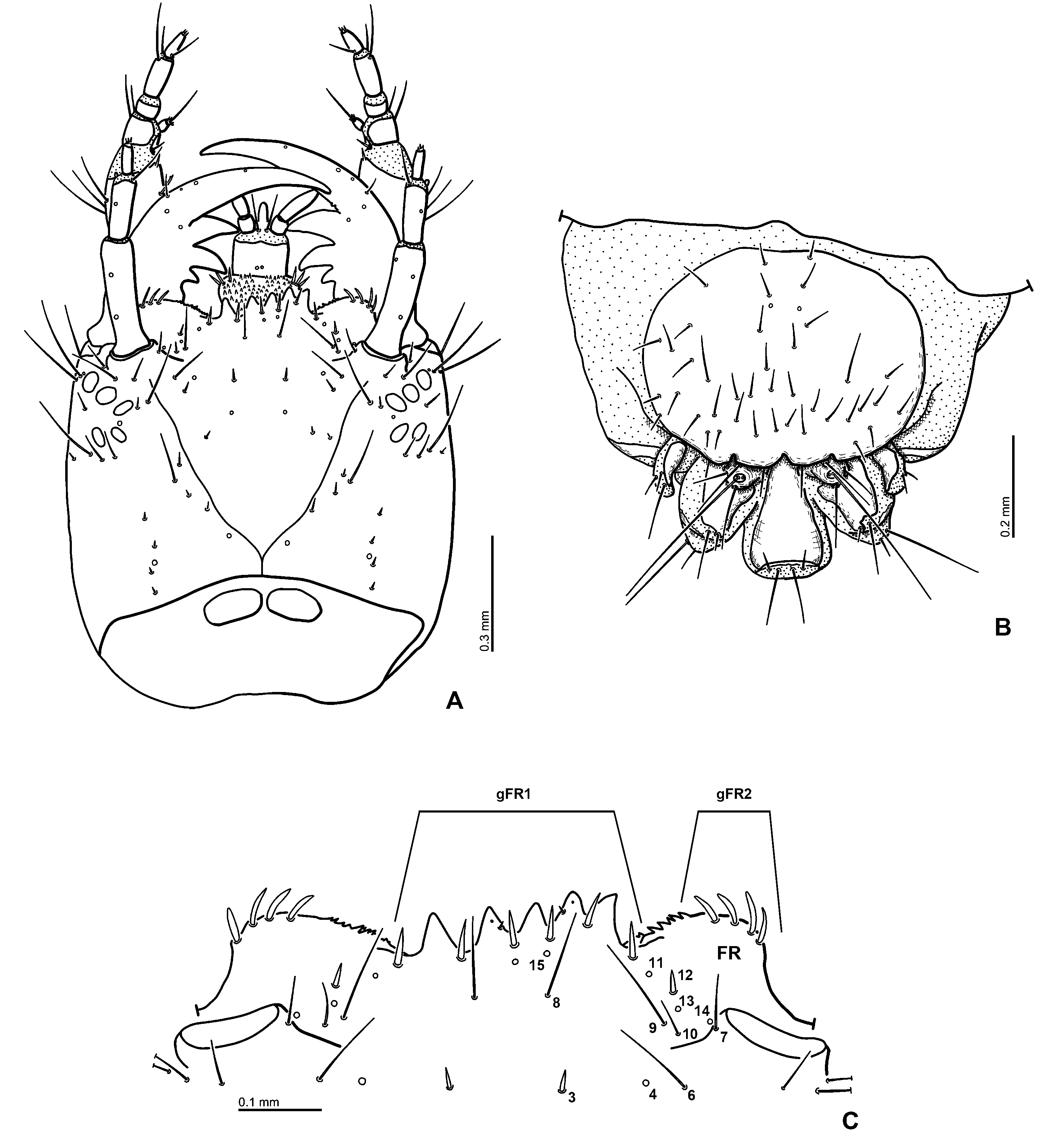
Head ( Figs. 48A, C View Fig ). Head capsule subquadrate. Frontal lines almost V-shaped, fused at base of head capsule, coronal line short. Six stemmata on each anterolateral portion of head capsule. Clypeolabrum slightly asymmetrical. Nasale with five teeth, two on right more closely aggregated and projecting further anteriad than two on left side. Epistomal lobes rounded, almost symmetrical, not projecting further than nasale, with small spine-like cuticular projection on inner margin.
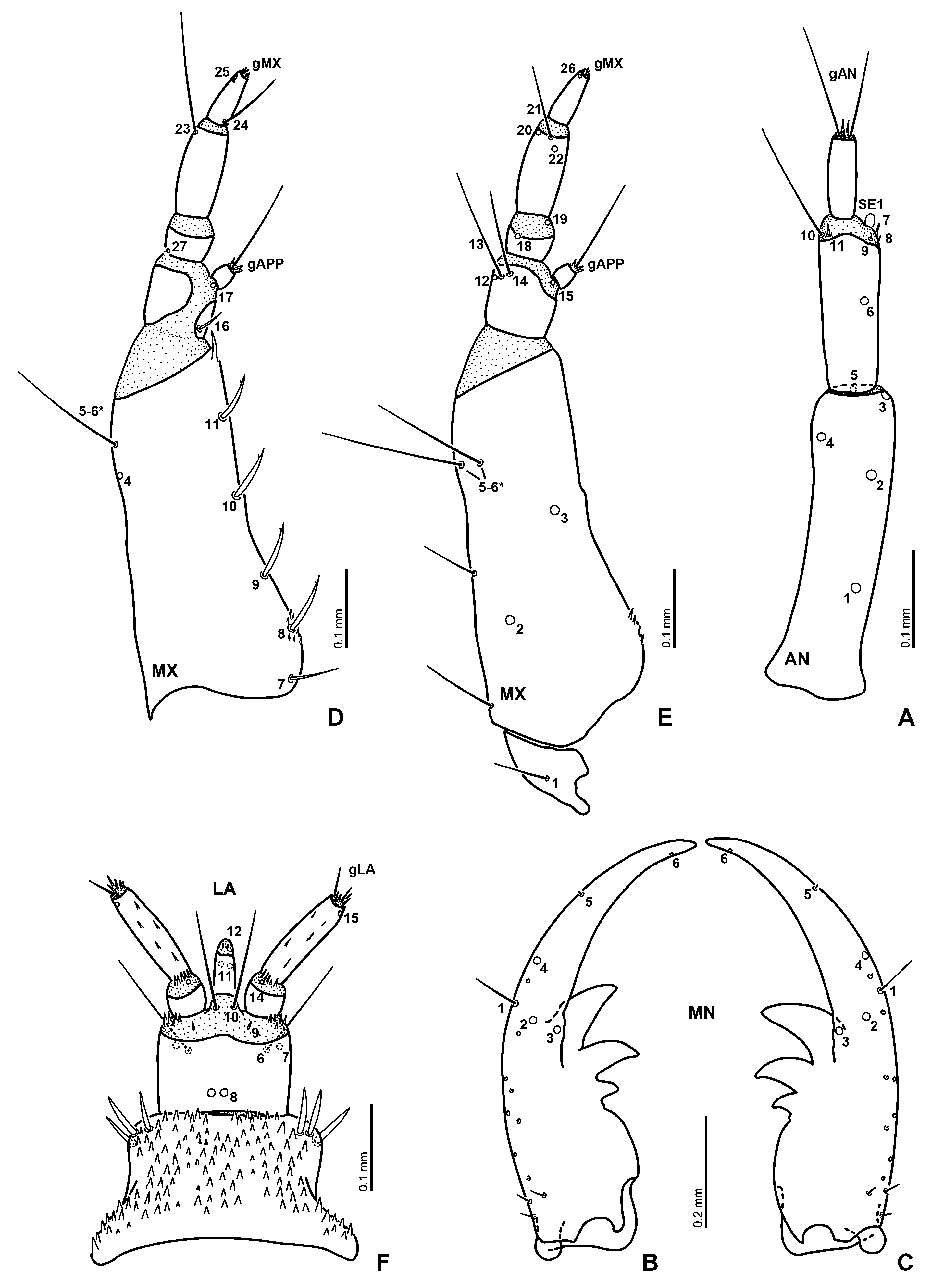
Antenna ( Fig. 49A View Fig ) 3-segmented, rather slender. Scape the longest, distinct longer than pedicel. Flagellum the shortest.
Mandibles ( Figs. 49 View Fig B–C) symmetrical, each with three inner teeth; distal two teeth large, basal one small; inner face of mandibular apex smooth.
Maxilla ( Figs. 49 View Fig D–E) 6-segmented (including cardo), distinctly longer than antenna. Cardo small, irregularly shaped. Stipes the longest, longer than palpomeres 1–4 combined; distal margin of stipes with spine-like cuticular projection on inner face; base of inner face bearing small cuticular projections. Maxillary palpus 4-segmented, palpomere 2 the shortest, palpomere 3 the longest, palpomere 1 slightly shorter than palpomere 4; palpomere 1 the widest, incompletely sclerotised on dorsal surface; inner process sclerotised.
Labium ( Fig. 49F View Fig ) well developed. Submentum fused to head capsule, large, subpentagonal, wider than mentum (e.g., Fig. 46B View Fig ). Mentum trapezoidal in dorsal view, wider than prementum, bearing small, strong cuticular spines dorsally. Prementum subquadrate, slightly wider than long. Ligula distinctly shorter than labial palpi, largely sclerotised. Membrane between sclerite of prementum and palpi bear short cuticular spines laterally. Labial palpi longer than prementum, covered with narrow cuticular spines on palpomere 2 and intersegmental membrane between palpomeres 1 and 2.
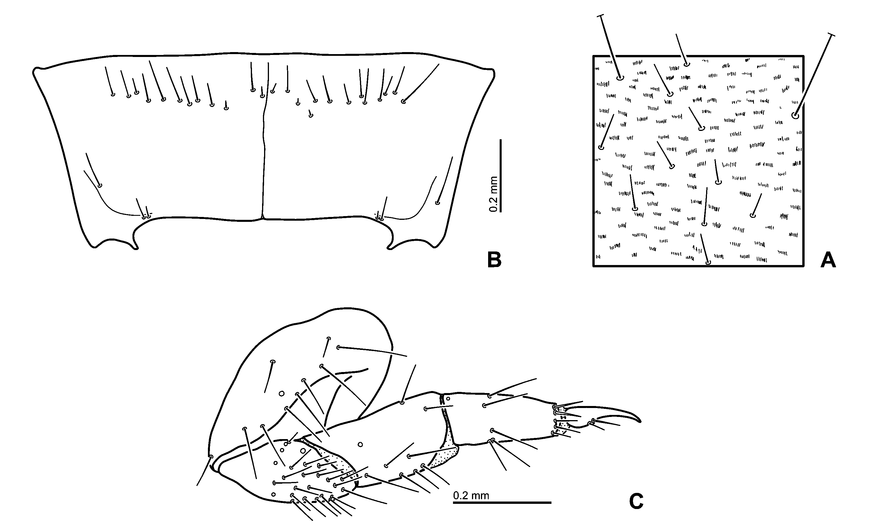
Thorax. Prothorax wider than head capsule ( Fig. 6C View Fig ). Proscutum formed by large plate subdivided by fine sagittal line, bearing densely arranged short to rather long setae and minute hair-like cuticular projections ( Fig. 50A View Fig ). Prosternum rectangular, with almost complete sagittal line ( Fig. 50B View Fig ). Mesothoracic tergum with two sclerites on each side, anterior one narrow, posterior one large; metathoracic tergum with two sclerites, anterior one large, posterior one small ( Fig. 6C View Fig ). Legs ( Fig. 50C View Fig ) rather short, weakly visible in dorsal view, 5-segmented, all pairs similar in shape.
Abdomen. Abdomen 10-segmented, tapering towards posterior end, segments 8 to 10 forming spiracular atrium ( Fig. 6C View Fig ); segments 1 to 7 similar in shape and size. Segment 1 with three dorsal sclerites on each side; anterior one oval, small; posterior two very small, weakly sclerotised, each with one long seta; segments 2 to 7 similar to segment 1 but anterior sclerites smaller ( Fig. 6C View Fig ).
Spiracular atrium ( Fig. 48B View Fig ): Segment 8 with large, oval dorsal plate; posterior edge of dorsal plate with three emarginations medially; dorsal surface of plate bearing numerous rather short setae; procercus incompletely sclerotised, with one rather long and two short setae. Segment 9 trilobed, partially sclerotised; median lobe almost as large as lateral lobes, with two long and two short apical setae; each lateral lobe with one long seta and three short to rather short apical setae; acrocerci undetectable; urogomphi short, one segmented; prostyli reduced.
Second instar. Very similar to third instar.
Head. Antenna ( Fig. 47A View Fig ) rather slender, proportionally shorter than in third instar. Scape slightly longer than pedicel.
Labium ( Figs. 47 View Fig B–C): Labial palpi slightly wider than in third instar. Anterior margin of mentum slightly wider than prementum.
Chaetotaxy of head. Second instar. Frontale altogether with 44 primary sensilla and two secondary sensilla ( Figs. 46A, C View Fig ). Central part with four pairs of sensilla divergent posteriad; FR 1 and one short secondary seta at midlength and close to frontal line; FR 2 pore-like situated
anteromesally to FR 1, between FR 1 and FR 3; FR 3 short and rather stout seta at midwidth and in anterior third of frontale. Two setae ( FR 5 and FR 6) and one pore-like sensillum FR 4 situated posteromesally to antennal socket, forming a triangular group; FR 5 rather short; FR 6 moderately long. Seta FR 7 rather short close to inner margin of antennal socket. Nasale with a group of six almost equidistant, stout and rather short setae (gFR1). Each epistomal lobe with a group of four short to moderately long setae (gFR2), two mesal setae distinct shorter than lateral ones. Pore-like sensillum FR 15 placed posteriorly to median setae of nasale; seta FR 8 situated posteriorly to FR 15. Two moderately long trichoid setae ( FR 9 and FR 10) situated between FR 7 and FR 8, slightly anteromesally to antennal socket. Four sensilla ( FR 11–14) on epistomal lobe, forming an irregular row; FR 11 on anteromesal margin of epistomal lobe, close to gFR1; FR 12 and FR 13 at midlength between FR 11 and FR 14; FR 14 situated close to inner margin of antennal socket, anteriorly to FR 7; FR 11 and FR 13–14 pore-like sensilla, FR 12 short and rather stout seta.
Parietale with 30 sensilla and seven secondary sensilla each ( Figs. 46 View Fig A–B). Dorsal surface with a group of five posterior sensilla ( PA 1–5) forming a slightly irregular longitudinal row at midwidth; PA 3 pore-like between PA 2 and PA 4; remaining sensilla short setae. PA 6 pore-like, situated anterolaterally to joint of frontal and coronal lines. Three short secondary setae between PA 6 and PA 7. PA 7 long seta, situated anteromesally to PA 5, equidistant from PA 6 and PA 8 as well as PA 5 and FR 1. Seta PA 8 long, behind antennal socket close to frontal line; one short secondary seta situated posterolaterally to PA 8. Seta PA 9 long, close to outer margin of antennal socket; one rather short secondary seta located behind antennal socket, mesally of PA 9. Two sensilla ( PA 10 and PA 11) behind antennal socket, between PA 9 and PA 12–14; PA 10 pore-like sensillum, PA 11 rather short seta. Three long setae ( PA 12–14) at about midlength of parietale, behind PA 10 and PA 11; one short secondary seta situated slightly laterally of PA 14.Anterior corner of epicranium with one pore-like sensillum ( PA 19) and three moderately long to long setae, PA 19 situated dorsally to remaining setae; PA 20 between PA 19 and PA 21; PA 22 situated ventrally to remaining sensilla; PA 20 and PA 21 moderately long setae, PA 22 long seta. Two pore-like sensilla ( PA 15 and PA 17) and two long setae ( PA 16 and PA 18) on anterior third of lateroventral surface of parietale; PA 16 between PA 15 and PA 17; PA 18 behind PA 15–17. Ventral surface with three pore-like sensilla ( PA 23–25) on anterior margin close to mandibular acetabulum; PA 23 on outer part, PA 24 and PA 25 on inner part; PA 24 between PA 23 and PA 25. Two long setae ( PA 26 and PA 28) and one pore-like sensillum ( PA 27) situated ventrally on median part of parietale; PA 27 between PA 26 and PA 28. Two pore-like sensilla ( PA 29 and PA 30) situated ventrally on basal third; PA 30 on lateral portion; PA 29 close to gular sulcus.
Antenna ( Fig. 47A View Fig ): Antennomere 1 with five pore-like sensilla ( AN 1–5); AN 1 on basal third; AN 2 on basal 0.67; AN 3–5 on distal margin, AN 3 and AN 4 on outer and inner face of sclerite respectively; AN 5 situated ventrally.Antennomere 2 with seven sensilla ( AN 6–11 and SE 1); AN 6 pore-like, located dorsally at midlength of sclerite; AN 7–11 on intersegmentary membrane between antennomeres 2 and 3; small setae AN 7–9 on outer part of antenna, behind and close to SE 1; long seta AN 10 and short seta AN 11 on outer face of antenna; SE 1 small, about as long as AN 7. Antennomere 3 with apical sensilla (gAN) on apical membranous area; gAN with two rather long and several short setae.
Mandible (e.g., Figs. 49 View Fig B–C) with two setae ( MN 1 and MN 5) and four pore-like sensilla ( MN 2–4 and MN 6), and with small secondary setae in basal half of outer face. MN 1 moderately long, at midlength of outer face of mandible. MN 2–4 on median part of dorsal surface; MN 2 at midlength between MN 1 and MN 3; MN 4 situated on outer face, anteriorly to MN 1. Seta MN 5 minute, on outer face of subapical part of mandible. MN 6 situated subapically on inner face of mandible.
Maxilla (e.g., Figs. 49 View Fig D–E) Cardo with one moderately long ventral seta ( MX 1). Stipes with a row of five rather short stout setae ( MX 7–11) situated dorsally along inner face; MX 7–11 almost equidistant; MX 8–11 with subapical tooth. Ventral surface of stipes with three porelike sensilla ( MX 2–4), two long setae ( MX 5–6), and three secondary setae; MX 2 on basal fourth of sclerite; MX 3 on apical two-fifths of sclerite; MX 4–6 on outer face of apical part of sclerite; three moderately long secondary setae on outer face, one situated basally, one at midlength of sclerite, one situated distally; distal one close to MX 4–6. Palpomere 1 with four sensilla on sclerite ( MX 12–14 and MX 16); one moderately short spiniform seta ( MX 16) situated dorsally on base of inner face; MX 12 pore-like, situated subapically on outer face of sclerite; MX 13–14 long setae situated subapically close to and mesally of MX 12; MX 13 between MX 12 and MX 14. Two pore-like sensilla ( MX 15 and MX 17) on membrane behind inner appendage, MX 17 dorsal, MX 15 ventral. Inner appendage with one rather long trichoid seta and rather to very short setae (gAPP). Palpomere 2 with two pore-like sensilla ( MX 18–19) and one minute seta ( MX 27); MX 18 situated ventrally on outer part of sclerite; MX 19 on inner face of intersegmental membrane between palpomeres 2 and 3; MX 27 on outer face of basal margin of sclerite. Palpomere 3 with two long setae ( MX 21 and MX 23) and two porelike sensilla ( MX 20 and MX 22); MX 20 and MX 23 situated distally on outer face of sclerite; MX 21 at midwidth of ventral surface, on borderline of sclerite and intersegmental membrane between palpomeres 3 and 4; MX 22 located posteriorly to MX 21. Palpomere 4 with one long seta ( MX 24) situated basally on inner face of sclerite, and with digitiform sensillum ( MX 25) and pore-like sensillum ( MX 26) situated subapically on outer face, MX 25 dorsal, MX 26 ventral. Apical membranous area of palpomere 4 with minute setae (gMX).
Labium ( Figs. 47 View Fig B–C): Submentum with two pairs of setae ( LA 1–2); LA 1 very long, in lateral corners, LA 2 short on anterior margin. Anterolateral face of mentum with three rather long, stout secondary setae, two on dorsal, one on ventral. Ventral surface of mentum with two primary sensilla ( LA 3 and LA 4) and one secondary seta on anterolateral part of sclerite; rather short seta ( LA 3), pore-like sensillum ( LA 4) and one rather long secondary seta forming a triangular group; LA 3 situated more mesally than remaining sensilla, LA 4 situated slightly lateroposteriorly to LA 3; one rather short, stout secondary seta on anterior corner of sclerite. Dorsal surface of prementum with a pair of pore-like sensilla ( LA 8) at midwidth of sclerite close its posterior margin. Ventral surface of prementum with three pairs of sensilla ( LA 5–7) on outer part; seta LA 5 short on posterior margin; LA 6 and LA 7 close to or on distal margin of sclerite; LA 6 long seta; LA 7 pore-like. Membrane between sclerite of prementum and palpi with a pair of pore-like sensilla ( LA 9) and moderately long setae ( LA 10) dorsally; LA 9 close to basal margin of sclerite; LA 10 at base of ligula. Ligula with two pairs of subapical pore-like sensilla ( LA 11 and LA 12); LA 12 situated dorsally on apical membranous area; LA 11 situated ventrally on sclerite. Palpomere 1 with one minute seta ( LA 13) situated ventrally on basal margin of sclerite; LA 14 pore-like, on dorsal surface of intersegmentary membrane between palpomeres 1 and 2. Palpomere 2 with one pore-like sensillum ( PA 15) situated subapically on outer face of sclerite. Apical membranous area of palpomere 2 with several short setae (gLA).
Third instar. Very similar to second instar larva.
Antenna: AN 4 situated in subapical part of antennomere 1 ( Fig. 47A View Fig ).
Habitat. Standing water. Larvae were found in water.
Identification. The larvae of the genus Hydrobius are known and therefore may be easily recognised ( RICHMOND 1920, BØVING & HENRIKSEN 1938, BERTRAND 1972, ARCHANGELSKY 1997, FIKÁČEK et al. 2008). Only one species of Hydrobius , H. pauper , is distributed in Japan and the larval material collected in the field may therefore be clearly assigned to this species. The Japanese record of Hydrobius fuscipes ( Linnaeus, 1758) by HANSEN (1999) is doubtful and H. fuscipes is not considered a part of the Japanese fauna.


| MN |
Museu Nacional, Universidade Federal do Rio de Janeiro |
No known copyright restrictions apply. See Agosti, D., Egloff, W., 2009. Taxonomic information exchange and copyright: the Plazi approach. BMC Research Notes 2009, 2:53 for further explanation.