
Gigantea gouvernoni, Jones & Sterrer, 2005
|
publication ID |
https://doi.org/ 10.11646/zootaxa.1001.1.3 |
|
persistent identifier |
https://treatment.plazi.org/id/F36987E4-2266-9711-FEE5-5986FC9C8066 |
|
treatment provided by |
Felipe |
|
scientific name |
Gigantea gouvernoni |
| status |
sp. nov. |
Gigantea gouvernoni View in CoL n. sp. ( Figs 9–19 View FIGURES 9–15 View FIGURES 16–19 , Color Plate 3)
Material examined: Holotype. Collected in the grounds of Government House , Bermuda on 20 August 2004 by WS. Found on underside of wall stones round a banana patch. Killed in hot water and initially preserved in 95% alcohol. A mature specimen, divided into two parts between the pharyngeal cavity and copulatory apparatus. The posterior portion containing the copulatory apparatus has been serially sectioned longitudinally (7 slides), remainder preserved. Specimen and slides deposited in the Natural History Museum, London, accession number: 2004.12.8.12.
Paratypes. Four specimens. One whole preserved specimen collected with holotype. Two further specimens collected from under stones in the grounds of Government House, Bermuda on 7 July 2003 by Olivier Gouvernon. One, a partially mature specimen is partially sectioned. Portions with copulatory apparatus (7 slides) and pharynx (9 slides) serially sectioned longitudinally. Small portion anterior to the pharynx sectioned transversely (2 slides). Remaining anterior portion preserved. Specimen and slides deposited in the Natural History Museum, London, accession number: 2004.12.8.13–15 .
Further specimens are deposited in the BAMZ.
Type locality: Grounds of Government House, Pembroke, Bermuda.
Etymology: After Olivier Gouvernon, who found the first specimens.
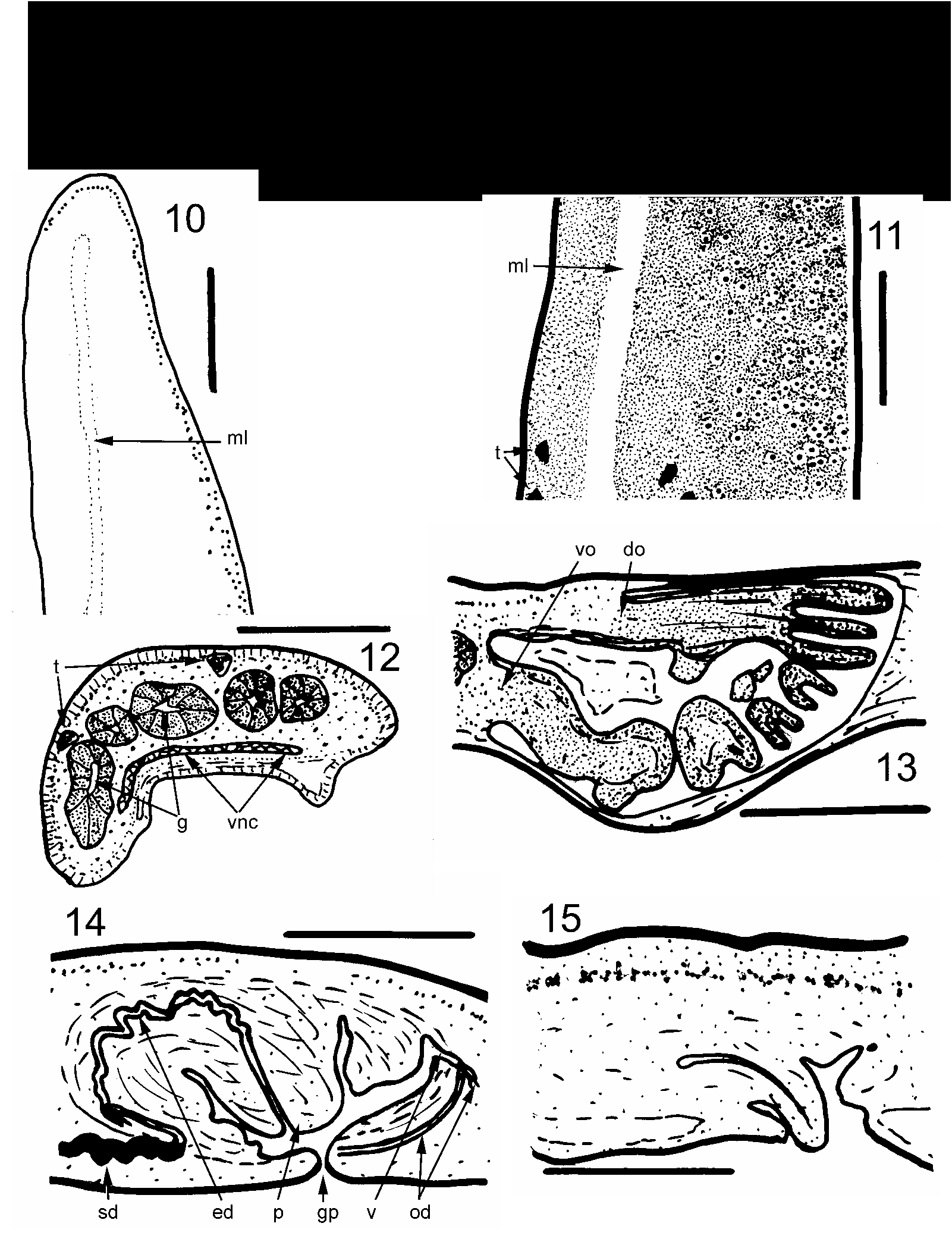
Description: The first specimens of this species were found in 2003 in the grounds of Government House, Bermuda. The extended living worms (Color Plate 3) are up to about 2.5 cm long, 4 mm wide and flattened. The body is widest at the level of the pharynx. The anterior third tapers towards the anterior end, and the posterior end is rounded. The dorsal ground color is dark brown with a narrow, middorsal, lighter brown line which broadens a little over the pharynx and the copulatory apparatus. There are fine, irregular, black mark ings, more in the anterior third so that the anterior is almost black. There are larger black spots irregularly arranged in two lateral rows from near the anterior end to the level of the pharynx ( Fig. 9 View FIGURES 9–15 ). These mark the positions of the dorsal testes. The ventral surface is uniformly light grey.
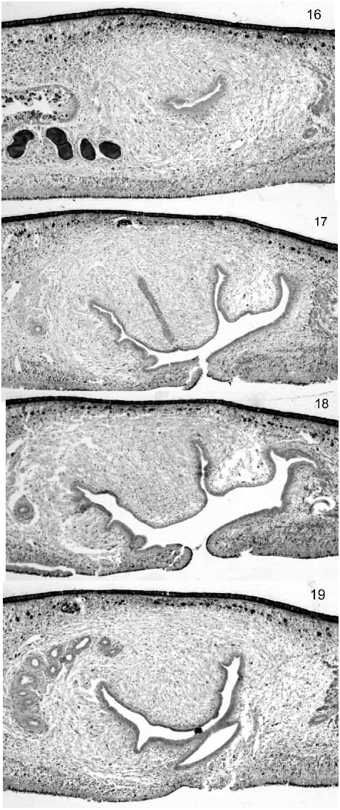
Dimensions of the preserved specimens are given in Table 1. The mouth is positioned on average 66% along the body, and the gonopore 80% along the body.Eyes are uniserial round the anterior tip, becoming multiserial in the translucent margin about 2 mm behind the anterior end ( Fig. 10 View FIGURES 9–15 ). They then spread dorsally, each surrounded by a small halo lacking the ground color pigment ( Fig. 11 View FIGURES 9–15 ). The zone of halo eyes diminishes at about the level of the most anterior black marks indicating the dorsal testes. Further posteriorly eyes are restricted to a narrow translucent margin. Anteriorly, there is a sensory margin just below the row of eyes. The creeping sole occupies the entire ventral surface which is somewhat concave in preserved specimens. Transverse sections (of the sectioned paratype) are 1 mm high and 3 mm wide ( Fig. 12 View FIGURES 9–15 ). Dorsally and laterally the epithelium is about 30 µm thick, columnar and contains very numerous elongate rhabdites. Nuclei are basal. Dorsal subcutaneous musculature is thinner (about 20 µm) than the overlying epithelium and illdefined so that circular and longitudinal fibres are not discernable. Ventrally, the epithelium is about 25 µm thick, has rhabdites about 12 µm long near the surface and is densely ciliated, the cilia are about 7 µm long. Ventral subcutaneous musculature is about 50 µm thick and is again illdefined, though longitudinal muscles appear to be in small bundles. Thus CMI is about 7% (this in a flattened species). In the sectioned paratype, the ventral musculature and epithelium are separated from the rest of the tissues. The ventral nerve plate extends across the body ventrally, but is not enlarged into ventral paired nerve cords. It is presumed that the ovaries are a single pair anteriorly, as in most terrestrial planarians (confirmation would require sectioning of the anterior portion). The pharynx ( Fig. 13 View FIGURES 9–15 ) is of the “glockenförmig” (bellshaped) type (see Graff 1899; Winsor et al. 1998). The ovovitelline ducts are not discernible in transverse sections of the sectioned paratype (not fully mature). In the holotype longitudinal sections they are fully developed and lie above and lateral to the ventral nerve plate. They pass posterior to the gonopore, turn dorsally and fuse to form a short vagina which in turn opens into the posterior common antrum ( Figs 14 View FIGURES 9–15 , 18 View FIGURES 16–19 ). Testes are dorsal and overlain by a small patch of dark pigment, clearly visible in transverse sections ( Fig. 12 View FIGURES 9–15 ). Sperm ducts are not visible in the paratype transverse sections. In the holotype, the sperm ducts expand posterior to the pharynx and widen to form convoluted sperm storage ducts ( Figs 14 View FIGURES 9–15 , 16 View FIGURES 16–19 ). These turn anteriorly and dorsally to fuse and discharge into a convoluted ejaculatory duct ( Figs 14 View FIGURES 9–15 , 19 View FIGURES 16–19 ). This straightens and discharges vertically through the blunt penis ( Figs 14 View FIGURES 9–15 , 17 View FIGURES 16–19 ). The musculature of the penis is relatively loose and similar to the rest of the copulatory bulb. Muscular layers are not clearly defined within the copulatory bulb. The penis of the sectioned paratype is conical and partly protruded through the gonopore ( Fig. 15 View FIGURES 9–15 ).
% of length.
Discussion: Since details of the copulatory apparatus are known, Pseudogeoplana Ogren & Kawakatsu 1990 (a collective genus for species with insufficient details of internal anatomy) is again inappropriate, though the present species does not match the description of any of the species listed in Pseudogeoplana . Clearly the present specimens have a broad body, and comparison with descriptions of other species suggests that the penis has a blunt papilla with no glandular ridges and that the female canal enters horizontally (or ventrally, depending on interpretation, but certainly not dorsally). The generic key in Ogren & Kawakatsu (1990) leads to either Gigantea Ogren & Kawakatsu 1990 or Liana E.M. Froehlich 1978. The original definition of Liana by Froehlich (1978) reads: “ Geoplanidae with elongated body and broad creeping sole of more than half the body width. Longitudinal cutaneous musculature strong dorsally and weak ventrally, where it has a portion sunk into the parenchyma, internally to the cutaneous nerve net. Sensory border, ventrolateral in position, with rare minute sensory pits, restricted to the anterior tip. Testes dorsal. Adhesive glandulomuscular organs absent. Copulatory apparatus without adenodactyls.” Ogren & Kawakatsu (1990) add: “penis papilla short and blunt; female canal approaches from horizontal or ventral aspect.” Liana contains a single species, L. guasa E.M. Froehlich 1978. This has a cylindrical pharynx and different arrangement of eyes to the Bermuda specimens, which also have weak dorsal subcutaneous muscle. Gigantea is defined as “of large, broad body; penis papilla present, typically with glandular ridges on the penis papilla (but ridges may be absent as in G. chiriquii ( Hyman 1962)) ; female canal horizontal or approaching from below; female antrum dilated.” Localities in Peru, Columbia, Costa Rica, Panama and Trinidad. The present specimens differ in some details from this (no glandular ridges, though these may be absent as above) and the female antrum does not appear dilated. Gigantea chiriquii (from Panama) has a “ruffled” pharynx (similar to G. gouvernoni ) and a blunt, short penis but which is horizontal ( Hyman 1962), rather than near vertical as in G. gouvernoni . However, we consider Gigantea to be the most appropriate genus for placement of these specimens, hence Gigantea gouvernoni .
Gigantea gouvernoni has been observed by WS feeding on snails. Presumably reproduction is by copulation and subsequent production of an egg capsule, as in all other terrestrial triclads, though there may be some asexual reproduction by fission.
Since all Geoplaninae originate from South or Central America ( Ogren et al. 1997), it is assumed that the Bermuda specimens of G. gouvernoni originated in that region. E. M. Froehlich (pers. comm.), who is familiar with the land flatworms from Brazil, does not recognise the Bermuda specimens (either Amaga expatria or Gigantea gouvernoni ) from photographs. However, LealZanchet (pers. comm.), who is also familiar with many Brazilian species, considers that the external appearance of G. gouvernoni is very similar to an undescribed species common in southern Brazil.
No known copyright restrictions apply. See Agosti, D., Egloff, W., 2009. Taxonomic information exchange and copyright: the Plazi approach. BMC Research Notes 2009, 2:53 for further explanation.
|
Kingdom |
|
|
Phylum |
|
|
Class |
|
|
Order |
|
|
Family |
|
|
Genus |