
Felisacus jacobsoni Poppius, 1914
|
publication ID |
https://doi.org/ 10.1206/0003-0090-403.1.1 |
|
persistent identifier |
https://treatment.plazi.org/id/296A879F-5643-7535-5D72-FB20FBBD0C28 |
|
treatment provided by |
Carolina |
|
scientific name |
Felisacus jacobsoni Poppius |
| status |
|
Felisacus jacobsoni Poppius View in CoL
Figures 5 View FIGURE 5 , 8M View FIGURE 8 , 12E, F View FIGURE 12 , 18 View FIGURE 18
Felisacus jacobsoni Poppius, 1914: 149 View in CoL (original description).
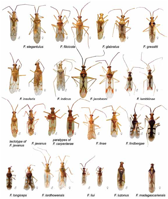
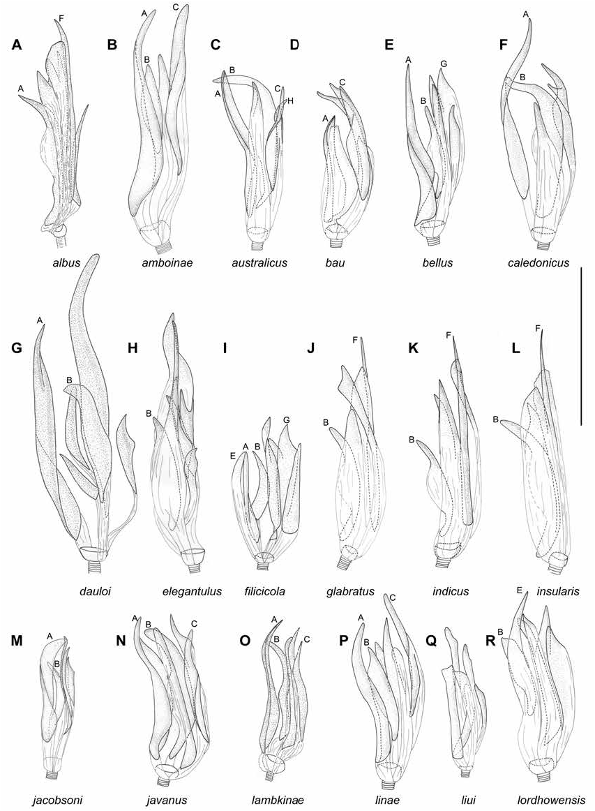
DIAGNOSIS: Recognized by the following combination of characters: dorsal surface of head brown anteriorly; pronotum brown along posterior margin; inner part of clavus opaque, brown; scent gland evaporative area brown; cuneus red with outer margin brown; marking along inner margin of corium brown, not reaching R+M anteriorly and posteriorly (fig. 5), cylindrical antennal segment I (as in Namyatova et al., 2016: fig. 8A), transverse depression on head extending laterally, vertex upraised (as in Namyatova et al., 2016: fig. 6D); labium reaching posterior margin of metasternum; vesica with five spicules, including spicules A and B, spicule A elongate and widened, spicule B small and not swollen (fig. 8M).
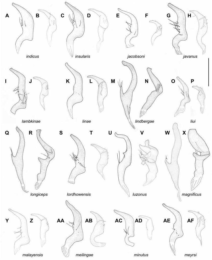
REDESCRIPTION: Male. Total length 4.4–4.7. COLORATION (fig. 5): Head: Mostly yellow, brown dorsally, labrum brown. Eye dark brown. Labium: Uniformly yellow. Antenna: Segment I brown with yellow base, segments II–IV dark brown. Thorax: Pronotum yellow with brown collar and markings on anterior part of pronotum laterally, posterior part of pronotum with paired brown markings near margin; scutellum yellow; pleura yellow, scent gland evaporative area brown, with whitish yellow apex and base. Hemelytron: Mostly translucent and colorless; inner part of clavus opaque, brown; marking along inner margin of corium brown, cuneus mostly red with outer margin translucent with yellow tinge and margins pale brown; membrane with grayish tinge and pale brown veins. Legs: Coxae whitish yellow; femora whitish yellow basally and yellow apically; tibiae yellow to pale brown, brown basally; tarsi brown. SUR- FACE AND VESTITURE: Corium smooth with shallow and scarce punctures. Dorsum and femora with suberect setae mostly subequal to antennal segment II diameter; antennal segment I with scarce suberect setae mostly shorter than antennal segment II diameter. STRUCTURE AND MEASUREMENTS: Body ca. 4.4–4.6× as long as pronotum width. Head: Depression, delimiting occipital region, present dorsally and laterally (as in Namyatova et al., 2016: fig. 4E); distance between depression and pronotum distinctly shorter than eye diameter; longitudinal sulcus on dorsal surface longer than eye diameter; distance from eye to pronotum slightly longer than eye diameter, not swollen laterally (as in Namyatova et al., 2016: fig. 4E); vertex ca. 1.4–1.7× as wide as eye upraised. Labium (as in Namyatova et al., 2016: figs. 6D, 9C): Almost reaching posterior margin of metasternum; segments I and II strongly shortened, combined length subequal to half of segment III; segment I shorter than wide; segment II slightly longer than wide, its dorsal surface elongate posteriorly; segment III slightly shorter than ventral side of head; segment IV twice as long as segment III. Antenna: Segment I cylindrical (as in Namyatova et al., 2016: fig. 8A), ca. 1.6× as long as head width, ca. 1.0–1.1× as long as pronotum width; segment II ca. 2.1–2.3× as long as head width, ca. 1.4× as long as pronotum width; segments III slightly longer than segment II; segment IV ca. 0.3× as long as segment IV. Thorax: Anterior part of pronotum slightly shorter than posterior part; posterior part of pronotum slightly upraised; posterior margin of pronotum concave, pronotum ca. 1.2–1.3× as wide as long and ca. 1.6× as wide as head; mesoscutum exposed. Hemelytron: Area behind clavus almost flat; inner margin of cuneus convex (as in Namyatova et al., 2016: fig. 13E), outer margin of cuneus almost ca. 3× as long as base. Genitalia: Right paramere (fig. 12E) apical part distinct; apex slightly concave; medial part less than twice as wide as basal part, bearing setae, with outer margin straight and inner margin convex; outer angle distinct, not widened; inner angle rounded, without setae; basal part ca. 0.2– 0.25× as long as rest of paramere. Left paramere (fig. 12F) L-shaped; apical part not flattened, with toothlike outgrowth on posterior side medially (as in fig. 11G) and without outgrowth on dorsal surface; middle part widened, without swelling or outgrowth(s); setae only on middle part near outer margin. Aedeagus conjunctiva weakly sclerotized; secondary gonopore placed at base of vesica in repose; sclerotization of ductus seminis around secondary gonopore shorter than wide; vesica with five spicules: spicule A is long and wide, spicule B short; three additional spicules present on left-hand side, all narrow and acute (fig. 8M).
Female. Total length 4.6–4.8. COLORATION (fig. 5): Similar to male, head often dark brown dorsally, sometimes with reddish-brown marking between eyes; pronotum yellow sometimes with brown stripe along posterior margin; tho- racic pleura sometimes with brown markings or mostly pale brown; tibiae sometimes uniformly yellow to pale brown; tarsal segments I–II sometimes whitish yellow. SURFACE AND VESTI-
TURE: As in male. STRUCTURE AND
MEASUREMENTS: Similar to male. body ca. 4.3–5.1× as long as pronotum width; vertex ca. 1.7–1.8× as wide as eye; antennal segment I ca. 1.5–1.7× as long as head width, ca. 0.9–1.1× as long as pronotum width; segment II ca. 2.1–2.2× as long as head width, ca. 1.3–1.4× as long as pronotum width; pronotum ca. 1.1–1.2× as wide as long and ca. 1.4–1.6× as wide as head. Genitalia (as in fig. 23F, G): Dorsal labiate plate wider than distance between apodemes of second valvula; mostly smooth, without distinct striations, with semicircular sclerite and distinct sclerotized rings laterally; lateral oviducts placed almost medially, very close to each other, spermathecal gland placed between lateral oviducts; dorsal labiate plate with distinct tubercles, without membranous lobe medially.
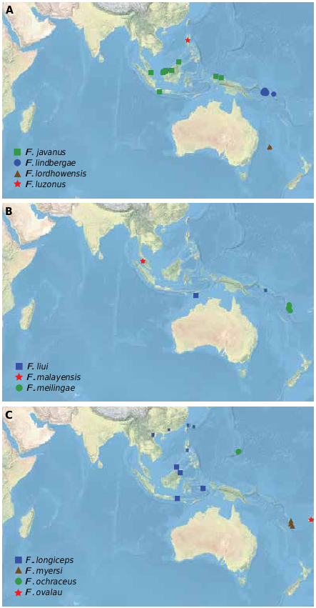
DISTRIBUTION: Java and Lombok Is. ( Indonesia) (fig. 18).
HOST PLANTS: Pteris sp.
DISCUSSION: Poppius (1914) described Felisacus jacobsoni from a single female, collected in Java and preserved in the FMNH. Some specimens in the collection of ZISP are from Lombok are very similar to the type specimen, and we assigned them to this species.
Felisacus jacobsoni is most similar to F. bellus , F. bau , and F. signis in coloration (fig. 4): Felisacus bau and F. signis differ in body size, which is less than 4 mm in both sexes. All these species differ in the shape of the vesical spicules (cf. fig. 8M with figs. 8D, E, 9K).
Felisacus jacobsoni can be confused with F. javanus (fig. 5), which is widely distributed in Indonesia, as the latter also has a bright red cuneus and brown markings. However, F. javanus is paler and its head and pronotum have brown markings, it is smaller, body length, which is 3.0– 3.7 mm in males and 3.5–4.1 mm in females, and has distinct vesical spicules with spicule B elongate and broad (fig. 8N).
MATERIAL EXAMINED: Lectotype: INDONE- SIA: East Java: Nongkojajar [Nongkodjadjar], 7.8965 ° S 112.8213 ° E, 1095 m, Jan 1911, E. Jacobson, 1♀ (00017877) ( MZH). Additional material: West Nusa Tenggara: Lombok Island, nr Senaru, 8.31956 ° S 116.405 ° E, 01 Sep 2012, F. Konstantinov, Pteris sp. (Pteridaceae) , det. Michael Lovave ( LAE herbarium, PNG), 23 (00386513, 00386514), 4♀ (00386515–00386518) ( ZISP).
Felisacus javanus (Reuter) Figures 5 View FIGURE 5 , 8N View FIGURE 8 , 12G, H View FIGURE 12 , 14Q View FIGURE 14 , 19 View FIGURE 19
Hyaloscytus elegantulus var. javanus Reuter, 1908: 187 (original description), stat. nov.
Felisacus carpenterae Hsiao, 1944: 385 View in CoL , new synonymy.
DIAGNOSIS: Recognized by the following combination of characters: main coloration of head and pronotum yellow to pale brown, without brown markings, cuneus with inner part red and outer part whitish yellow, margins red (fig. 5), cylindrical antennal segment I (as in Namyatova et al., 2016: fig. 8A), transverse depression on head delimiting occipital region extending laterally, vertex upraised (as in Namyatova et al., 2016: fig. 6D) body length in male 3–3.7, in female 3.5–4.1; labium reaching posterior margin of mesosternum or slightly surpassing it; cuneus ca. 2.5× as long as base; ventral wall of genital capsule ca. 1.5× as long as dorsal wall (fig. 14Q); medial part of right paramere slightly wider than basal part, shorter than basal and apical parts combined, its outer margin concave (fig. 12G); vesica with five spicules, including spicules A, B, and C; spicule A longer than spicule B, spicule B long and swollen, spicule C long and distinctly convolute (fig. 8N).
REDESCRIPTION: Male. Total length 3.0–3.7. COLORATION (fig. 5): Head: Yellow to pale brown, dorsal surface and clypeus sometimes with reddish tinge. Eye dark brown to black, sometimes with pale brown spots or reddish tinge. Labium: Whitish yellow to pale brown, segment III sometimes with reddish tinge ventrally. Antenna: Segment I pale brown, some- times with reddish tinge, segment II reddish brown, segments III dark brown. Thorax: Pronotum yellow with anterior margin brown, sometimes whitish yellow posteriorly, sometimes with reddish tinge laterally; humeral angle or marking nearby often yellow to pale brown; mesoscutum and scutellum whitish yellow to pale brown, punctures between them pale brown, scutellum with longitudinal brownish stripe; thoracic pleura yellow to pale brown, sometimes with reddish tinge; scent gland evaporative area yellow, sometimes red apically. Hemelytron: Mostly translucent; clavus sometimes opaque, inner part of clavus whitish yellow to yellow, sometimes brown apically, with yellow, reddish or pale brown margins; outer part of clavus colorless or whitish yellow, with apex yellow to brown; corium whitish yellow to yellow, area along inner margin of corium yellow to pale brown at middle part and reddish posteriorly; embolium yellow with pale brown margins and often with reddish margin; cuneus with inner part red and outer part whitish yellow, margins and often its inner part red; membrane with yellow or grayish tinge, cell red. Legs: Coxa whitish yellow to yellow; femora whitish yellow to yellow basally and yellow to pale brown apically, sometimes with reddish tinge; tibia yellow to pale brown, sometimes somewhat paler basally, sometimes with reddish tinge basally; tarsi pale brown, segment I often whitish yellow to yellow. SURFACE AND VES- TITURE: Corium smooth, with shallow and scarce punctures. Dorsum with setae shorter or subequal to antennal segment II diameter; antennal segment I and femora clothed with suberect setae mostly shorter than width of antennal segment II diameter. STRUCTURE AND MEA- SUREMENTS: Body ca. 4.0–4.4× as long as pronotum width. Head: Depression delimiting occipital region present dorsally and laterally (as in Namyatova et al., 2016: fig. 4E); distance between depression and pronotum distinctly shorter than eye diameter; longitudinal sulcus on dorsal surface of head longer than eye diameter; distance from eye to pronotum longer than eye diameter, not swollen laterally (as in Namyatova et al., 2016: fig. 4E); vertex ca. 1.3–1.9× as wide as eye, upraised (as in Namyatova et al., 2016: fig. 6D). Labium (as in Namyatova et al., 2016: figs. 6D, 9C): Reaching posterior margin of mesosternum or slightly surpassing it; segments I and II strongly reduced, combined subequal to half of segment III; segment I shorter than wide; segment II slightly longer than wide, its dorsal surface elongate posteriorly; segment III as long as ventral side of head; segment IV ca. 1.5× as long as segment III. Antenna: Segment I cylindrical (as in Namyatova et al., 2016: fig. 8A), ca. 1.4– 1.6× as long as head width, ca. 1.0–1.2× as long as pronotum width; segment III slightly longer than segment II. Thorax: Anterior part of pronotum only slightly shorter than posterior part; collar delimited; posterior part slightly upraised; posterior margin concave, pronotum ca. 1.1– 1.2× as wide as long and ca. 1.4–1.5× as wide as head; mesoscutum exposed. Hemelytron: Vein along behind clavus almost flat; inner margin of cuneus convex (as in Namyatova et al., 2016: fig. 13E), outer margin of cuneus ca. 2.5× as long as base. Genitalia: Genital capsule (fig. 14Q) ventral wall ca. 1.5× as long as dorsal wall, with posterior margin of ventral wall semioval, smooth, without outgrowth(s), its apex inclined leftward, not curved; sides of genital capsule not modified; right paramere socket slightly acute, left paramere socket rounded; distance between paramere sockets subequal to half of genital capsule width at base. Right paramere (fig. 12G) distinctly curved in apical half; apex almost straight; medial part only slighter wider than basal part, with row of setae, with outer margin slightly concave and inner margin widened; outer angle distinct, slightly swollen; inner angle rounded, not bearing setae; basal part of paramere ca. 0.15– 0.2× as long as rest of paramere. Left paramere (fig. 12H) L-shaped; apical part flattened, with tooth on posterior side medially (as in fig. 11G) and without outgrowth on dorsal surface; middle part widened, without swelling or outgrowth; setae only on middle part near outer margin. Aedeagus (general view as in Namyatova et al., 2016: fig. 22I) conjunctiva weakly sclerotized, secondary gonopore placed at base of vesica in repose; sclerotization of ductus seminis around secondary gonopore shorter than wide; vesica with five long spicules, vesica with five spicules, including spicules A, B, and C; spicule A long and wide, spicule B long and swollen, spicule C long and distinctly convolute (fig. 8N).
Female. Total length 3.5–4.1. COLORATION (fig. 5): Head: Similar to male, but sometimes with stripe above antennal fossae, markings between antennal fossae and stripe or marking behind eye red. Labium and antenna: As in male. Thorax: Similar to male, sometimes pale brown with brown anterior margin, scent gland evaporative area sometimes yellow with pale brown margin. Hemelytron: Similar to male, embolium rarely pale brown. Legs: Similar to male, rarely uniformly pale brown. Abdomen: Whitish yellow with reddish dorsum. SURFACE AND VESTITURE: As in male. STRUCTURE AND MEASUREMENTS: Structure as in male; body ca. 4.0–4.4× as long as width of pronotum; vertex ca. 1.3–2.0× as wide as eye; antennal segment I ca. 1.3–1.5× as long as head width, ca. 0.9–1.0× as long as pronotum width; antennal segment II ca. 1.7–1.9× as long as head width, ca. 1.1–1.3× as long as pronotum width; pronotum ca. 1.2–1.4× as wide as long and ca. 1.5–1.6× as wide as head. Genitalia (as in Namyatova et al., 2016: fig. 23F, G): Dorsal labiate plate wider than distance between apodemes of second valvula; mostly smooth, without distinct striations, with semicircular sclerite and distinct sclerotized rings laterally; lateral oviducts placed almost medially, very close to each other, spermathecal gland placed between lateral oviducts; dorsal labiate plate with distinct tubercles, without membranous lobe medially.
DISTRIBUTION: Indonesia (Java, West Papua), Malaysian Borneo (Sabah, Sarawak), Singapore (fig. 19).
HOST PLANTS: Nephrolepis biserrata (Davalliaceae) .
DISCUSSION: Felisacus javanus was described from a series of specimens from Java ( Reuter, 1908), and are preserved in the MZH. We examined the male and female of the type series. Feli- sacus carpenterae was described from Singapore ( Hsiao, 1944) and its type specimens are preserved in the USNM, and we examined male and female paratypes. On the basis of very similar external morphology and identical male genitalia, we synonymize F. carpenterae with F. javanus . In contrast, F. carpenterae signis Hsiao, 1944 , differs from the nominotypical subspecies and represents a separate species (see the discussion of F. signis ). See generic discussion for the explanation of the nomenclatural confusion concerning F. javanus in literature.
Felisacus javanus is most similar to F. amboinae , F. filicicola , F. ochraceus , and F. solomonicus in coloration and structure. Felisacus amboinae , F. filicicola , and F. solomonicus can be separated by the right paramere with the outer margin straight (cf. fig. 12G with figs. 11C, AD, 13R) and the shape of the vesical spicules (cf. fig. 8N with figs. 8B, I, 9L). Felisacus ochraceus is similar to F. javanus in the shape of the right paramere and the configuration of the spicules (figs. 9F, 13C). In contrast, F. ochraceus has spicule B longer than spicule A.
Felisacus javanus and F. lambkinae are near identical in the shape and configuration of the vesical spicules (cf. fig. 8N with 8O), body size and structure of the right paramere, with both having the medial region elongate, and the outer margin is concave (cf. fig. 12G with 12I). Felisacus lambkinae differs from F. javanus by the anterior part of the pronotum being brown, the inner part of the clavus is pale brown to brown (fig. 5), and the labium reaches or slightly surpasses the posterior margin of the metasternum.
Felisacus javanus can be confused with F. jacobsoni as the two species can be collected together and are similar to each other in coloration, including the pale brown ground color, presence of brown markings, with the cuneus at least partly red. However, in contrast to F. javanus , specimens of F. jacobsoni have a longer body, with males 4.4–4.7 mm and females 4.6– 4.8 mm in length and, the labium reaches the posterior margin of the mesosternum. Felisacus jacobsoni differs slightly in coloration, having the inner part of the clavus, scent gland evaporative area, and anterior part of the head brown and in vesical spicules (fig. 8M).
MATERIAL EXAMINED: Lectotype ( F. javanus ): INDONESIA: Java: collector unknown, 1902, O.M. Schmiedeknecht, Lectotype, 13 (00018430) ( MZH). Paralectotypes ( F. javanus ). INDONE- SIA: Java: collector unknown, 1902, O.M. Schmiedeknecht, paralectotype, 13 (00018435), 4♀ (00018431–00018434) ( MZH). Paratypes ( F. carpenterae ). SINGAPORE: Singapore: Singapore, 1.291 ° N 103.848 ° E, no date provided, C.F. Baker, 13 (00338826), 1♀ (00338825) ( USNM). Additional material: INDONESIA: West Papua: Biak Island , W of Airport, 1.3425 ° S 136.1875 ° E, 12 Aug 2012, F. Konstantinov, 2♀ (00271384, 00271385) ( ZISP). Biak Island nr Taman Burung , 1.17603 ° S 136.18756 ° E, 10 Aug 2012, F. Konstantinov, Nephrolepis biserrata (Sw.) Schott (Davalliaceae) , det. Michael Lovave ( LAE herbarium, PNG), 113 (00271364–00271374), 3 juvenile (00271375–00271377), 6♀ (00271378– 00271383) ( ZISP). Manokwari , nr Taman Gunung , Meja , 0.84497 ° S 134.07342 ° E, 21 Aug 2012, F. Konstantinov, 53 (00271389–00271393), 3♀ (00271395–00271397), 1 sex unknown (00271394) ( ZISP). Owi Island nr Biak Island , 1.23817 ° S 136.20294 ° E, 12 Aug 2012, F. Konstantinov, Nephrolepis biserrata (Sw.) Schott (Davalliaceae) , det. Michael Lovave ( LAE herbarium, PNG), 33 (00271386–00271388), 1♀ (00271432) ( ZISP). MALAYSIA: Sabah: West Coast Residency , Ranau , 8 mi N Poring Hot Springs, 6.11407 ° N 116.72104 ° E, 500 m, 08 Oct 1958 – 11 Oct 1958, T.C. Maa, 1♀ (00042310) ( BPBM). Sarawak: Bau, 1.4167 ° N 110.15 ° E, 29 Aug 1958 – 30 Aug 1958, T.C. Maa, 13 (00042341), 1♀ (00042311) ( BPBM). Merirai Valley nr. Kapit, 1.91009 ° N 113.63583 ° E, 01 Jul 1958 – 06 Jul 1958, T.C. Maa, 13 (00021523) ( BPBM). Rajang (Rejang) Delta, Sarikei Dist. , 2.42873 ° N 111.44251 ° E, 15 Jul 1958 – 26 Jul 1958, T.C. Maa, 13 (00021526), 1♀ (00021526) ( BPBM) GoogleMaps ; 15 Jul 1958 – 25 Jul 1958, T.C. Maa, 43 (00042337, 00021519–00021521), 2♀ (00021524, 00021525) ( BPBM). Sadong, Kampong Tapuh , 1.50361 ° N 110.74222 ° E, 04 Jul 1958 – 09 Jul 1958, T.C. Maa, 1♀ (00021535) ( ANIC), 33 (00021522, 00021533, 00021534) ( BPBM) GoogleMaps .


No known copyright restrictions apply. See Agosti, D., Egloff, W., 2009. Taxonomic information exchange and copyright: the Plazi approach. BMC Research Notes 2009, 2:53 for further explanation.
|
Kingdom |
|
|
Phylum |
|
|
Class |
|
|
Order |
|
|
Family |
|
|
Genus |
Felisacus jacobsoni Poppius
| Namyatova, Anna A. & Cassis, Gerasimos 2016 |
Felisacus carpenterae
| Hsiao, T. 1944: 385 |
Felisacus jacobsoni
| Poppius, B. 1914: 149 |
Hyaloscytus elegantulus var. javanus
| Reuter, O. M. 1908: 187 |