
Felisacus lambkinae, Namyatova & Cassis, 2016
|
publication ID |
https://doi.org/ 10.1206/0003-0090-403.1.1 |
|
persistent identifier |
https://treatment.plazi.org/id/296A879F-567D-7533-5D41-FEAFFBB80E7B |
|
treatment provided by |
Carolina |
|
scientific name |
Felisacus lambkinae |
| status |
sp. nov. |
Felisacus lambkinae , sp. nov.
Figures 5 View FIGURE 5 , 8O View FIGURE 8 , 12I, J View FIGURE 12 , 14R View FIGURE 14 , 18 View FIGURE 18
DIAGNOSIS: Recognized by anterior part of pronotum brown; inner part of clavus brown to pale brown; marking along inner margin of corium brown, not reaching R+M; cuneus reddish with colorless and translucent outer part and brown outer margin; cylindrical antennal segment I (as in Namyatova et al., 2016: fig. 8A), transverse depression on head extending laterally, vertex upraised; small size, body length in male 3.1–3.2 and in female 3.2–3.5; labium reaching posterior margin of metasternum or slightly surpassing it; vertex in male ca. 1.2–1.3× as long as eye diameter; cuneus ca. 2.5× as long wide; medial part of right paramere almost as wide as basal part, its length subequal to basal and apical parts combined; its outer margin concave and inner margin convex (fig. 12I); vesica with five spicules, including spicules A, B, and C (fig. 8O).
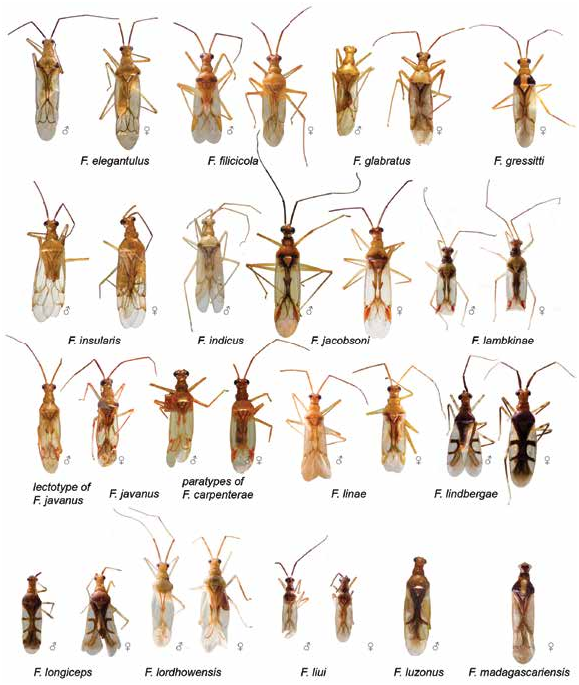
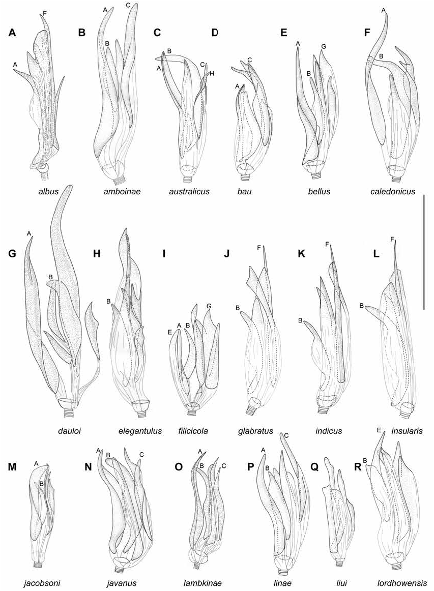
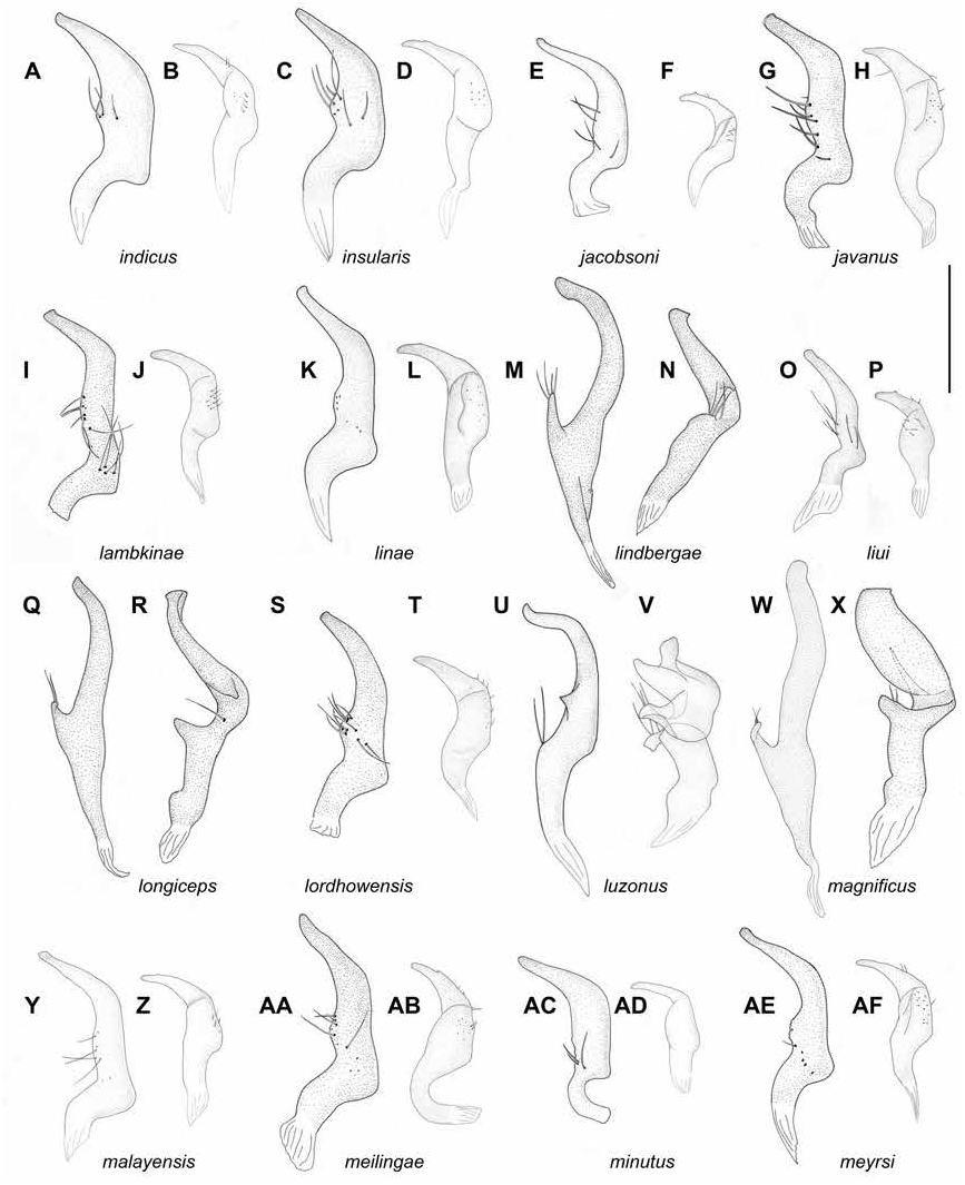
DESCRIPTION: Male. Total length 3.1–3.2. COLORATION (fig. 5): Head: Dorsal surface brown medially, yellow to pale brown laterally, sometimes with reddish tinge, longitudinal stripe dark brown; anterior side yellow to brown; frons brown, areas below antennal fossae whitish yellow, clypeus yellow to pale brown, brown basally, sometimes with reddish tinge; tubercle around antennal fossa whitish yellow with pale brown markings; lateral side whitish yellow to pale brown, buccula, mandibular and maxillary plates whitish yellow; ventral side whitish yellow to pale brown, sometimes brown anteriorly. Eye pale brown. Labium: Segments I–II whitish yellow, segment III whitish yellow, yellow ventrally, segment IV yellow. Antenna: Segment I yellow to pale brown; segment II pale brown to brown, sometimes with reddish tinge, segments III–IV brown to dark brown. Thorax: Anterior part of pronotum and punctures between anterior and posterior parts brown, anterior margin dark brown, posterior part of pronotum whitish yellow with brown marking near posterior angle; scutellum and mesoscutum whitish yellow to pale brown; mesoscutum sometimes somewhat darker than scutellum; punctures between scutellum and mesoscutum pale brown; thoracic pleura brown to dark brown; scent gland evaporative area uniformly whitish yellow. Hemelytron: Clavus translucent, its inner part pale brown to brown with margins brown, sometimes with reddish tinge, outer part of clavus colorless, pale brown to brown apically; corium translucent with marking along inner margin of corium brown, narrow, not reaching R+M; embolium opaque, whitish yellow, pale brown or reddish apically, with brown margins, opaque; cuneus mostly opaque, reddish with colorless and translucent outer part and brown outer margin; membrane translucent with grayish tinge; membrane cell pale brown veins. Legs: Coxae whitish yellow; femora whitish yellow basally, yellow apically, tibiae mostly yellow or pale brown, often whitish yellow apically, fore- and middle tarsi whitish yellow to pale brown, hind tarsus whitish yellow to yellow. Abdomen: Whitish yellow ventrally and pale brown with reddish tinge dorsally. SURFACE AND VESTITURE: Corium smooth, with shallow and scarce punctures. Dorsum, antennal segment I and femora with suberect setae shorter than antennal segment II diameter; abdomen clothed with suberect setae of different length. STRUCTURE AND MEASUREMENTS: Body ca. 4.4–4.6× as long as pronotum width. Head: Depression, delimiting occipital region distinct dorsally and laterally (as in Namyatova et al., 2016: fig. 4E); distance between depression and pronotum distinctly shorter than eye diameter; longitudinal sulcus on dorsal surface longer than eye diameter; distance from eye to pronotum slightly longer than eye diameter, not swollen laterally (as in Namyatova et al., 2016: fig. 4E); vertex ca. 1.2–1.3× as wide as eye, upraised (as in Namyatova et al., 2016: fig. 6D). Labium (as in Namyatova et al., 2016: figs. 6D, 9C): Reaching posterior margin of metasternum or slightly surpassing it; segments I and II strongly reduced; combined subequal to half of segment III; segment I as long as wide; segment II slightly longer than wide, its dorsal surface elongate posteriorly; segment III slightly longer than ventral side of head; segment IV twice as long as segment III. Antenna: Segment I cylindrical (as in Namyatova et al., 2016: fig. 8A), ca. 1.2–1.6× as long as head width, ca. 0.9–1.1× as long as pronotum width; segment II ca. 1.9× as long as head width, ca. 1.3× as long as pronotum width; segment III slightly longer than segment II; segment IV ca. 0.25× as long as segment IV. Thorax: Anterior and posterior parts of pronotum subequal in length; collar delimited; posterior part slightly upraised; posterior margin straight or slightly concave, pronotum ca. 1.2–1.3× as wide as long and ca. 1.4–1.5× as wide as head; mesoscutum exposed. Hemelytron: Marking along inner margin of corium almost flat; inner margin of cuneus convex (as in Namyatova et al., 2016: fig. 13E), outer margin of cuneus almost ca. 2.5× as long as base. Abdomen: Genital capsule rotated left at right angle relative to the rest of abdomen. Genitalia: Genital capsule (fig. 14R) ca. 1.5× as long as wide; ventral wall ca. 1.5× as long as dorsal wall, its posterior margin smooth, semioval, without outgrowth, not curved, its apex inclined leftward; sides of genital capsule not modified; right paramere socket slightly angulate and left socket rounded; distance between paramere sockets ca. 0.7× as long as genital capsule width at base. Right paramere (fig. 12I) distinctly curved in apical half; apex slightly concave; medial part slightly wider than basal part, bearing setae, with outer margin concave and inner margin convex; outer angle distinct; inner angle rounded, without setae; basal part of paramere ca. 0.15–2× as long as rest of paramere. Left paramere (fig. 12J) widened, only slightly curved; apical part straight, with toothlike outgrowth on posterior side medially (as in fig. 11G) and without outgrowth on dorsal surface; middle part of paramere widened, without swelling or outgrowth; setae only on middle part near outer margin. Aedeagus (general view as in Namyatova et al., 2016: fig. 22I) conjunctiva weakly sclerotized; secondary gonopore placed at base of vesica in repose; sclerotization of ductus seminis around secondary gonopore shorter than wide; vesica with five spicules, including spicules A, B, and C (fig. 8O).
Female. Total length 3.2–3.5. COLORATION (fig. 5): Head: Similar to male, but dorsal surface sometimes yellow to pale brown with reddish tinge, brown anteriorly; frons yellow to brown, sometimes with reddish tinge; clypeus yellow sometimes with reddish tinge, sometimes brown basally; ventral side whitish yellow to pale brown. Labium and antenna: As in male. Thorax: Similar to male, but anterior part of pronotum sometimes pale brown with reddish tinge and brown anterior margin; thoracic pleura yellow to brown, embolium translucent. Abdomen: Whitish yellow with segment X and dorsal surface red. SUR- FACE AND VESTITURE: As in male. STRUC- TURE AND MEASUREMENTS: Structure as in male; body ca. 4.1–4.4× as long as pronotum width; vertex ca. 1.8–1.9× as wide as eye diameter; antennal segment I ca. 1.4–1.6× as long as head width, ca. 0.9–1.0× as long as pronotum width; segment II ca. 1.7–1.9× as long as head width, ca. 1.1–1.3× as long as pronotum width; pronotum ca. 1.2–1.3× as wide as long and ca. 1.5–1.7× as wide as head. Genitalia (as in Namyatova et al., 2016: fig. 23F, G): Dorsal labiate plate wider than distance between apodemes of second valvula; mostly smooth, without distinct striations, with semicircular sclerite and distinct sclerotized rings laterally; lateral oviducts placed almost medially, very close to each other, spermathecal gland placed between lateral oviducts; dorsal labiate plate with distinct tubercles, without membranous lobe medially.
DISTRIBUTION: Christmas Is. (fig. 18).
HOST PLANTS: Unknown.
ETYMOLOGY: The species is named after Christine Lambkin, curator of the Entomology in Queensland Museum (Brisbane), who has assisted us in this work including the loan of material.
DISCUSSION: Felisacus lambkinae is not very similar in coloration to any other Felisacus . How-
ever, it has near identical vesical spicules, with F. javanus (cf. fig. 8N with 8O), sharing spicules A, B, and C, with all of them similar in shape. These species also have similar body size and a red cuneus, but F. javanus differs by the anterior part of the pronotum being yellow to pale brown, the inner part of the clavus is mostly whitish yellow to yellow, with reddish or pale brown margins, and the labium reaches the posterior margin of the mesosternum or slightly surpasses it.
MATERIAL EXAMINED: Holotype: AUSTRA- LIA: Territory of Christmas Island : Grants Well. Christmas Is., 10.48333 ° S 105.65 ° E, 27 Apr 1989, J.C. Cardale, 13 (00033781) ( ANIC). Paratypes: AUSTRALIA: Territory of Christmas Island: Grants Well. Christmas Is., 10.48333 ° S 105.65 ° E, 15 Apr 1989, J.C. Cardale, 23 (00033781, 00033783), 1♀ (00033784) ( ANIC) GoogleMaps ; 27 Apr 1989, J.C. Cardale, 4♀ (00033785–00033788) ( ANIC) .


| ANIC |
Australian National Insect Collection |
No known copyright restrictions apply. See Agosti, D., Egloff, W., 2009. Taxonomic information exchange and copyright: the Plazi approach. BMC Research Notes 2009, 2:53 for further explanation.