
Cheliplana triductibus, Steenkiste, Niels Van, Volonterio, Odile, Schockaert, Ernest & Artois, Tom, 2008
|
publication ID |
https://doi.org/ 10.5281/zenodo.184571 |
|
DOI |
https://doi.org/10.5281/zenodo.6230415 |
|
persistent identifier |
https://treatment.plazi.org/id/443987C2-BB33-266E-FF4D-2E786D44FD15 |
|
treatment provided by |
Plazi |
|
scientific name |
Cheliplana triductibus |
| status |
sp. nov. |
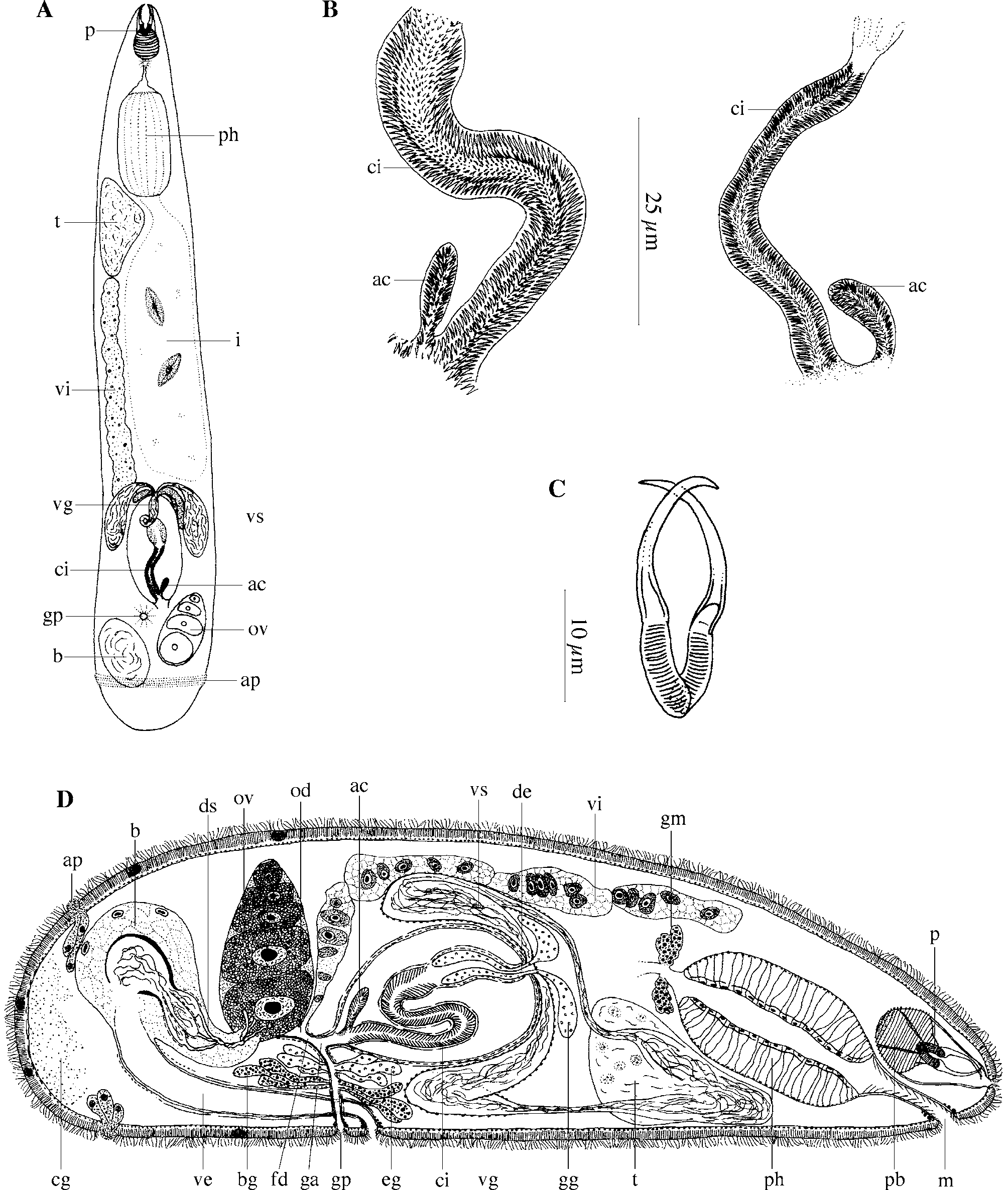
Cheliplana triductibus n.sp.
( Fig. 4 View FIGURE 4 )
Locality. Playa Ramírez, Departamento de Montevideo, Uruguay (34°54’58.55”S, 56°10’11.70”W). Mid-eulittoral, rather coarse sand from a sheltered area with a large amount of fine fraction (12/08/04): type locality.
Material. Observations on live animals. Three whole mounts, one designated holotype ( SMNH 7496), the rest paratypes (HU no. 401–402) and seven serially-sectioned specimens, three of which designated paratypes (HU no. 403–405).
Etymology. The species epithet refers to the three sclerotized spermatic ducts and muscular ducts connecting the ovary with the bursa. Tres (Lat.): three. Ductus (Lat.): duct.
Description. The body length of the animal varies between 0.7– 1 mm. Habitus and internal organisation strongly resemble those of C. varicauda Brunet, 1971 (see Brunet 1971).
The epidermis is syncytial and ± 2 µm thick, with cilia ± 2 µm long. The basal membrane is ± ½ of the epidermis height thick. Caudally, a girdle of adhesive papillae surrounds the body.
The proboscis is about 1/8 to 1/10 of the body length long. The two small proboscis halves are 10–12 μm long. Both are armed with a simple, 17–20 μm long hook. Accessory hooks are lacking. The postrostral bulb is ± 2/3 of the length of the proboscis (± 20 μm).
The pharynx is situated in the anterior part of the body. The wall of the prepharyngeal cavity is covered with spines. The pharynx is typically cylindrical and its length is about 1/5 of the body length (± 140–220 μm). The mouth is situated rostrally, at the ventral side, and is surrounded by circular muscles.
The gonads are unpaired. The testis is situated in the anterior part of the body, ventrally from the pharynx. The ovary lies at about ¾ of the body length at the left-hand side of the body. The vitellarium is situated dorsally, and extends from just behind the pharynx up to the oviduct. The gonopore lies at 70 %, just behind the external opening of the vagina externa, and is surrounded by a weak sphincter. The common genital atrium is slender, tubular and proximally surrounded by a circular muscle layer. Two types of glands empty into the common genital atrium: eosinophilic glands and, more distally, basophilic glands. Distally from the opening of the basophilic glands, the common genital atrium is surrounded by longitudinal muscles.
Two vasa deferentia leave the testis, and run caudally until halfway past the copulatory organ. Each of them then widens to form a seminal vesicle, which turns 180° and runs back in anterior direction. The seminal vesicles and the vasa deferentia are lined with an anucleated, membranous epithelium, and the seminal vesicles are surrounded by a circular muscle layer. Proximally, both vasa deferentia join to form the ejaculatory duct, which enters the copulatory bulb, and receives prostate secretion (conjuncta-type copulatory organ; terminology of Karling 1956a). The prostate vesicle is surrounded by an inner circular and an outer longitudinal muscle layer. In live animals, the prostate vesicle and ejaculatory duct clearly showed a single winding. The prostate glands are eosinophilic, with the nuclei outside of the prostate vesicle. The prostate vesicle and the ejaculatory duct open into a slender cirrus. The cirrus and the prostate vesicle are surrounded by a muscular septum, which is lined with an inner circular and an outer longitudinal muscle layer (conjuncta-duplex type copulatory organ; terminology of Karling 1956a). The cirrus is 52–76 μm long, with a diameter of about 8 μm.
It is lined with a large number of rows of small, sclerotized teeth, which are all oriented in a distal direction. Just before the cirrus enters the atrium through its anterior wall, it receives a small, armed accessory cirrus, 12–17 μm long and 3–4 μm broad. The cirrus, the accessory cirrus and the common genital atrium are surrounded by a longitudinal muscle layer.
The short oviduct receives the vitelloduct through its dorsal wall, and together they form the very short female duct. This female duct enters the common genital atrium a little caudally from the place where the male genital system enters. A circular muscle layer surrounds the female duct, the oviduct and the most distal part of the vitelloduct. The female duct is lined with an anucleated, membranous epithelium. Caudally from the ovary a large bursa occurs, which contains several compartments with sperm. The female bursa is connected to an external vagina, which is a relatively long duct, lined with a high, anucleated epithelium. Its proximal part is surrounded by circular muscles. This part also contains spermatozoa. The distal part of the vagina is surrounded by longitudinal muscles. The vaginal opening is surrounded by a strongly-developed sphincter. Within the dorso-anterior part of the bursa, three muscular sperm-containing compartments can be observed. From each of these compartments a sclerotized spermatic duct departs towards the ovary. In their middle parts these three ducts are wound around each other, so that they cannot be discerned separately. Near the ovary they diverge again, and enter the ovary as three separate ducts near to the place where the oviduct departs. The spermatic ducts are surrounded by a parenchymatous tissue, which is a continuation of the parenchymatous tissue of the bursa. A uterus is absent.
Discussion. See discussion Cheliplana uruguayensis n.sp.
| SMNH |
Saskatchewan Museum of Natural History |
No known copyright restrictions apply. See Agosti, D., Egloff, W., 2009. Taxonomic information exchange and copyright: the Plazi approach. BMC Research Notes 2009, 2:53 for further explanation.