
Cheliplana elkhornica Karling, 1989
|
publication ID |
https://doi.org/ 10.11646/zootaxa.4970.3.2 |
|
publication LSID |
lsid:zoobank.org:pub:FEABE248-E1EA-48F5-A1AF-0077FE40C257 |
|
DOI |
https://doi.org/10.5281/zenodo.4766738 |
|
persistent identifier |
https://treatment.plazi.org/id/03E0878B-1866-FF80-62BE-1EAFFBB4CF2D |
|
treatment provided by |
Plazi |
|
scientific name |
Cheliplana elkhornica Karling, 1989 |
| status |
|
Cheliplana elkhornica Karling, 1989
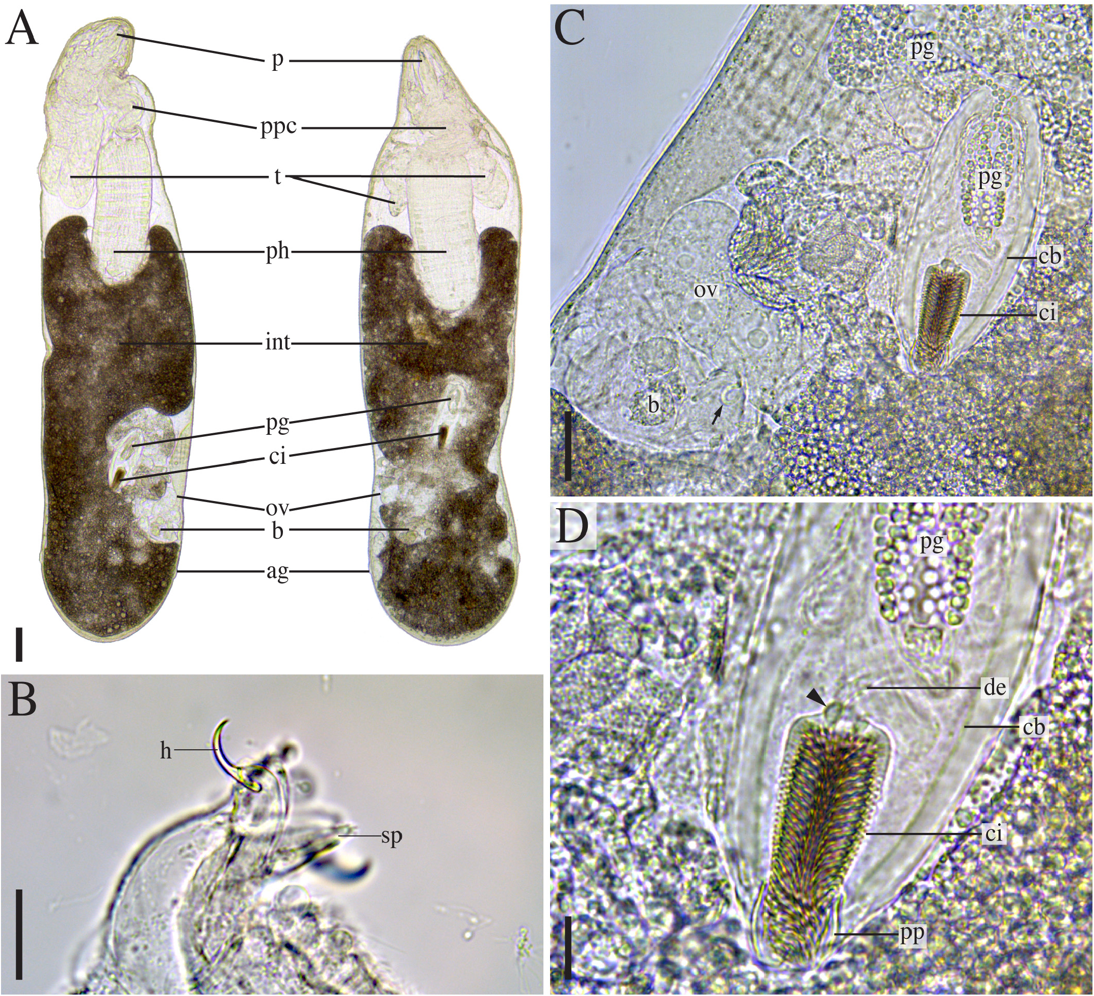
Fig. 7 View FIGURE 7
Material examined. New material. CANADA • 2 whole mounts; British Columbia, Calvert Island, North Beach ; 51°39’53”N, 128°08’47”W; superficial sediment with coarse fraction in run off and intertidal pool near boulders; MI4184–MI4185 GoogleMaps .
Reference material 1 whole-mounted specimen from California (holotype, SMNH Type 6800) .
Known distribution. Monterey Bay, California, United States ( Karling 1989).
Remarks. Our observations on the holotype and the new material from Canada mostly correspond to what was reported by Karling (1989). Live specimens are ~ 1.5 mm long ( Fig. 7A View FIGURE 7 ). The haptic girdle (hg, Fig. 7A View FIGURE 7 ) is comprised of ~30 papillae. Proboscis hooks are smooth and are 20–25 μm (California) or 20–23 μm ( Canada) long (h, Fig. 7B View FIGURE 7 ). Proboscis hook supports are ~30 μm long in the Canadian specimens. Blunt side pieces with at least two very short protrusions were observed in one Canadian specimen (sp, Fig. 7B View FIGURE 7 ).
Karling (1989) describes only one testis above the pharynx. However, in one of the Canadian specimens (t, Fig. 7A View FIGURE 7 , right specimen, dorsal view), paired testes could be observed. The other specimen from Canada appears to have only one testis, but it is clearly bilobed (t, Fig. 7A View FIGURE 7 , left specimen, lateral view). A connection between the two lobes or testes may be present, or the orientation of the specimen may play a role in the appearance of the testes. The seminal vesicles are paired. The cylindrical copulatory bulb (cb, Fig. 7C–D View FIGURE 7 ) is ~140 μm (California) or 190–250 μm ( Canada) long and is surrounded by strong longitudinal muscles. The prostatic glands contain a very coarse-grained secretion and enter the copulatory bulb proximally, where they form an elongate prostatic vesicle (pg, Fig. 7C–D View FIGURE 7 ). A short ejaculatory duct is separated from the cirrus by a muscular constriction (arrowhead, Fig. 7D View FIGURE 7 ). The cirrus measures ~75 μm (California) to 80–82 μm ( Canada) and is lined with spines of 4–8 μm (California) up to 10 μm ( Canada) (ci, Fig. 7A,C,D View FIGURE 7 ). The spines are arranged symmetrically, except for the distal part where their orientation appears slightly twisted (Fig. 15B–C in Karling 1989). A short, slightly sclerotised penis papilla (20–27 μm in the Canadian specimens) folds back over the distal part and can be everted (pp, Fig. 7D View FIGURE 7 ).
The female reproductive system was described by Karling (1989) as ‘principally like those in C. californica ’. No vagina externa was observed. Anterior to the genital pore, Karling (1989) reports a small structure, which he interpreted as a temporary vagina. A bursa is situated alongside the ovary (b, ov, Fig. 7A View FIGURE 7 ). In the Canadian specimens, a slightly sclerotised tube was visible between the bursa and ovary (arrow, Fig. 7C View FIGURE 7 ). However, Karling did not observe a spermatic duct. Possibly, this structure is the ‘temporary vagina’ Karling referred to. The holotype and other whole-mounted specimens did not allow for more detailed observations.
| SMNH |
Department of Paleozoology, Swedish Museum of Natural History |
No known copyright restrictions apply. See Agosti, D., Egloff, W., 2009. Taxonomic information exchange and copyright: the Plazi approach. BMC Research Notes 2009, 2:53 for further explanation.
|
Kingdom |
|
|
Phylum |
|
|
Class |
|
|
Order |
|
|
Family |
|
|
Genus |