
Pterosphenus biswasi, Rage & Bajpai & Thewissen & Tiwari, 2003
|
publication ID |
https://doi.org/ 10.5281/zenodo.4650665 |
|
persistent identifier |
https://treatment.plazi.org/id/03A08794-FFF0-FF8D-CA20-FE80E0ADDE2A |
|
treatment provided by |
Felipe |
|
scientific name |
Pterosphenus biswasi |
| status |
sp. nov. |
Pterosphenus biswasi n. sp. ( Figs 4 View FIG ; 5A View FIG )
HOLOTYPE. — 1 trunk vertebra ( RUSB 2784-4 ).
ETYMOLOGY. — Named for Dr. S. K. Biswas, in recognition of his work on the geology of Kutch.
TYPE LOCALITY. — HD Pit in Panandhro Mine, Kutch District, India.
REFERRED MATERIAL. — 2 vertebrae: 1 from HD Pit ( RUSB 2565-1) and 1 from Channel Pit ( RUSB 2790-21).
HORIZON. — Naredi Formation, Ypresian, Lower Eocene.
DIAGNOSIS. — Species of Pterosphenus distinguished from Pt. schucherti , Pt. schweinfurthi , and Pt. muruntau by its markedly less deeply concave anterior border of the zygosphene. Differs from Pt. schucherti and Pt. schweinfurti in having the zygapophyseal plane located slightly higher. Differs from Pt. sheppardi by its longer and more oblique paradiapophyses, and the anteroposteriorly longer basis of its hypapophysis. Distinguished from Pt. kutchensis n. sp. by its less laterally compressed vertebrae, the concave anterior border of its zygosphene, its markedly shorter paradiapophyses, separated bases of the paradiapophyses, and the presence of an anterior hypapophysis.
DESCRIPTION OF HOLOTYPE
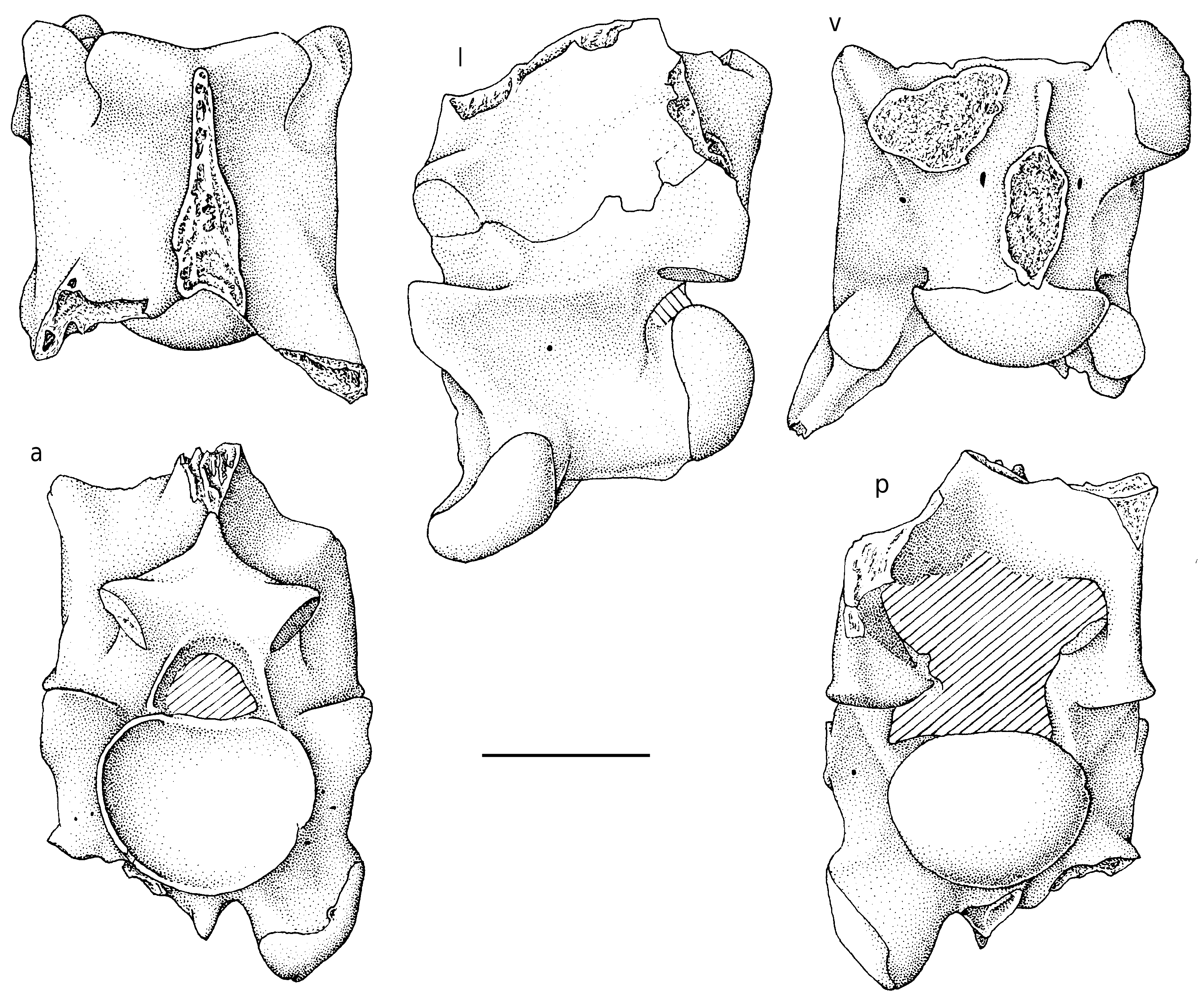
The holotype is a large, massive trunk (presumably mid-trunk) vertebra ( Fig. 4 View FIG ). The measurements are as follows: length of centrum from cotylar rim to tip of condyle: 18.9 mm; width through prezygapophyses: 19.7 mm; width of interzygapophyseal constriction: 18.2 mm; diameter of cotyle: 12.8 mm; width of zygosphene: 13.2 mm.
In anterior view, the vertebra is clearly compressed laterally and high. The prezygapophyses are small; their articular facets are slightly inclined above the horizontal and they lie slightly above the level of the floor of the neural canal. The zygosphene is thick and hardly wider than the cotyle; its dorsal border forms the base
d
of the anterior edge of the neural spine, which gives a subtriangular shape to the frontal aspect of the zygosphene. The cotyle appears to be slightly depressed dorsoventrally and its dorsal part is truncated. The section of the neural canal is small, markedly narrower than the zygosphene and cotyle. The pterapophyses are incomplete; the base of the right one shows that they were high. The paradiapophyses are situat- ed low and distant from the centrum. Below the cotyle, a space that represents about one third the diameter of the cotyle, separates the bases of the paradiapophyses. A small anterior hypapophysis is present beneath the cotyle, between the paradiapophyses. The vertebra lacks para- cotylar foramina but irregular small foramina open in the anterior face of the prezygapophyseal buttresses.
In dorsal aspect, the vertebra appears to be more or less squarish, not clearly longer than wide. The prezygapophyseal facets are small, not elongate, and directed more anteriorly than laterally. The interzygapophyseal constriction is hardly expressed. The lateral borders of the interzygapophyseal ridges are slightly convex laterally. The zygosphene does not form clearly defined lateral lobes; its anterior border is weakly concave. Anteriorly, the neural spine reaches the anterior face of the zygosphene; it grows thicker posteriorly. The basal parts of the pterapophyses that are preserved form blunt, poorly defined keels. The median notch in the posterior border of the neural arch is shallow and obtuse, but its bottom is clearly triangular. The roof of the zygantrum is not extended.
In lateral view, the vertebra is short and high. The neural spine and the hypapophysis are broken off. The zygosphenal facet is small, subcircular, and directed more dorsally than anteriorly. The interzygapophyseal ridge is strong and prominent. The anterolateral ridge of the prezygapophyseal buttress originates on the anterodorsal margin of the paradiapophysis. The articular facet of the paradiapophysis is elongate and markedly oblique (about 45° from the vertical); there is no distinction between the dia- and parapophyseal areas. The axis of the condyle is horizontal. A small lateral foramen opens below the interzygapophyseal ridge.
In posterior view, the lateral flanks of the neural arch are vertical. The cotyle is slightly depressed. The area of zygantral foramina is obscured by matrix.
In ventral view, the centrum appears cylindrical. It lacks subcentral ridges. The base of the hypapophysis is elongate. Posteriorly, it reaches the condyle; anteriorly, it is prolonged by a thin keel the anterior part of which forms the anterior hypapophysis. Elongate subcentral foramina are present.
OTHER SPECIMENS AND VARIATION
Only two other specimens are available. One large vertebra (RUSB 2565-1) is very worn; a smaller vertebra (RUSB 2790-21) is damaged. They are referred to Pt. biswasi n. sp. on the basis of markedly separated bases of paradiapophyses (i.e. they clearly differ from Pt. kutchensis n. sp. from the same locality), slightly concave anterior border of zygosphene, and an anterior hypapophysis below the cotyle (the latter feature cannot be checked in RUSB 2565-1). In both vertebrae, as in the holotype, the anterior edge of the neural spine is continuous with the anterior face of the zygosphene; as a result, the latter face is subtriangular. The presence or absence of foramina is not verifiable in these two specimens.
COMMENTS
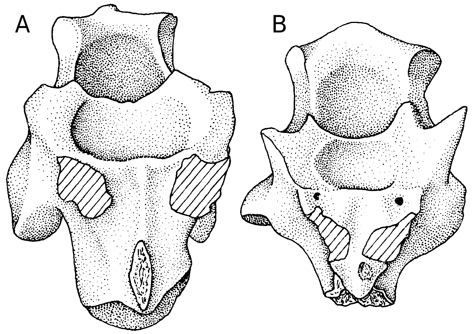
A problem arises from the fact that there are two species at Panandhro Mine, a small and a large one. Therefore, it may be argued that the palaeophiid described above as Pt. kutchensis n. sp. is only represented by juvenile individuals of Pt. biswasi n. sp. However, as shown above, the vertebrae referred to Pt. kutchensis n. sp. include some juveniles but mainly adult specimens. Moreover, at least one of the characters that distinguish the two species cannot be interpreted as an ontogenetic change: the paradiapophyses originate from a common base in Pt. kutchensis n. sp. whereas the bases are markedly separated in Pt. biswasi n. sp. as in all other snakes. Such an ontogenetic change has never been reported. In addition, vertebrae of similar size belonging to these two species (i.e. a large vertebra of Pt. kutchensis n. sp. and a small one of Pt. biswasi n. sp.) display the conditions of the paradiapophyses typical for these two species: the paradiapophyses arise from a single base in the large vertebra of Pt. kutchensis n. sp. while the bases of the two paradiapophyses are clearly separated on the vertebra of similar size belonging to Pt. biswasi n. sp. ( Fig. 5 View FIG ). This clearly demonstrates that this difference is not of ontogenetic nature and that there are two distinct species.
Pt. biswasi n. sp. is easily distinguished from Pt. kutchensis n. sp. Apart from its larger size and separate bases of paradiapophyses, it differs from Pt. kutchensis n. sp. in having an anterior hypapophysis, less laterally compressed vertebrae, shorter paradiapophyses, and a concave anterior border of zygosphene.
The distinction between Pt. biswasi n. sp. and the other species of Pterosphenus is less marked. It differs from all other species in having a shallow concave anterior border of zygosphene, while it is deeply concave, even notched, in Pt. schucherti , Pt. schweinfurthi , and Pt. muruntau (not observable in Pt. sheppardi ). Pt. biswasi n. sp. further differs from Pt. schucherti and Pt. schweinfurthi by its zygapophyseal plane that is located slightly higher (mainly shown by the postzygapophyseal facets) and from Pt. sheppardi by its more elongate and more oblique paradiapophyses, and the anteroposteriorly longer base of its hypapophysis.
No known copyright restrictions apply. See Agosti, D., Egloff, W., 2009. Taxonomic information exchange and copyright: the Plazi approach. BMC Research Notes 2009, 2:53 for further explanation.
|
Kingdom |
|
|
Phylum |
|
|
Class |
|
|
Order |
|
|
Family |
|
|
Genus |