
Pterosphenus kutchensis, Rage & Bajpai & Thewissen & Tiwari, 2003
|
publication ID |
https://doi.org/ 10.5281/zenodo.4650665 |
|
persistent identifier |
https://treatment.plazi.org/id/03A08794-FFFC-FF8A-CA50-FE00E6B1DC2B |
|
treatment provided by |
Felipe |
|
scientific name |
Pterosphenus kutchensis |
| status |
sp. nov. |
Pterosphenus kutchensis n. sp. ( Figs 2 View FIG ; 3 View FIG ; 5B View FIG )
HOLOTYPE. — 1 trunk vertebra ( RUSB 2721-1 ).
ETYMOLOGY. — From Kutch, name of the District in which is situated the type locality.
TYPE LOCALITY. — HD Pit in Panandhro Mine, Kutch District, India.
REFERRED MATERIAL. — 105 vertebrae: 85 from HD Pit ( RUSB 2564-1 to 2564-26; RUSB 2721-2 to 2721-57; RUSB 2784-1 to 2784-3); 20 from Channel Pit ( RUSB 2790-1 to 2790-20).
HORIZON. — Naredi Formation, Ypresian, Lower Eocene.
DIAGNOSIS. — Pterosphenus that differs from all other snakes in having paradiapophyses that extend further anteroventrally than in any other snake. These paired structures originate from a common base, or may rarely be separated but with their bases closely appressed against each other. Differs from other species in the genus in lacking anterior hypapophyses, in having the anterior edge of the neural spine separat- ed from the anterior border of the zygosphene by a narrow step in most vertebrae, and in having a nonconcave anterior border of the zygosphene. Further differs from Pt. sheppardi in having higher pterapophyses.
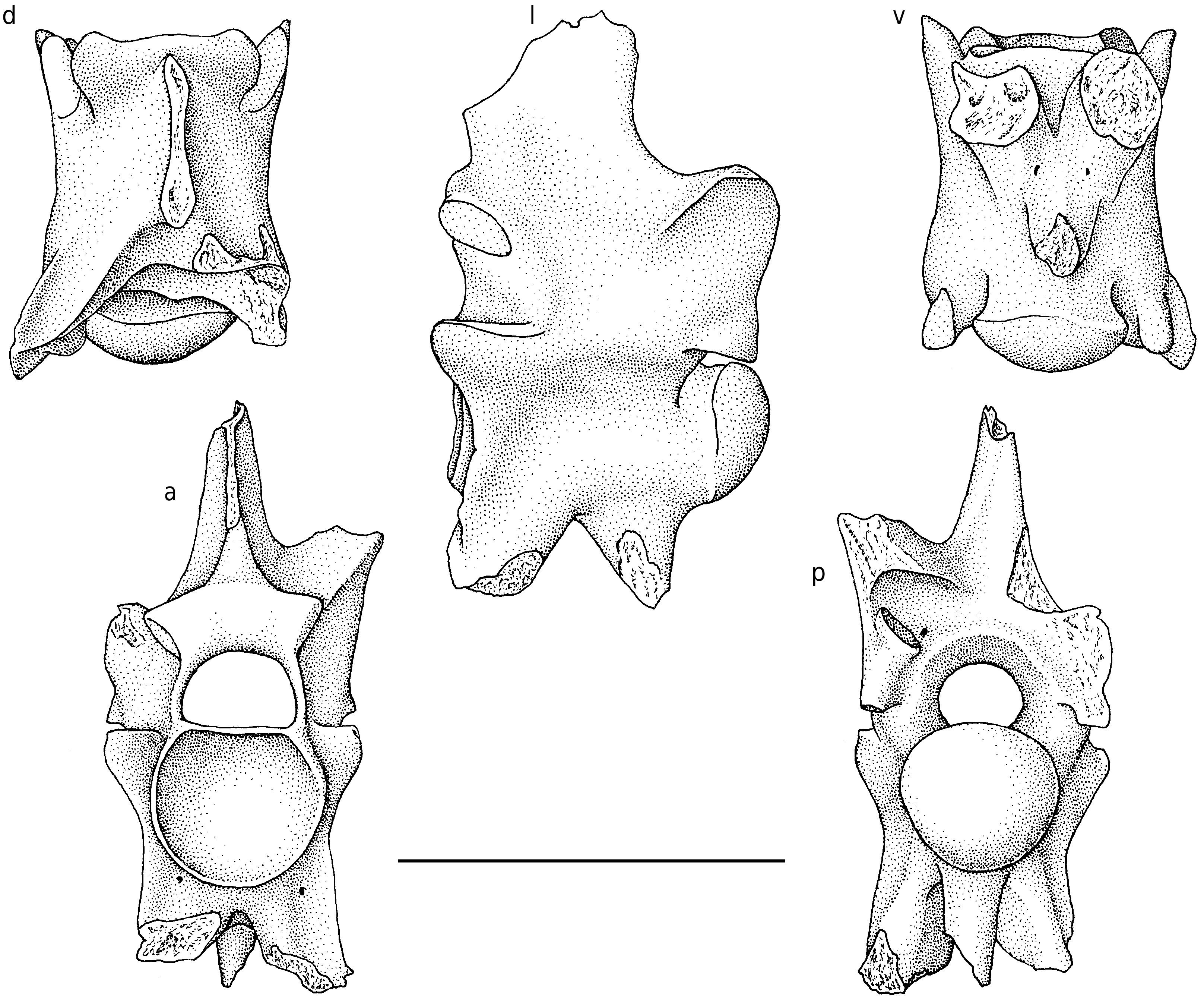
DESCRIPTION OF HOLOTYPE
The holotype ( Fig. 2 View FIG ) is a relatively small trunk vertebra, presumably from the mid-trunk region. Its measurements are as follows: length of centrum from cotylar rim to tip of condyle: 8.3 mm; width through prezygapophyses: 6.6 mm; minimum width of interzygapophyseal constriction: 5.4 mm; diameter of cotyle: 4.4 mm; width of zygosphene: 4.7 mm.
In anterior view, the vertebra is markedly compressed laterally and high. The prezygapophyses are very reduced; their articular facets are horizontal and level with the floor of the neural canal. The zygosphene is thick and slightly wider than the cotyle. The dorsal border of the zygosphene is slightly arched dorsally. The base of the anterior edge of the neural spine is rather thick but it narrows dorsally; the dorsal part of the neural spine is broken off. The cotyle is subcircular but its dorsal part is truncated. The neural canal is relatively small. The pterapophyses are damaged but the left one shows that they were high. The paradiapophyses show a very unusual morphology: they are thick, very long (although their distal parts are broken off), and they are not separated from each other in the sagittal plane, i.e. they have a common base. As a result, the vertebra lacks an anterior hypapophysis. The anterior face of each paradiapophysis bears a wide and shallow groove. A small foramen opens in each of these grooves, close to the cotyle. The hypapophysis is compressed laterally. Paracotylar foramina are absent.
In dorsal aspect, the vertebra appears narrow and relatively elongate. The prezygapophyseal articular facets are small, elongate, and directed obliquely, almost anteriorly. On each side, the vertical ridge formed by the prezygapophyseal buttress slightly projects beyond the articular facet. The interzygapophyseal constriction is weakly expressed. The lateral borders of the interzygapophyseal ridges are nearly straight. The zygosphene comprises two lateral lobes that do not strongly project anteriorly; between them, the anterior border is feebly convex. The neural spine approaches the anterior border of the zygosphene but it does not reach it. The remaining part of the left pterapophysis appears as a low, but well defined keel. The median notch in the posterior border of the neural arch is wide and obtuse, it appears as a broad embayment. As in all palaeophiids, the zygantral roof is reduced.
In lateral view, the vertebra is markedly higher than long, despite the fact that the dorsal part of the neural spine and the ventral parts of the paradiapophyses and hypapophysis are broken off. The height of the neural spine cannot be estimat- ed. The zygosphenal facets are small, ovaloid and oblique. There is no marked interzygapophyseal ridge. The prezygapophysis lacks a prezygapophyseal process, but it forms a vertical ridge that extends from the tip of the articular facet to the anterolateral border of the paradiapophysis. The paradiapophysis is directed ventrally and slightly anteriorly. The articular facet for the rib is lacking, but an eroded area on the distal part of the remaining portion might correspond to the dorsal part of the diapophyseal surface. Anyway, at least most of the articular facet was on the missing part, i.e. it occupied a very ventral position, far from the centrum. The incomplete hypapophysis is vertical and not located very posteriorly. The axis of the condyle is horizontal. There is no perceivable lateral foramen.
In posterior view, as in anterior aspect, the laterally compressed morphology is striking. Beneath the pterapophyses the lateral flanks of the neural arch are subvertical. Only the left zygantral foramen appears to be present. The centrum is somewhat triangular in cross-section.
The ventral view displays the unusual position of the paradiapophyses the bases of which are not separated in the sagittal plane. As a consequence of the subtriangular cross-section of the centrum, subcentral ridges are lacking. Anterior to the condyle, the centrum forms a neck that is clearly narrower than the condyle. Two subcentral foramina open between the bases of the hypapophysis and paradiapophyses.
OTHER VERTEBRAE AND VARIATION
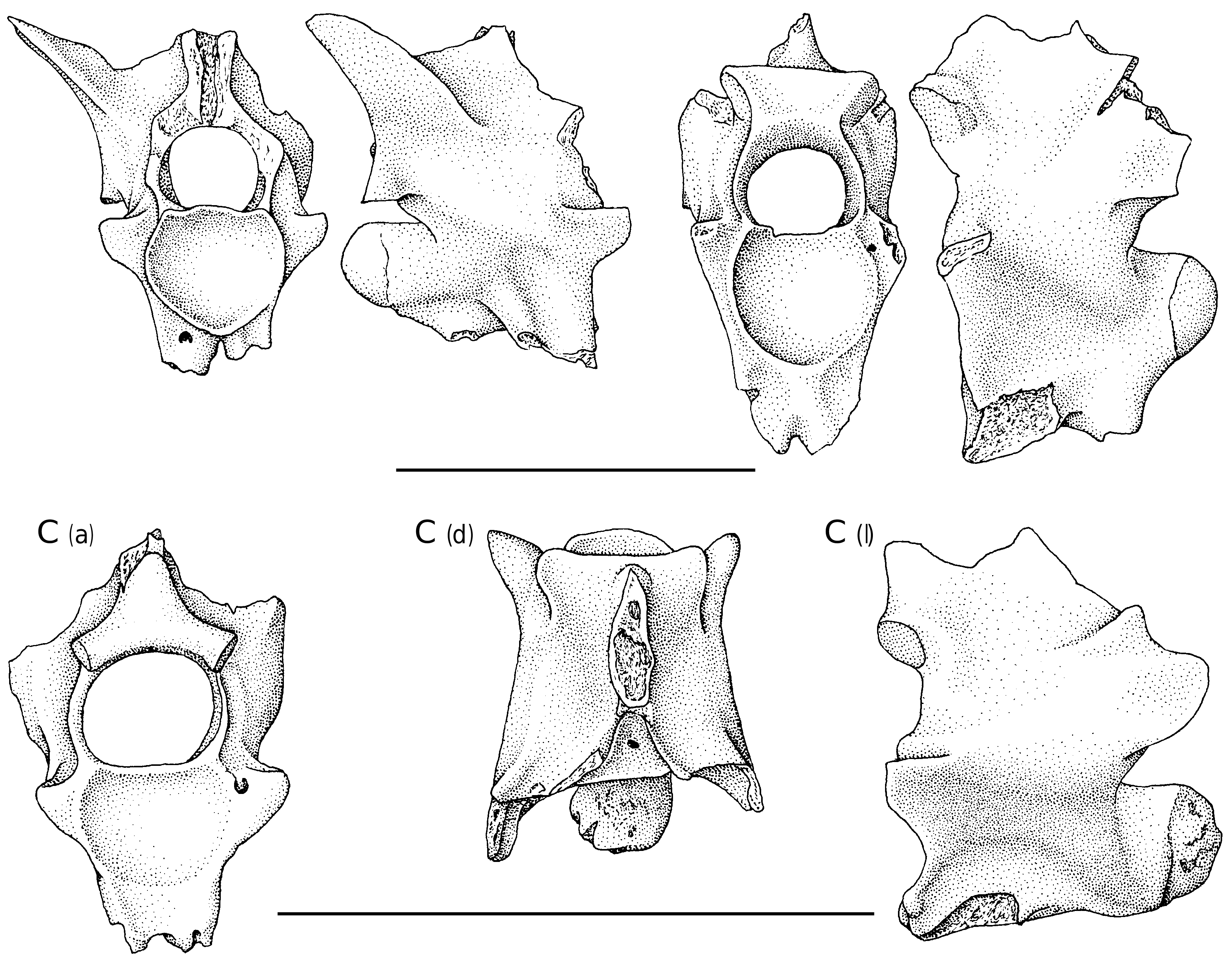
No caudal vertebrae are known. Two vertebrae each preserve a complete pterapophysis. In lateral aspect, this process appears as a triangular lamina the anterior border of which is sharp. In RUSB 2790-1, the pterapophysis is directed dorsolaterally ( Fig. 3A View FIG ) whereas in RUSB 2784-1 it is more vertical.
A few vertebrae of juvenile individuals are known ( Fig. 3C View FIG ). They are of interest because they prove that the “large” vertebrae of Pt. kutchensis n. sp., that are small for the genus Pterosphenus , belong to adults. The vertebrae of juveniles show the features that are usual in all snake families: neural canal relatively wider than in adults, zygosphene and lateral walls of vertebrae thinner, cotyle more depressed dorsoventrally, and zygosphene entirely overhanging (i.e. anterior parts of lateral walls of the neural canal not completed).
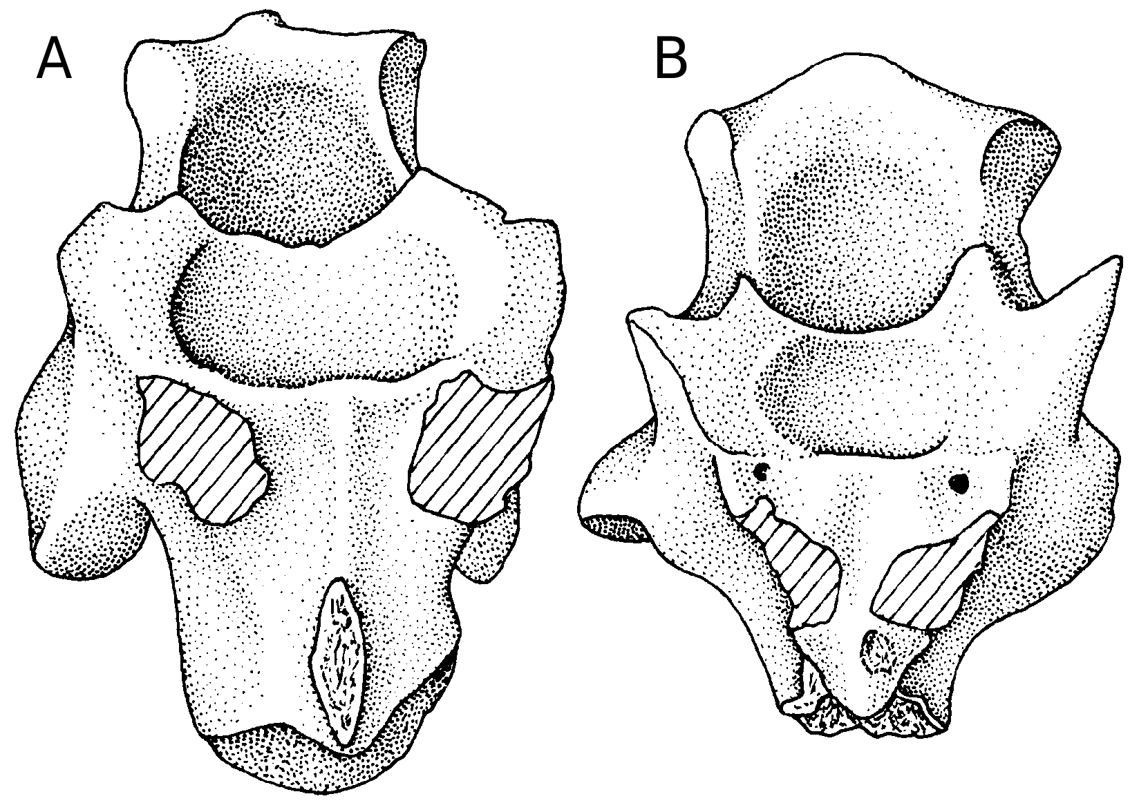
Variation in the trunk vertebrae is minimal. In most vertebrae, as in the holotype, the anterior edge of the neural spine is separated from the anterior border of the zygosphene by a narrow surface; however, in a few vertebrae the top of the anterior border of the zygosphene is prolonged without a break into the anterior edge of the neural spine. The latter condition is seen in other species of Pterosphenus . In Pt. kutchensis n. sp., the variation of this feature does not appear to be related to the position of vertebrae in the vertebral column. In some vertebrae, that are more laterally compressed than the holotype, the common base of the paradiapophyses is deeper; it appears as a thick process beneath the cotyle ( Fig. 3B View FIG ). It is not possible to determine whether such vertebrae are more anterior or more posterior than those exemplified by the holotype. In a few, damaged vertebrae, it is possible that the common base of the paradiapophyses is very shallow or the paradiapophyses are separated but closely appressed against each other. Zygantral foramina are often lacking whereas their presence is constant in non-palaeophiid snakes. But, irrespective of the presence or absence of the usual zygantral foramina, a sagittal foramen sometimes pierces the posterior wall of the neural arch between the two zygantral fossae, below the neural spine. This
A (a) A (l)
B (a) B (l)
condition of the zygantral foramina seems common in Palaeophiidae . Paracotylar, lateral, and subcentral foramina are rarely and irregularly present. The foramen that opens in the anterior groove of each paradiapophysis, close to the cotyle, is nearly always present.
The size ranges from juveniles (centrum length: about 4.3 mm) to largest adults (centrum length: 10.5 mm).
COMMENTS
This snake poses a peculiar problem. The long paradiapophyses are more or less reminiscent of pleurapophyses, i.e. processes present only in caudal vertebrae. Since, on the available vertebrae, paradiapophyseal articular facets are not obser- vable we are led to conclude that either these facets were on the distal parts of the paradiapophyses that are always broken off (which is quite possible because the facets are borne by spongy bone) or that the processes are pleurapophyses. But, if these processes are pleurapophyses, then all vertebrae come from the caudal region, which is not possible. Caudal vertebrae are, by far, more rarely found than vertebrae from the trunk region. Moreover, these vertebrae do not come from a single individual; they have been found in two sites (HD Pit and Channel Pit) and the vertebrae are of different sizes. Besides, caudal vertebrae of Palaeophis are known, and as in nearly all snakes they have typical pleurapophyses and paired haemapophyses ( Rage 1983a). The verte- brae of Pt. kutchensis n. sp. lack the latter processes but they have all a hypapophysis. The caudal vertebrae of nearly all snakes have paired haemapophyses; they are replaced by a haemal keel in a very few snakes ( Szyndlar & Böhme 1996). In the caudal region, hypapophyses occur only in the anterior caudal vertebrae of two living genera; moreover, they appear as deep keels rather than true hypapophyses ( Szyndlar & Rage 2003). Consequently, the presence of true hypapophyses on all vertebrae demonstrates that they come from the trunk region. Caudal vertebrae of Pt. kutchensis n. sp. have not been found.
These vertebrae show characteristic features of the Palaeophiinae , more especially of the genus Pterosphenus (see above).
They differ from all other species of Pterosphenus in having a non-concave anterior border of the zygosphene in dorsal aspect and longer, deeper paradiapophyses. Moreover, the two paradiapophyses originate from a common base, or at least (in a few vertebrae) the bases of the two paradiapophyses are perhaps very narrowly separated, which is unique in snakes. This condition plus the marked ventral orientation of the paradiapophyses and the narrowness of the vertebrae lead to a reduction of the width but it increases the depth of the animal. This certainly corresponds to a very strong adaptation to aquatic life. This lateral compression is stronger in Pt. kutchensis n. sp. than in other species of Pterosphenus ; therefore, as far as this feature is concerned, Pt. kutchensis n. sp. appears to be the most advanced palaeophiid. As a consequence of the position of the paradiapophyses, the anterior hypapophysis that is characteristic of other species of Pterosphenus is absent in Pt. kutchensis n. sp. In addition, in most vertebrae of Pt. kutchensis n. sp. there is a step between the anterior border of the zygosphene and the base of the anterior edge of the neural spine. This character recalls Palaeophis although the step is clearly narrower than in the latter genus. This step does not occur in the other species of Pterosphenus . This feature probably represents a plesiomorphic state within palaeophiids.
It may be added that the pterapophyses of Pt. kutchensis n. sp. are higher than those of Pt. shep- pardi, but this difference might be a result of intracolumnar variation. Finally, it should be noted that Pt. kutchensis n. sp. is the smallest and one of the two earliest species of Pterosphenus .
No known copyright restrictions apply. See Agosti, D., Egloff, W., 2009. Taxonomic information exchange and copyright: the Plazi approach. BMC Research Notes 2009, 2:53 for further explanation.
|
Kingdom |
|
|
Phylum |
|
|
Class |
|
|
Order |
|
|
Family |
|
|
Genus |