
Ischnopelta luteicornis ( Walker, 1867 )
|
publication ID |
https://doi.org/ 10.11646/megataxa.6.2.3 |
|
DOI |
https://doi.org/10.5281/zenodo.5753404 |
|
persistent identifier |
https://treatment.plazi.org/id/03828787-2C09-FFA3-FCD5-FA76FC92075D |
|
treatment provided by |
Plazi |
|
scientific name |
Ischnopelta luteicornis ( Walker, 1867 ) |
| status |
|
Ischnopelta luteicornis ( Walker, 1867) ( Figs. 5G View FIGURE 5 , 31–32 View FIGURE 31 View FIGURE 32 )
Discocephala luteicornis Walker, 1867: 185 ; Lethierry & Severin, 1893: 84; Kirkaldy, 1909: 215; Rolston, 1990: 24.
Ischnopelta luteicornis: Becker & Grazia, 1992: 203 ; Grazia et al., 2015: 712.
Lectotype. Male. BRAZIL. The Natural History Museum (British Museum) ( NHMUK), London, United Kingdom.
Paralectotypes. 2 females. BRAZIL, Pará, Santarém. The National History Museum (British Museum) ( NHMUK), London, United Kingdom .
Material examined: BRAZIL, Pará, Santarém , 1 male, (homotype, det. H. Ruckes, 1960), [-2.4359, - 54.7156], ( UFRG); 1 female, 3. IV GoogleMaps .1956, Elias & Roppa Col., (homotype, det. VI.1995), [-2.4359, -54.7156], ( MNRJ) GoogleMaps .
Description. The overall somatic morphology is as described for I. scutellata , except for the following features. Body densely punctured, brownish. Head. Labrum inserted anteriorly to half the distance between the anterior margin of the eyes and the apex of mandibular plates. Antennae dark yellowish and unpunctured; segments ratio: I<II<III-?-?.
Thorax. Scutellum reaching the apical angles of urosternite VI. Hemelytra: corium as long a scutellum; minute spot at apex of radial vein. Pro-, meso-, and metapleura dark yellowish. Setae on posterodorsal margin of protibiae as long as the others.
Abdomen. Urosternite VII unarmed.
Male. Median portion of posterior margin of urosternite VII concave; urosternite VII reaching anteriorly the imaginary line connecting the spiracles of sternite V.
Genitalia. Pygophore with dorsal rim concave ( Fig. 31C View FIGURE 31 , dr); ventral rim sinuous, moderately excavated ( Fig. 31D View FIGURE 31 , vr). Posterolateral angles 1.5 times longer than the the rest of the pygophore, base perpendicular and apex oblique to the frontal plane, convergent from the base ( Fig. 31C–E View FIGURE 31 , pla). Setae short and sparse on posterior half of ventral and lateral surfaces of pygophore, and on outer surface of the posterolateral angles. Segment X slightly longer than wide, not reaching the apex of posterolateral angles and parameres; slightly oval; surface covered by setae, sclerotized on the laterals, basal portion and apical margin membranous ( Figs. 31C–E, X; 31L–M View FIGURE 31 ). Parameres falciform, flat, slightly shorter than the posterolateral angles; subparallel to the frontal plane; outer margin strongly convex; inner margin sinuous, distal portion strongly excavated and with an apical aculeiform process, convergent and directed ventrolaterally; apical margin convex; ventral surface with an oblique crest delimiting a small apical area ( Fig. 31D and G View FIGURE 31 , vcp); setae covering the crest and the area posterior to it ( Figs. 31D View FIGURE 31 , pa; 31F–I). Cup-like sclerites externally visible and with convergent apices ( Fig. 31D View FIGURE 31 , cls). Phallus: proximal portion of vesica broader, dorsally flat and slightly expanded ventrally; median portion flat, broader anteriorly, directed ventroposteriorly; distal portion subcylindrical; secondary gonopore ventral and beveled ( Fig. 31J–K View FIGURE 31 ).
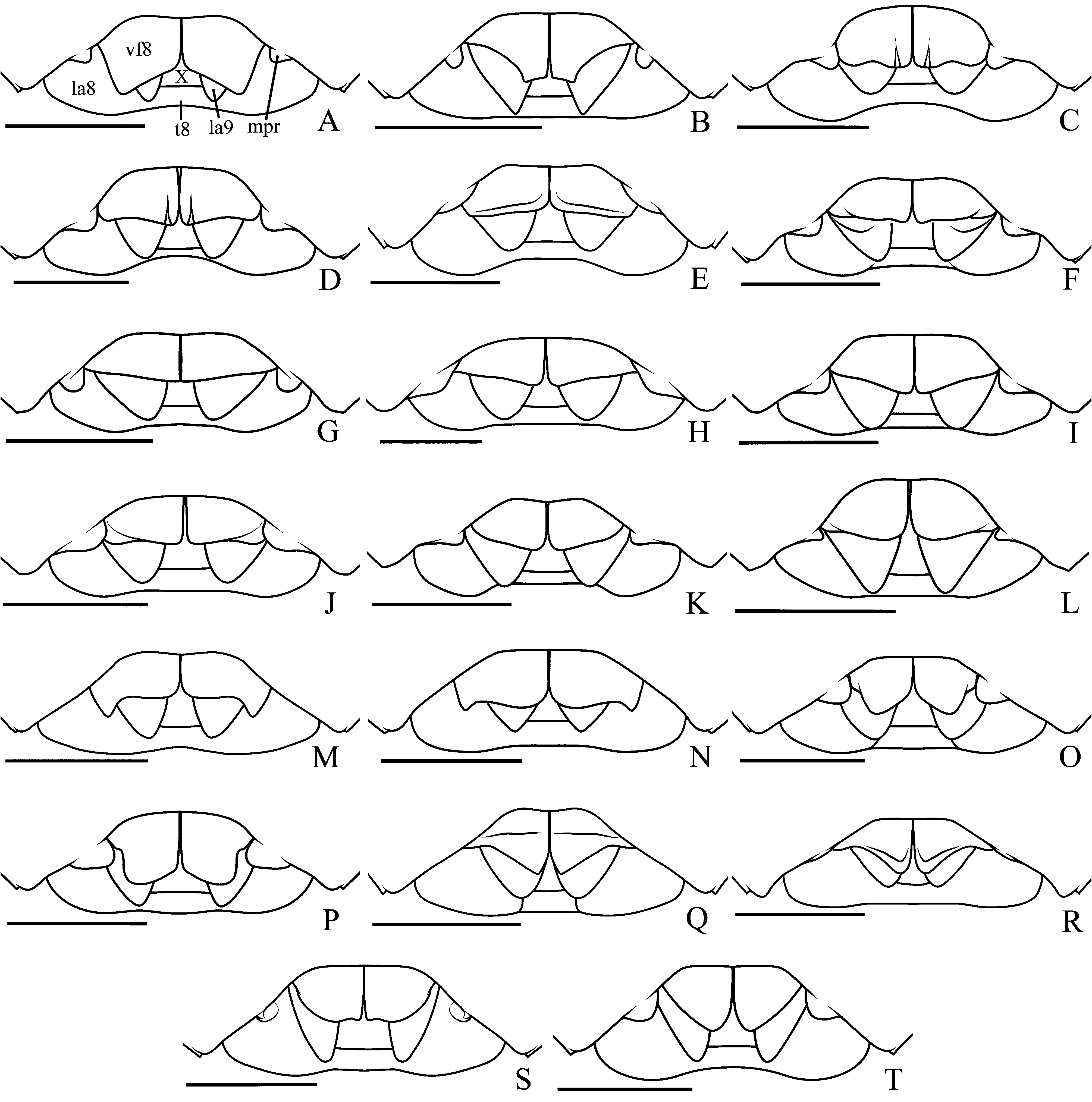
Female. Membrane of hemelytra not reaching the posterior margin of mediotergite VIII, posterior margin convex; median portion of posterior margin of mediotergite VIII subrectilinear; median portion of posterior margin of urosternite VII concave; projections on the lateral 1/3 of posterior margin of urosternite VII thick and perpendicular to the urosternite surface ( Fig. 32C View FIGURE 32 , mpr). Genitalia. Valvifers VIII wider than long; posterior margin subrectilinear and slightly oblique to the median line; sutural margins subrectilinear and folded dorsally; surface dark yellowish, with punctures and brown blotches; setae sparse on distal half of sutural margins and on median half of posterior margin; longitudinal grooves narrow and shallow on basal portion ( Figs. 5G View FIGURE 5 ; 32C View FIGURE 32 , vf8). Apices of valvifers IX partially visible; lateral margin subrectilinear; setae on mid-basal portion of ventral surface ( Fig. 32D View FIGURE 32 , vf9). Laterotergites IX not reaching the posterior margin of mediotergite VIII; lateral margin convex; setae on mid-basal portion of lateral margin and ventral surface ( Fig. 32C–D View FIGURE 32 , la9). Thickening of vaginal intima hexagonal; slightly wider than long; distal portion more sclerotized, mid-basal subrectangular area membranous; distal margin slightly concave ( Fig. 32D View FIGURE 32 , vi). Vesicular area anterior to the collar 1/8 of posterior portion; median duct anterior to the collar with slight proximal widening ( Fig. 32D View FIGURE 32 , mdp), median duct posterior to the collar with proximal widening, inner duct curved, almost coiled, in the proximal widening ( Fig. 32D View FIGURE 32 , id). Distal ductus receptaculi of same caliber as the proximal ( Fig. 32D View FIGURE 32 , drd, drp). Pars intermedialis cylindrical ( Fig. 32D View FIGURE 32 , pi); distal annular crest larger than the proximal one, both directed to the ductus receptaculi ( Fig. 32D View FIGURE 32 , dac, pac). Capsula seminalis elongated, subconical and with a filiform laterobasal projection ( Fig. 32D View FIGURE 32 , cs, pr).
Measurements: Table 13.
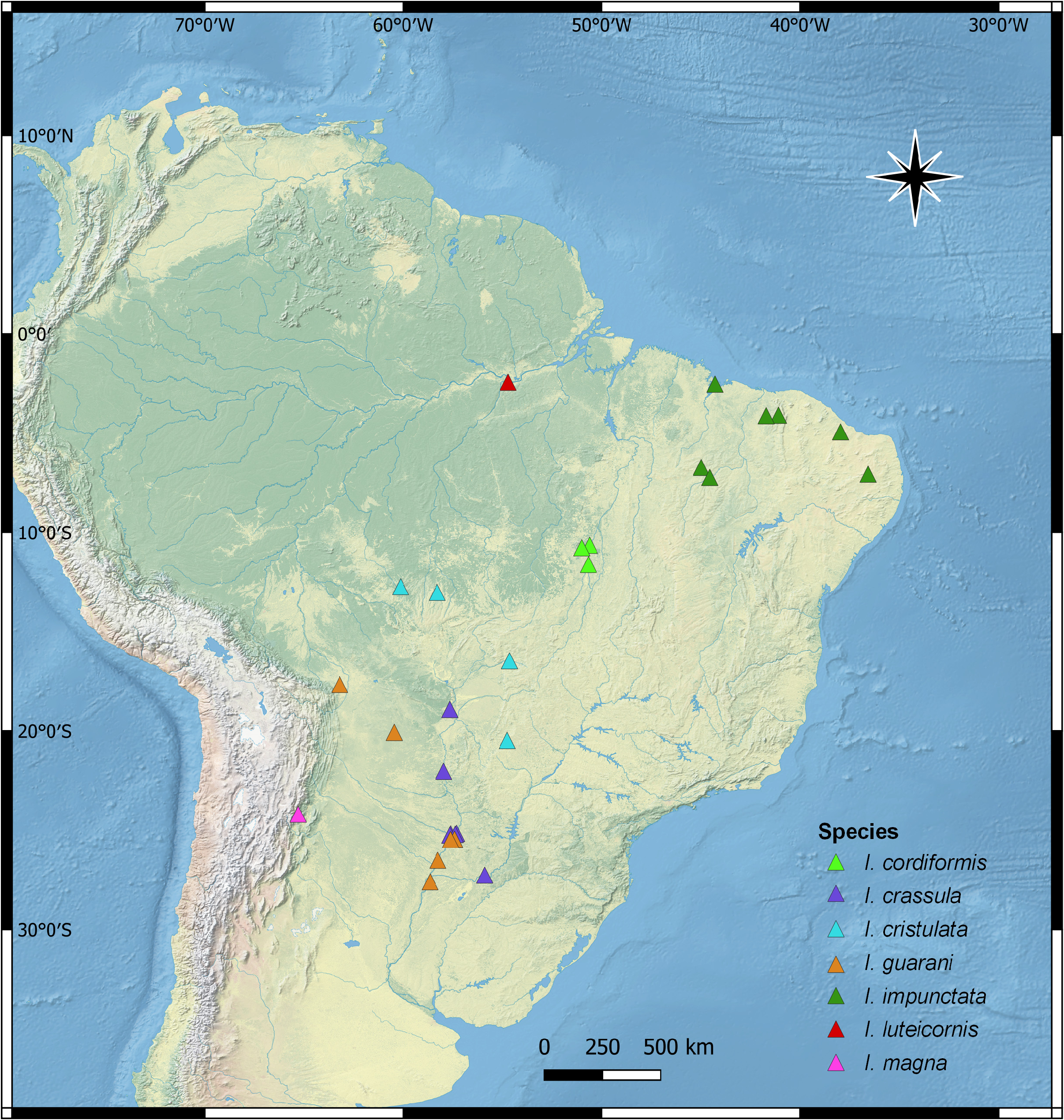
Distribution. Brazil (Pará) ( Fig. 7 View FIGURE 7 ).
Comments. Among the species grouped with I. coralinae sp. n. (see comments for the latter), females of Ischnopelta luteicornis are the only whose projections of the posterior margin of urosternite VII are thick and perpendicular to the surface of the urosternite ( Fig. 32C View FIGURE 32 , mpr). The males of I. luteicornis are the only in this group whose urosternite VII reaches anteriorly the imaginary line connecting the spiracles of urosternite V ( Fig. 31B View FIGURE 31 ).


No known copyright restrictions apply. See Agosti, D., Egloff, W., 2009. Taxonomic information exchange and copyright: the Plazi approach. BMC Research Notes 2009, 2:53 for further explanation.
|
Kingdom |
|
|
Phylum |
|
|
Class |
|
|
Order |
|
|
Family |
|
|
Genus |
Ischnopelta luteicornis ( Walker, 1867 )
| Rosso, Pedro & Campos, Luiz Alexandre 2021 |
Ischnopelta luteicornis:
| Grazia, J. & Panizzi, A. R. & Greve, C. & Schwertner, C. F. & Campos, L. A. & Garbelotto, T. A. & Fernandes, J. A. M. 2015: 712 |
| Becker, M. & Grazia, J. 1992: 203 |
Discocephala luteicornis
| Rolston, L. H. 1990: 24 |
| Kirkaldy, G. W. 1909: 215 |
| Lethierry, L. & Severin, G. 1893: 84 |
| Walker, F. 1867: 185 |