
Ischnopelta scutellata ( Signoret, 1851 )
|
publication ID |
https://doi.org/ 10.11646/megataxa.6.2.3 |
|
DOI |
https://doi.org/10.5281/zenodo.5753392 |
|
persistent identifier |
https://treatment.plazi.org/id/03828787-2C35-FF89-FCD5-FCD6FB75067E |
|
treatment provided by |
Plazi |
|
scientific name |
Ischnopelta scutellata ( Signoret, 1851 ) |
| status |
|
Ischnopelta scutellata ( Signoret, 1851) ( Figs. 2 View FIGURE 2 ; 5A View FIGURE 5 ; 9–10 View FIGURE 9 View FIGURE 10 )
Discocephala scutellata Signoret, 1851: 334 .
Discocephala (Ischnopelta) scutellata: Stål, 1868: 18 ; Stål, 1872: 6; Lethierry & Severin, 1893: 84.
Ischnopelta scutellata: Berg, 1891: 238 ; Kirkaldy, 1909: 215; Rolston, 1990: 20; Grazia et al., 2015: 712.
Holotype. VENEZUELA. Muséum National d’Histoire Naturelle ( MNHN), Paris, France (examined).
Material examined. 20 males and 28 females. BRAZIL, Mato Grosso, Santa Teresinha (close to the outfall of Tapirapé river), 1 male, 14. I.1963, Borys Malkin, [-10.616111, -50.613056], ( CAS); Campo Novo do Parecis, Utiariti (Papagaio river), 4 males and 7 females, 22-31.X.1966, Lenko & Pereira ( K. Lenko Col.), [-13.0215, -58.2870], ( UFRG); Chapada dos Guimarães, 1 female, homotype (det. H. Ruckes, 1961), [-15.433333, -55.75], ( AMNH, Acc:23739); 3 males and 3 females, 01. II.1961, J. & B. Bechyné, [-15.433333, - 55.75], ( MPEG); 1 male, March, [-15.433333, -55.75], ( USNM); Cuiabá, 3 males and 1 female, 14. II.1961, J. & B. Bechyné, [-15.5960, -56.0970], ( MPEG); Tocantins, Palmas (Fazenda Céu, Serra do Lageado), 1 female, XI.1992, Exp. MCN / MZSP, [-10.1669, -48.3328], 6-96 ( MCNZ); Distrito Federal, Brasília , Planaltina (32 km N Brasília ), 1 male and 2 females, 17–21.XI.1997, T. J. Henry, [-15.4548, -47.6130], ( USNM); Goiás, Corumbá de Goiás (Fazenda Monjolinho), 1 female, 14. VI.1942, F. Lane, [-15.9275, -48.8103], ( UFRG); Minas Gerais, Cardeal Mota (4 km SW Cardeal Mota and Rio Cipó, Rod. MG 10), 2 males and 2 females, 6.XI.1997, T. J. Henry & A. Paula, [-19.3564, -43.655], ( USNM); Paracatu, 1 female, VI.1960, Exp. Formosa, [-17.2211, -46.8741], ( MNRJ); Pirapora, 1 female, XI.1975, M. Alvarenga, [- 17.3374, -44.9271], ( AMNH); Goiás, Jataí, (Fazenda Cachoeirinha), 2 males and 3 females, X.1962, Exp. Dep. Zool., [-17.8872, -51.7182], ( UFRG); (Fazenda Nova Orlandia), 2 males and 2 females, 1964, Martins, Morgante & Silva, [-17.8872, -51.7182], ( UFRG); São Paulo, Pereira Barreto (old village from Lussanvira, Zone of the old Estrada de Ferro Noroeste Brazil— N. O. B.), 1 male and 1 female, 4.X.1938, Instituto Oswaldo Cruz, [-20.651389, -51.072222], ( FIOC); Ribeirão Preto, 1 female, 11.XII.1995, A. M. de Faria, [-21.1794, -47.7999], (UNIFESP), 1 female, III.1996, A. M. de Faria, [-21.1794, -47.7999], (UNIFESP).
Description. Male and female respectively 1.8 and 1.9 times longer than wide; dorsal surface somewhat glossy and dark yellowish; ventral surface pale yellow.
Head two times wider than long; anterior margin slightly emarginated. Clypeus 0.4 times the length of head. Distance between ocelli 0.3 times the distance between the eyes, on the line connecting the inner angles of the eyes. Maxillary plates and ventral ocular peduncles paleyellow; punctures on bucculae scarce, denser on ocular peduncles. Bucculae low, not concealing the first labial segment. Labium slightly surpassing the metacoxae. Labrum inserted halfway between the anterior margin of the eyes and the apex of mandibular plates. Antennae light brown, with irregular reddish striated blotches on segments II and III; segments ratio: I=II<III<IV<V.
Thorax. Pronotum as long as the head; width at the anterolateral angles as wide as the head. Scutellum surpassing the apical angles of urosternite VI, 1.8 times longer than wide at base; post-frenal lobe 1.6 times longer than frenal; post-frenal lobe narrowly rounded at apex. Hemelytra: corium slightly shorter than scutellum; in some specimens the radial vein is continued by a reddish line; apical margin of hemelytral membrane convex. Pro-, meso-, and metapleura pale-yellow, moderately punctured. Evaporatorium reaching the lateral margin of mesopleura. Legs dark yellowish, femora with punctures and reddish striated blotches on distal half, tibiae moderately punctured, setae on posterodorsal margin of protibiae longer than the others.
Abdomen dark yellowish; urosternites weakly punctured on median third, more densely on lateral thirds. Dark spots at the lateral of urosternites narrow, the anterior one longer than the posterior; minute spine present at apical angles of urosternite VII.
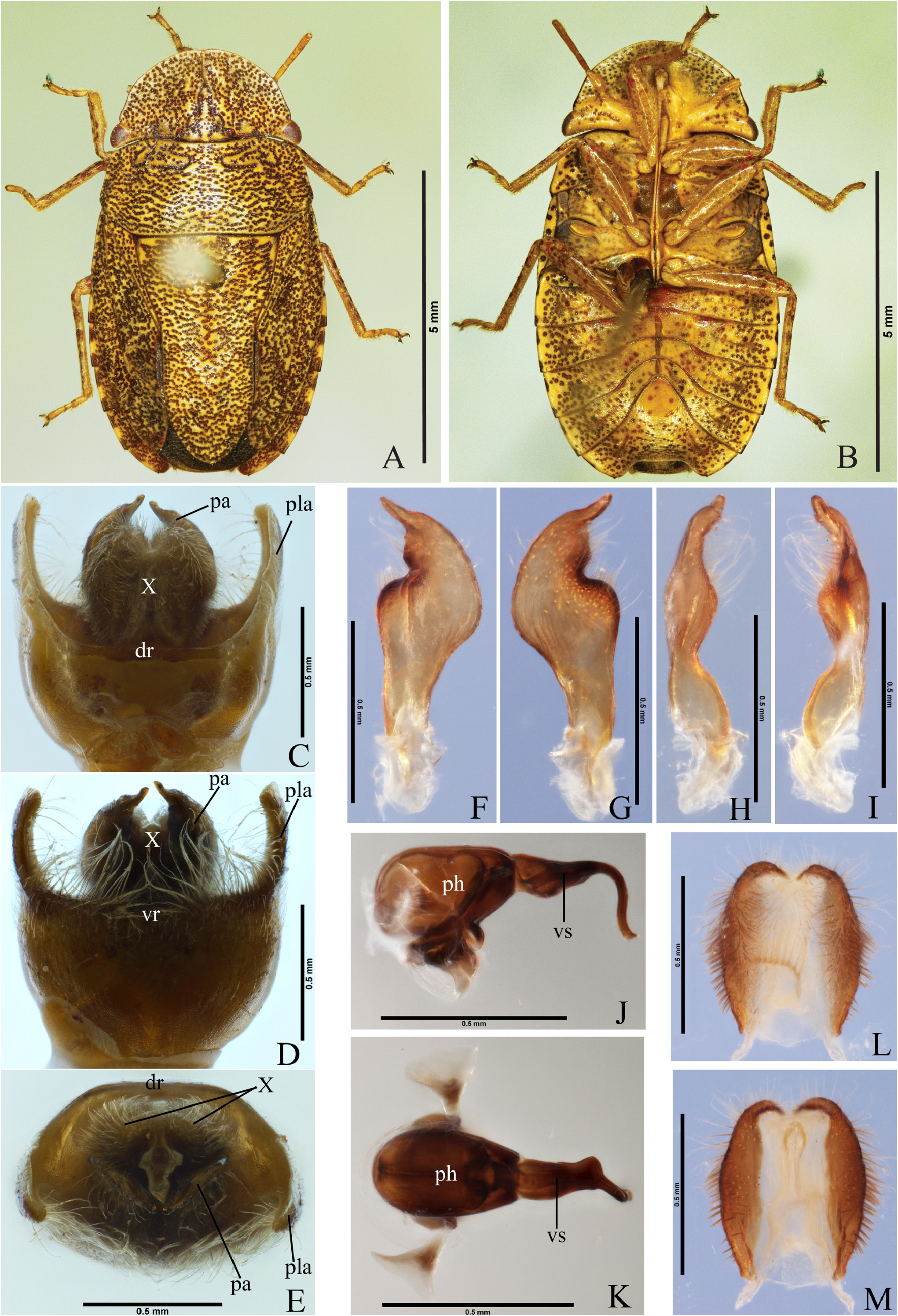
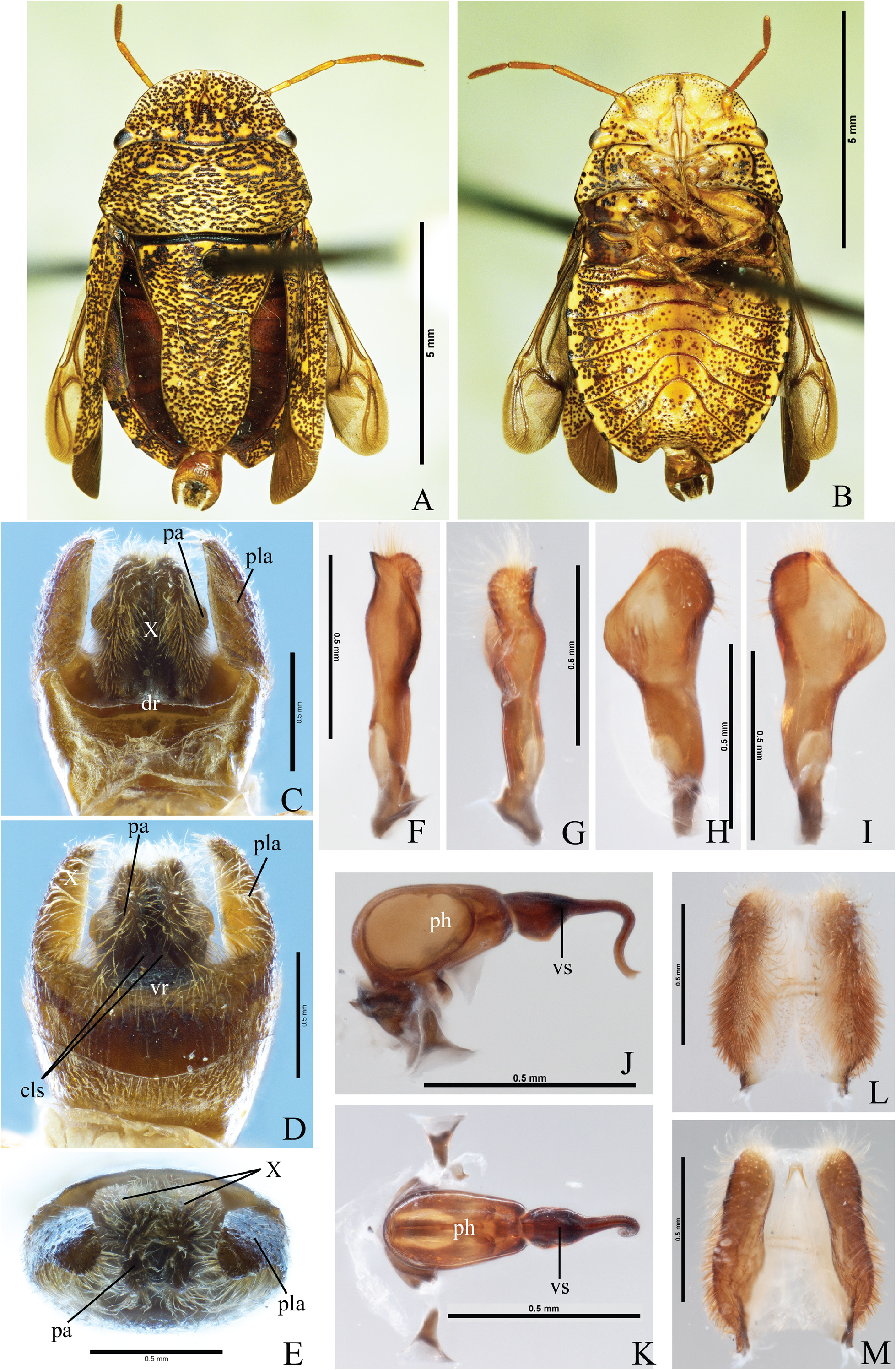
Male. Median portion of the posterior margin of urosternite VII subrectilinear; urosternite VII surpassing anteriorly the imaginary line connecting the spiracles of urosternite V. Genitalia. Dorsal rim of pygophore subrectilinear ( Fig. 9C View FIGURE 9 , dr); ventral rim shallowly concave ( Fig. 9D View FIGURE 9 , vr). Posterolateral angles 1.3 times longer than the rest of the pygophore, perpendicular to the frontal plane, slightly bent ventrally, divergent from the base and slightly convergent at apex ( Fig. 9C–E View FIGURE 9 , pla). Setae short and sparse on the posterior half of the ventral and lateral surfaces of the pygophore, and on the lateral surface of the posterolateral angles; setae long and dense on the ventral rim, and on the ventral margin of the posterolateral angles. Segment X longer than wide, not reaching the apex of the posterolateral angles and parameres; oval, and deeply emarginated apically; lateral and apical margins sclerotized and covered by setae; basal margin and mid-longitudinal surface membranous ( Figs. 9C and E, X; 9L–M View FIGURE 9 ). Parameres falciform, flat, as long as the posterolateral angles; distal portion oblique to the frontal plane; outer margin convex, inner margin sinuous, with strong excavation on the distal half; apex aculeiform, convergent, ventroposterioly directed; setae covering the posterior half of the ventral surface ( Fig. 9F–I View FIGURE 9 ). Cup-like sclerites little developed. Phallus: vesica broader on proximal half, bearing ventral and lateral expansions, followed by a lateral curvature; distal half ( Signoret, 1851) specimens evaluated (n). curved ventrally; secondary gonopore ventrally directed and beveled ( Fig. 9J–K View FIGURE 9 ).
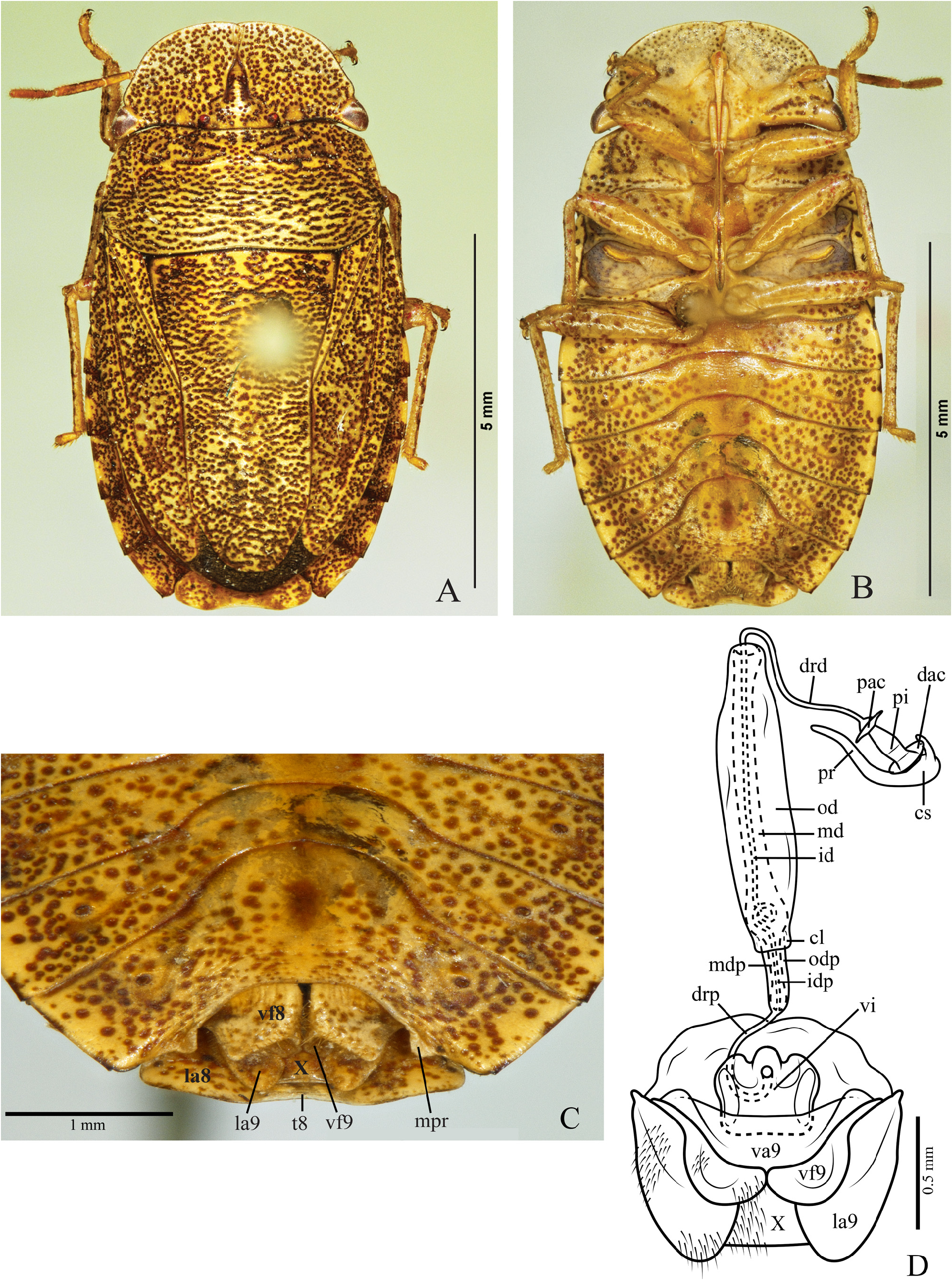
Female. Membrane of hemelytra not reaching the posterior margin of mediotergite VIII; median portion of the posterior margin of mediotergite VIII and of sternite VII subrectilinear; projections on the lateral1/3of posterior margin of sternite VII laminate, semicircular and slightly oblique in relation to the sternite surface ( Fig. 10C View FIGURE 10 , mpr).
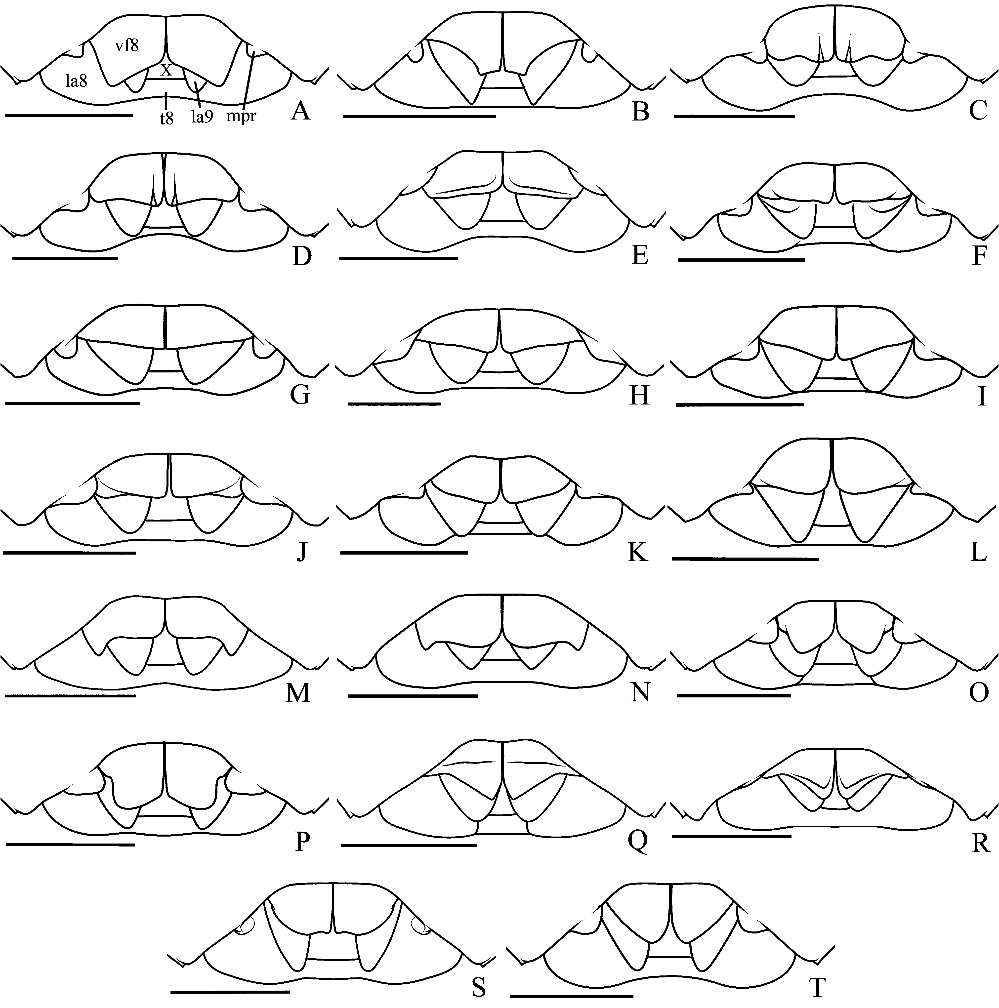
Genitalia. Valvifers VIII as long as wide, sutural angles subrectilinear, lateral angles slightly concave; sutural margins subrectilinear and dorsally folded; surface dark yellowish with brown punctures and setae on the distal half of the sutural margins; longitudinal grooves narrow and shallow at the basal portion ( Figs. 5A View FIGURE 5 ; 10C View FIGURE 10 , vf8). Valvifers IX almost completely covered by the valvifers VIII; lateral margin convex; setae on the mid-basal portion of the ventral surface ( Fig. 10C–D View FIGURE 10 , vf9). Laterotergites IX not reaching the posterior margin of mediotergite VIII; lateral margin convex; setae on the median portion of the lateral margin and mid-basal portion of the ventral surface ( Fig 10C–D View FIGURE 10 , la9). Thickening of vaginal intima barrel-shaped, slightly wider than long; distal portion more sclerotized; lateral margins convex; distal margin sinuous with 1+1 processes on the laterals, ventrodistal cone with membranous subcircular apex ( Fig. 10D View FIGURE 10 , vi). Vesicular area: anterior portion to the collar 1/5 of the posterior portion; median duct anterior to the collar slightly widened ( Fig. 10D View FIGURE 10 , mdp); median duct posterior to the collar with proximal and distal widening ( Fig. 10D View FIGURE 10 , md); inner duct coiled in the proximal widening ( Fig. 10D View FIGURE 10 , id). Distal ductus receptaculi 0.54 times the length of vesicular area posterior to the collar ( Fig. 10D View FIGURE 10 , drd, drp). Pars intermedialis barrel shaped, longer than capsula seminalis ( Fig. 10D View FIGURE 10 , pi); annular crests convergent, diameter of the proximal crest slightly smaller than the distal one ( Fig. 10D View FIGURE 10 , dac, pac). Capsula seminalis with two filiform lateral projections, one long and sinuous and the other short and slightly curved, both directed to the pars intermedialis ( Fig. 10D View FIGURE 10 , cs, pr).
Measurements: Table 1 View TABLE 1 .
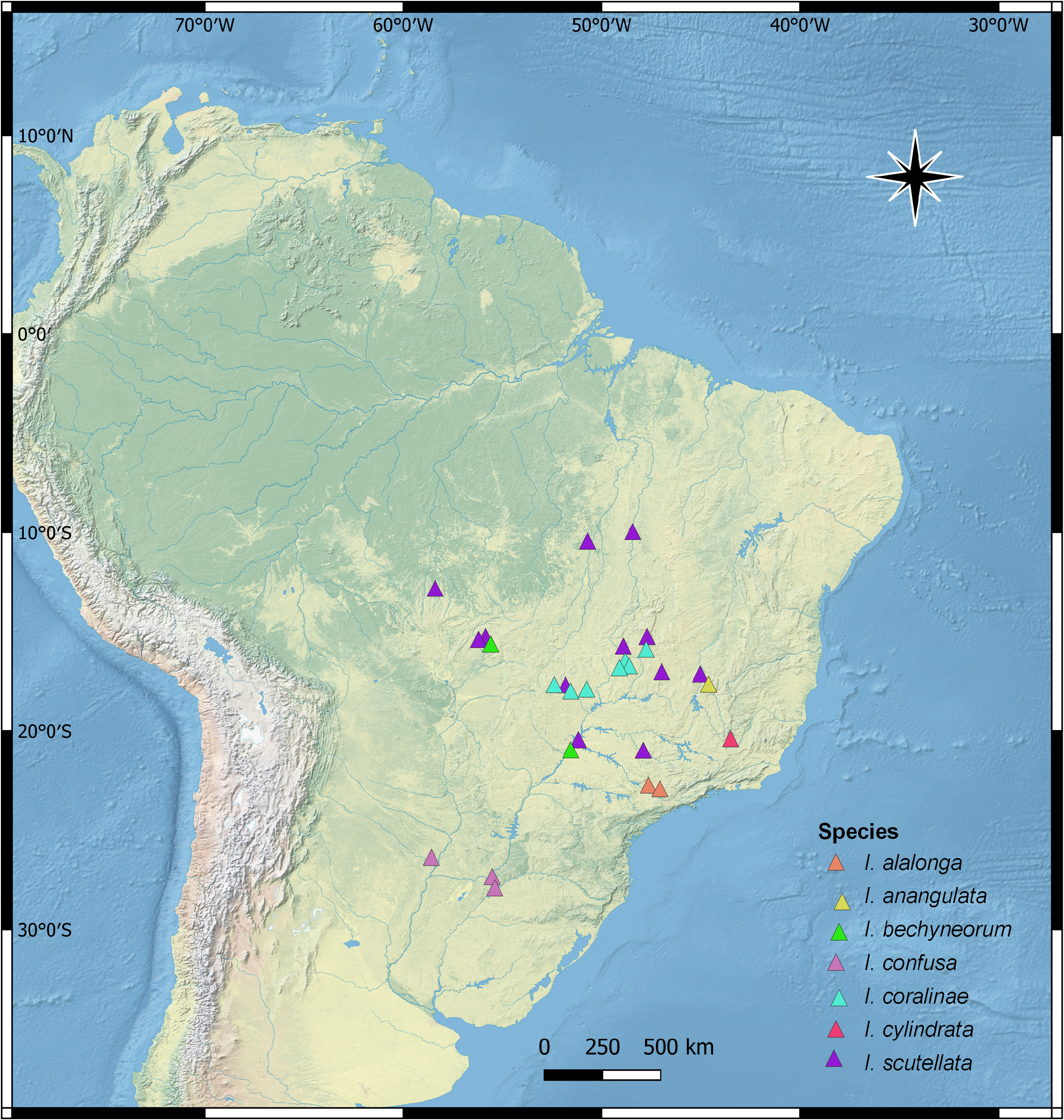
Distribution. Venezuela, Brazil (Tocantins, Mato Grosso, Goiás, Minas Gerais, Brasília (DF) , São Paulo) ( Fig. 6 View FIGURE 6 ).
Comments.Males of this species are easily recognized by the parameres aculeiform at apex, convergent and ventroposteriorly directed (observed in posterior view of the pygophore) ( Fig. 9E View FIGURE 9 , pa). Female identification is possible through the analysis of the posterior margin of valvifers VIII shaped as an open “V” with sutural angles subrectilinear and laterals slightly concave ( Figs. 5A View FIGURE 5 , vf8; 10C, vf8).
Ischnopelta alalonga Rosso & Campos , sp. n. ( Figs. 5O View FIGURE 5 ; 11–12 View FIGURE 11 View FIGURE 12 )
Etymology. The epithet refers to the length of the wings, which in this species surpass the posterior margin of mediotergite VIII. Latin: ala = wing + longus = long.
Type locality. BRAZIL, São Paulo, Piracicaba [- 22.7274, -47.6448] GoogleMaps .
Holotype. Male. BRAZIL, São Paulo, Piracicaba, 3.IX.1986, F. D. Bennett. Deposited at Museu de Zoologia da Universidade de São Paulo ( MZSP)], São Paulo ( SP), Brazil.
Paratypes. 2 males and 14 females. BRAZIL, São Paulo, Piracicaba , 1 female, 3.IX.1986, F . D. Bennett, [- 22.7274, -47.6448], ( J. E. Eger, Personal collection) GoogleMaps ; 3 females, 11. III .1987, F . D. Bennett, [-22.7274, -47.6448], ( J. E. Eger, Personal collection) GoogleMaps ; 1 female, 28. I .1988, D. H. Haback & F . D. Bennett, [-22.7274, -47.6448], ( J. E. Eger, Personal collection) GoogleMaps ; 1 male and 1 female, 20. II .1988, F . D. Bennett, [-22.7274, -47.6448], ( J. E. Eger, Personal collection); Campinas ( Campus UFU) GoogleMaps , 1 male and 2 females, 19.XI.1990, [-22.9095, -47.0674], ( MZSP) GoogleMaps ; 6 females, 19.XI.1990, [-22.9095, -47.0674], ( UFRG) GoogleMaps .
Description. The overall somatic morphology is as described for I. scutellata , except for the following features. Head. Bucculae slightly higher than the first labial segment. Antennae yellow dorsally, and dark yellowish ventrally; segments ratio: I>II<III<IV<V.
Thorax. Pro-, meso- and metasternum not punctured. Evaporatorium not reaching the outer margin of mesopleura. Setae on posterodorsal margin of protibiae as long as the others.
Abdomen. Dark spots at the lateral of urosternites subtriangular, wide.
Male. Apical margin of membrane of hemelytra convex; urosternite VII not reaching anteriorly the imaginary line connecting the spiracles of urosternite V. Genitalia. Dorsal rim of pygophore sinuous ( Fig. 11C View FIGURE 11 , dr); ventral rim slightly concave ( Fig. 11D View FIGURE 11 , vr). Posterolateral angles 1.6 times longer than the rest of the pygophore, base and apex respectively perpendicular and oblique to the frontal plane, convergent from the base, dorsal margin folded into the pygophore ( Fig. 11C–E View FIGURE 11 , pla). Setae short on the posterior half of ventral and lateral surfaces of the pygophore, and on the outer and inner surfaces of the posterolateral angles; setae long and dense on the lateral portions of the ventral rim, and on the ventral and apical margins of the posterolateral angles. Segment X longer than wide, surpassing the parameres, but not reaching the apex of the posterolateral angles, subrectilinear and strongly emarginated apically; lateral margins sclerotized and densely covered with long setae; mid-longitudinal region membranous and with sparse setae ( Figs. 11C and E, X; 11L–M View FIGURE 11 ). Parameres clubshaped, swollen, and perpendicular to the frontal plane; inner and outer surfaces sinuous, distal portion of inner surface slightly concave, with transverse lines and a minute apical process; ventral surface sinuous; dorsal surface narrow, distal half strongly convex longitudinally; setae covering the apex ( Figs. 11D View FIGURE 11 , pa; 11F–I). Cup-like sclerites externally visible and with apices rounded and slightly convergent ( Fig. 11D View FIGURE 11 , cls). Phallus: proximal portion of vesica laterally biconcave, ventrally expanded; median portion subcylindrical, gradually narrowed and bent ventrally; distal portion subcylindrical and sinuous; secondary gonopore ventroposterior and beveled ( Fig. 11J–K View FIGURE 11 ).
Female. Hemelytral membrane surpassing the posterior margin of mediotergite VIII, posterior margin convex; median portion of posterior margin of urosternite VII, and projections on its lateral 1/3 ( Fig. 12C View FIGURE 12 , mpr) as described for I. scutellata . Genitalia. Valvifers VIII wider than long; posterior margin strongly sinuous, sutural portion slightly convex; lateral portion sinuous, slightly oblique to the midline and with slender cut on the lateral; sutural margins subrectilinear and folded dorsally; surface dark yellowish with brown punctures and setae on the distal half of the sutural margins and on the posterior margin; longitudinal grooves narrow and shallow at the basal portion ( Figs. 5O View FIGURE 5 ; 12C View FIGURE 12 , vf8). Valvifers IX exposed; lateral margin subrectilinear; setae on mid-basal portion of ventral surface ( Fig. 12C–D View FIGURE 12 , vf9). Laterotergites IX not reaching the posterior margin of mediotergite VIII; lateral margin convex; setae on median portion of lateral margin and on mid-basal portion of the ventral surface ( Fig. 12C–D View FIGURE 12 , la9). Thickening of vaginal intima subcircular, slightly wider than long; proximal margin concave and wider than the distal one; distal margin weakly emarginated; mid-ventral area with membranous elliptical cone, dorsal longitudinal ridges divergent distally and reaching the margins ( Fig. 12D View FIGURE 12 , vi). Vesicular area: anterior portion to the collar 1/6.5 of the posterior portion; median duct anterior to the collar with slight proximal widening ( Fig. 12D View FIGURE 12 , mdp); median duct posterior to the collar with proximal and distal widening ( Fig. 12D View FIGURE 12 , md); inner duct coiled in the proximal widening ( Fig. 12D View FIGURE 12 , id). Distal ductus receptaculi 0.40 times the length of the vesicular area posterior to the collar ( Fig. 12D View FIGURE 12 , drd, drp). Pars intermedialis wider distally ( Fig. 12D View FIGURE 12 , pi); proximal annular crest perpendicular to pars intermedialis, the distal one facing the pars intermedialis and almost twice the size the proximal ( Fig. 12D View FIGURE 12 , dac, pac). Capsula seminalis globose, with a long and sinuous laterobasal projection directed to the pars intermedialis; & Campos, sp. n. specimens evaluated (n).
in some specimens, a lateral projection of variable size may occur ( Fig. 12D View FIGURE 12 , cs, pr).
Measurements: Table 2.
Distribution. Brazil (São Paulo) ( Fig. 6 View FIGURE 6 ).
Comments. Ischnopelta alalonga sp. n. ( Figs. 11A–B View FIGURE 11 ; 12A–B View FIGURE 12 ), although similar to Ischnopelta crassula sp. n. ( Figs. 23A–B; 23A–B View FIGURE 23 ), presents the corium slightly shorter than the scutellum, and the lateral abdominal stripe unpunctured, better delimited and with dark-brown blotches, whilst in I. crassula the corium and scutellum are subequal, and the lateral abdominal stripe presents, besides the blotches, few irregularly distributed punctures. The dorsal rim of the pygophore is sinuous in I. alalonga ( Fig. 11C View FIGURE 11 , dr) and slightly concave in I. crassula ( Fig. 22C View FIGURE 22 , dr), the parameres with more developed apical process, and the area with differentiated texture in the inner surface is longer and narrow in I. alalonga ( Figs. 11F–I View FIGURE 11 ; 22F–I View FIGURE 22 ); the proximal portion of vesica is laterally biconvex in I. alalonga ( Fig. 11K View FIGURE 11 , vs), while in I. crassula it is wider at the base and gradually narrows up to the curvature ( Fig. 22K View FIGURE 22 , vs). The female hemelytral membrane surpasses the posterior margin of mediotergite VIII in I. alalonga ( Fig. 12A–B View FIGURE 12 ), but not in I. crassula ( Fig. 23A–B View FIGURE 23 ). The thickening of the vaginal intima forms a median and elliptical membranous cone, and the dorsal longitudinal ridges are sinuous and divergent distally reaching the margins in I. alalonga ( Fig. 12D View FIGURE 12 , iv), while in I. crassula the cone is subtriangular, and the ridges are sinuous and not reaching the margins ( Fig. 23D View FIGURE 23 , iv).


| MNHN |
Museum National d'Histoire Naturelle |
| I |
"Alexandru Ioan Cuza" University |
| CAS |
California Academy of Sciences |
| K |
Royal Botanic Gardens |
| UFRG |
Instituto de Biologia |
| H |
University of Helsinki |
| AMNH |
American Museum of Natural History |
| J |
University of the Witwatersrand |
| B |
Botanischer Garten und Botanisches Museum Berlin-Dahlem, Zentraleinrichtung der Freien Universitaet |
| MPEG |
Museu Paraense Emilio Goeldi |
| USNM |
Smithsonian Institution, National Museum of Natural History |
| MCN |
McNeese State University |
| MZSP |
Sao Paulo, Museu de Zoologia da Universidade de Sao Paulo |
| MCNZ |
Porto Alegre, Museu de Ciencias Naturais da Fundacao Zoo-Botanica do Rio Grande do Sul |
| N |
Nanjing University |
| T |
Tavera, Department of Geology and Geophysics |
| VI |
Mykotektet, National Veterinary Institute |
| F |
Field Museum of Natural History, Botany Department |
| MG |
Museum of Zoology |
| A |
Harvard University - Arnold Arboretum |
| MNRJ |
Museu Nacional/Universidade Federal de Rio de Janeiro |
| M |
Botanische Staatssammlung München |
| O |
Botanical Museum - University of Oslo |
| FIOC |
Fundacao Instituto Oswaldo Cruz |
| SP |
Instituto de Botânica |
| E |
Royal Botanic Garden Edinburgh |
| UFU |
Ural Federal University "B. N. Yeltsin" |
No known copyright restrictions apply. See Agosti, D., Egloff, W., 2009. Taxonomic information exchange and copyright: the Plazi approach. BMC Research Notes 2009, 2:53 for further explanation.
|
Kingdom |
|
|
Phylum |
|
|
Class |
|
|
Order |
|
|
Family |
|
|
Genus |
Ischnopelta scutellata ( Signoret, 1851 )
| Rosso, Pedro & Campos, Luiz Alexandre 2021 |
Ischnopelta scutellata:
| Grazia, J. & Panizzi, A. R. & Greve, C. & Schwertner, C. F. & Campos, L. A. & Garbelotto, T. A. & Fernandes, J. A. M. 2015: 712 |
| Rolston, L. H. 1990: 20 |
| Kirkaldy, G. W. 1909: 215 |
| Berg, C. 1891: 238 |
Discocephala (Ischnopelta) scutellata: Stål, 1868: 18
| Lethierry, L. & Severin, G. 1893: 84 |
| Stal, C. 1872: 6 |
| Stal, C. 1868: 18 |
Discocephala scutellata
| Signoret, M. V. 1851: 334 |