
Wellstenhelia calliope, Karanovic & Kim, 2014
|
publication ID |
https://doi.org/ 10.11646/zootaxa.3783.1.1 |
|
publication LSID |
lsid:zoobank.org:pub:E6155BDC-AEAE-475D-BC83-61B3B863344C |
|
DOI |
https://doi.org/10.5281/zenodo.4910568 |
|
persistent identifier |
https://treatment.plazi.org/id/6878D460-FFA8-FFC4-64D0-F8C201D1FEFC |
|
treatment provided by |
Felipe |
|
scientific name |
Wellstenhelia calliope |
| status |
sp. nov. |
Wellstenhelia calliope sp. nov.
( Figs. 2–11 View FIGURE 2 View FIGURE 3 View FIGURE 4 View FIGURE 5 View FIGURE 6 View FIGURE 7 View FIGURE 8 View FIGURE 9 View FIGURE 10 View FIGURE 11 )
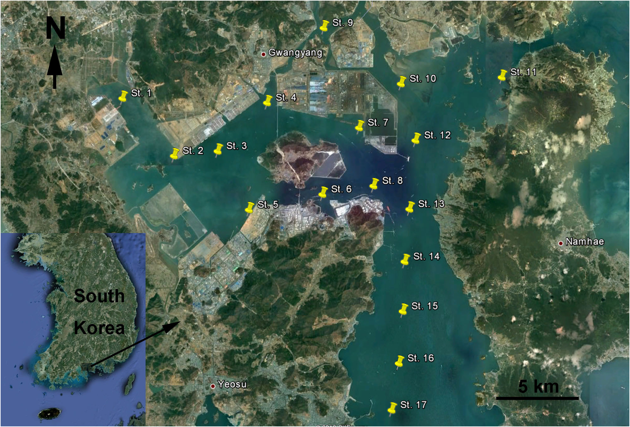
Type locality. South Korea, South Sea , Gwangyang Bay, sampling station 5, muddy sediments, 34.852500°N 127.684722°E ( Fig. 1 View FIGURE 1 ) GoogleMaps .
Specimens examined. Female holotype dissected on one slide (collection number NIBRIV0000232672), holotype’s right antennula destroyed for DNA sequence (amplification successful, Code 0122), male allotype dissected on one slide (collection number NIBRIV0000232673), two males paratypes and one female paratype together on one SEM stub (collection number NIBRIV0000232674), two male paratypes and one copepodid paratype together in ethanol (collection number NIBRIV0000232675); one male destroyed for DNA sequence (amplification unsuccessful), type locality, 30 July 2012, leg. K. Kim.
Etymology. The species is named after Calliope (Ancient Greek: Καλλιόπη), one of nine Muses from Greek mythology, who was a patron of epic poetry and song. The species name is a noun in apposition (in the nominative case), despite the Recommendation 31A of the ICZN (1999) about avoidance of personal names as nouns in appositions, because there is no case for it being confusing or misleading. Nine Muses refer to the nine new species described in this paper.
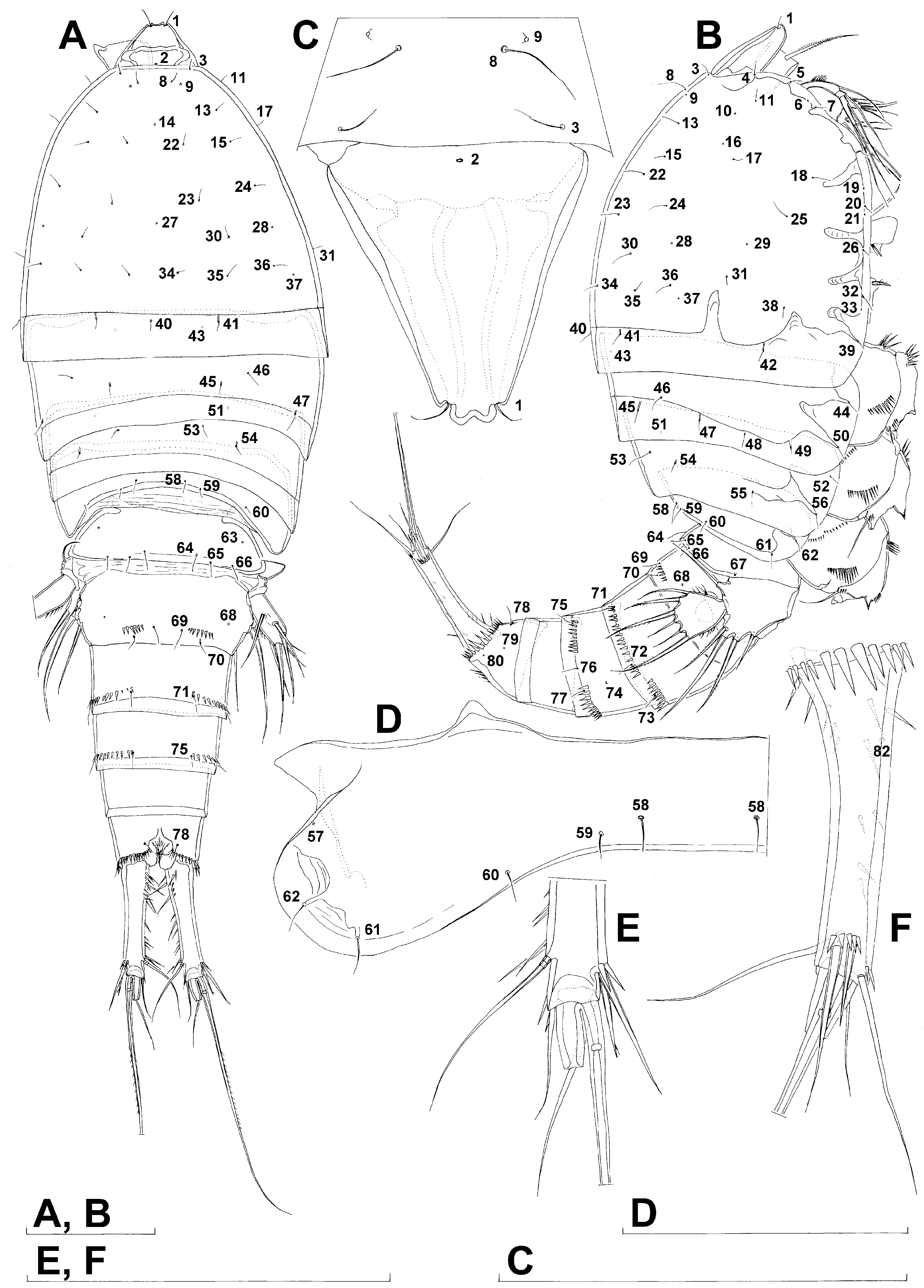
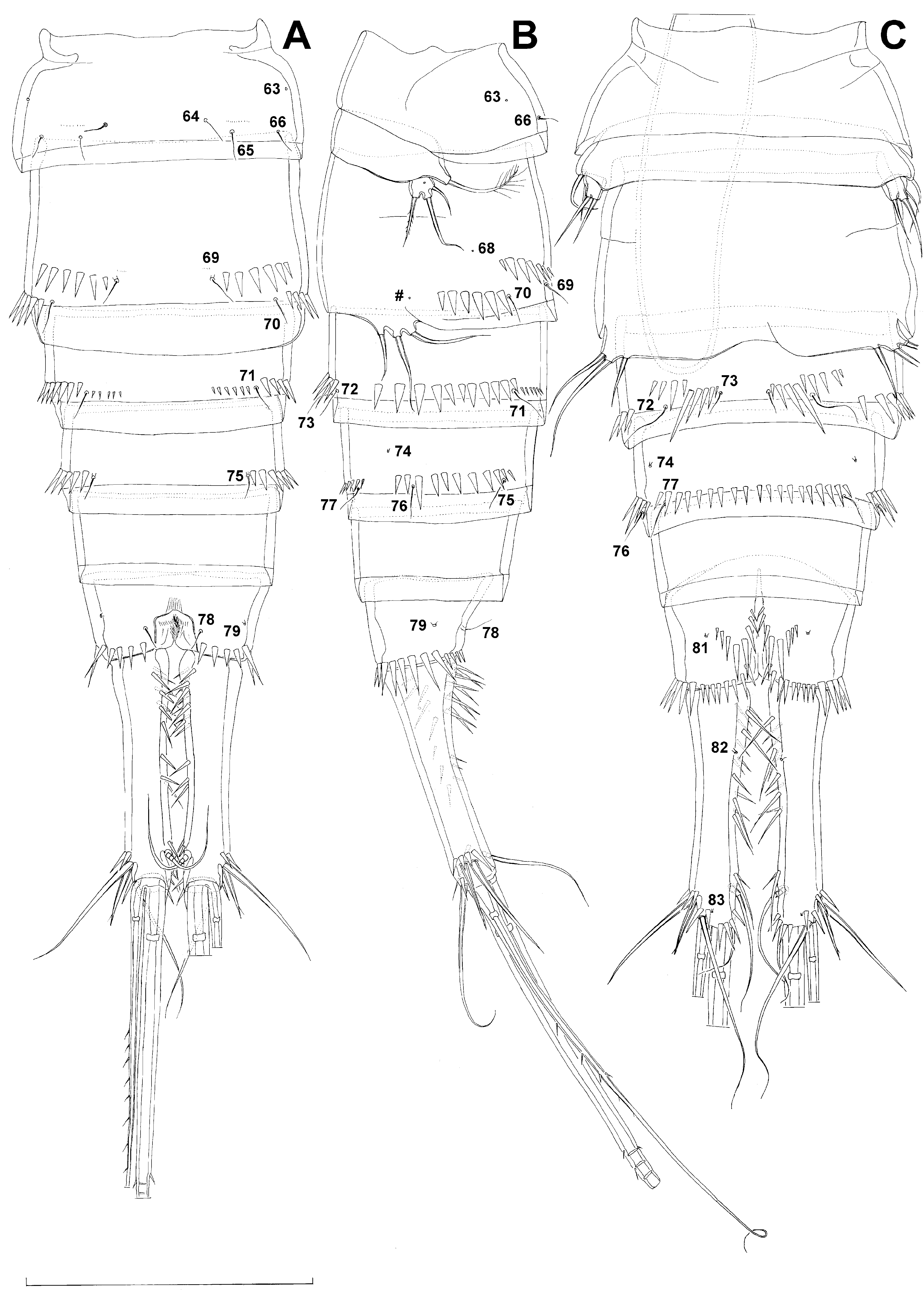
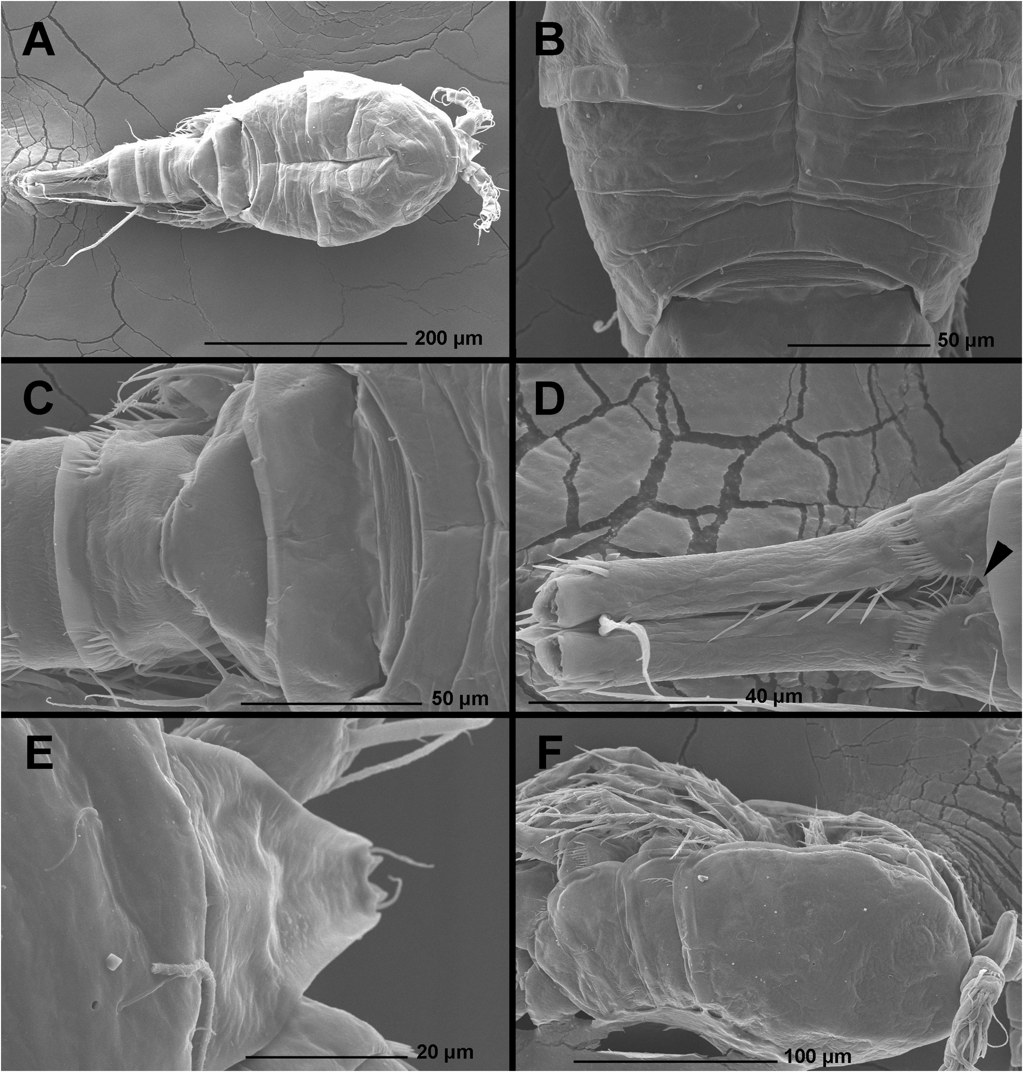
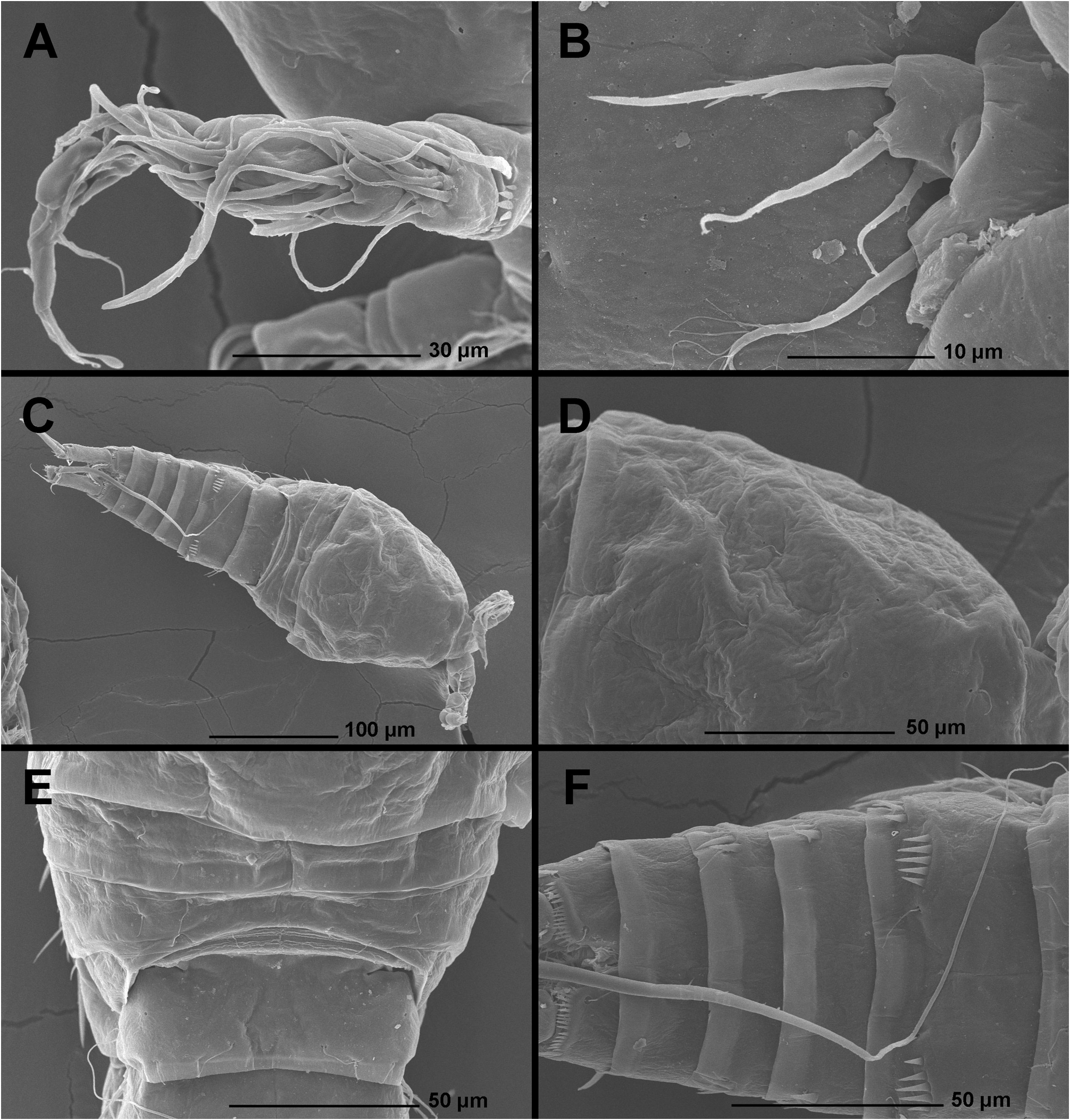
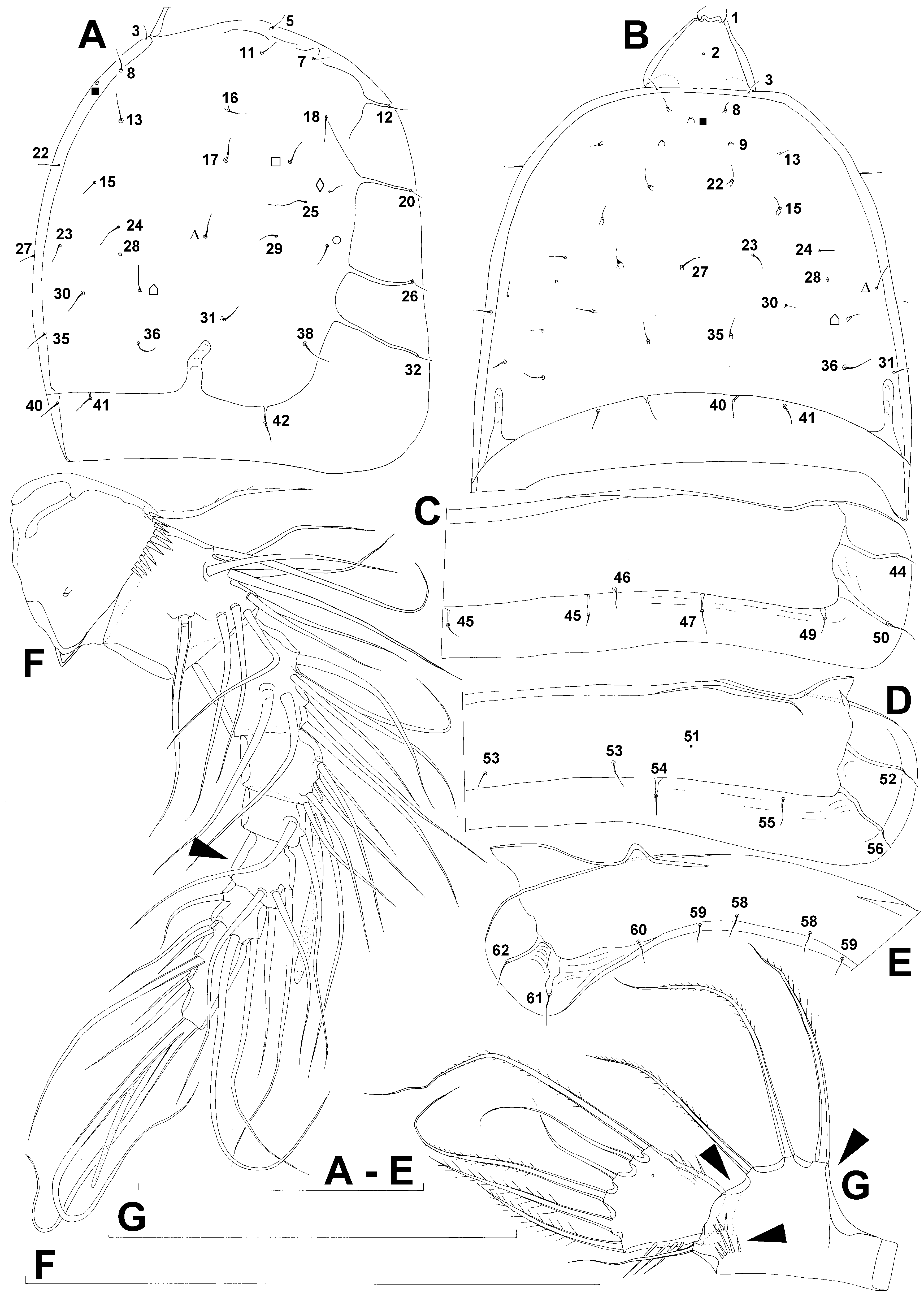
Description. Female (based on holotype and one paratype). Total body length, measured from tip of rostrum to distal margin of caudal rami 742 and 755 µm respectively. Colour of preserved specimens yellowish; live specimens not observed. Nauplius eye not visible. Prosome comprising cephalothorax with completely fused first pedigerous somite, and three free pedigerous somites; urosome comprising first urosomite (= fifth pedigerous somite), genital double-somite (fused genital and third urosomites) and three free urosomites (last one being anal somite). Short sclerotized joint between prosome and urosome only discernible on ventral side. Habitus ( Figs. 2A, B View FIGURE 2 , 10A View FIGURE 10 ) robust, spindle shaped in dorsal view, widest at posterior end of cephalothorax and tapering posteriorly, boundary between prosome and urosome conspicuous; prosome/urosome length ratio 1.05, but prosome much wider and more voluminous. Body length/width ratio about 3; cephalothorax 1.7 times as wide as genital doublesomite. Free pedigerous somites without lateral or dorsal expansions, pleurons only partly covering coxae of swimming legs in lateral view. Integument of all somites relatively weakly sclerotized, generally very smooth, without cuticular windows but covered with a sparse pattern of extremely minute and deep pits, only visible at highest magnifications on scanning electron microscope (such as in Fig. 11B View FIGURE 11 ). Hyaline fringe of all somites broad and smooth, except for fourth pedigerous somite with narrow fringe dorsally, and for anal somite without hyaline fringe. Surface ornamentation of somites and caudal rami consisting of 78 paired and five unpaired pores and sensilla (numbered with Arabic numerals consecutively from anterior to posterior end of body, and from dorsal to ventral side in Figs. 2 View FIGURE 2 , 3 View FIGURE 3 , 4 View FIGURE 4 ), and several rows of spinules on urosomites only.
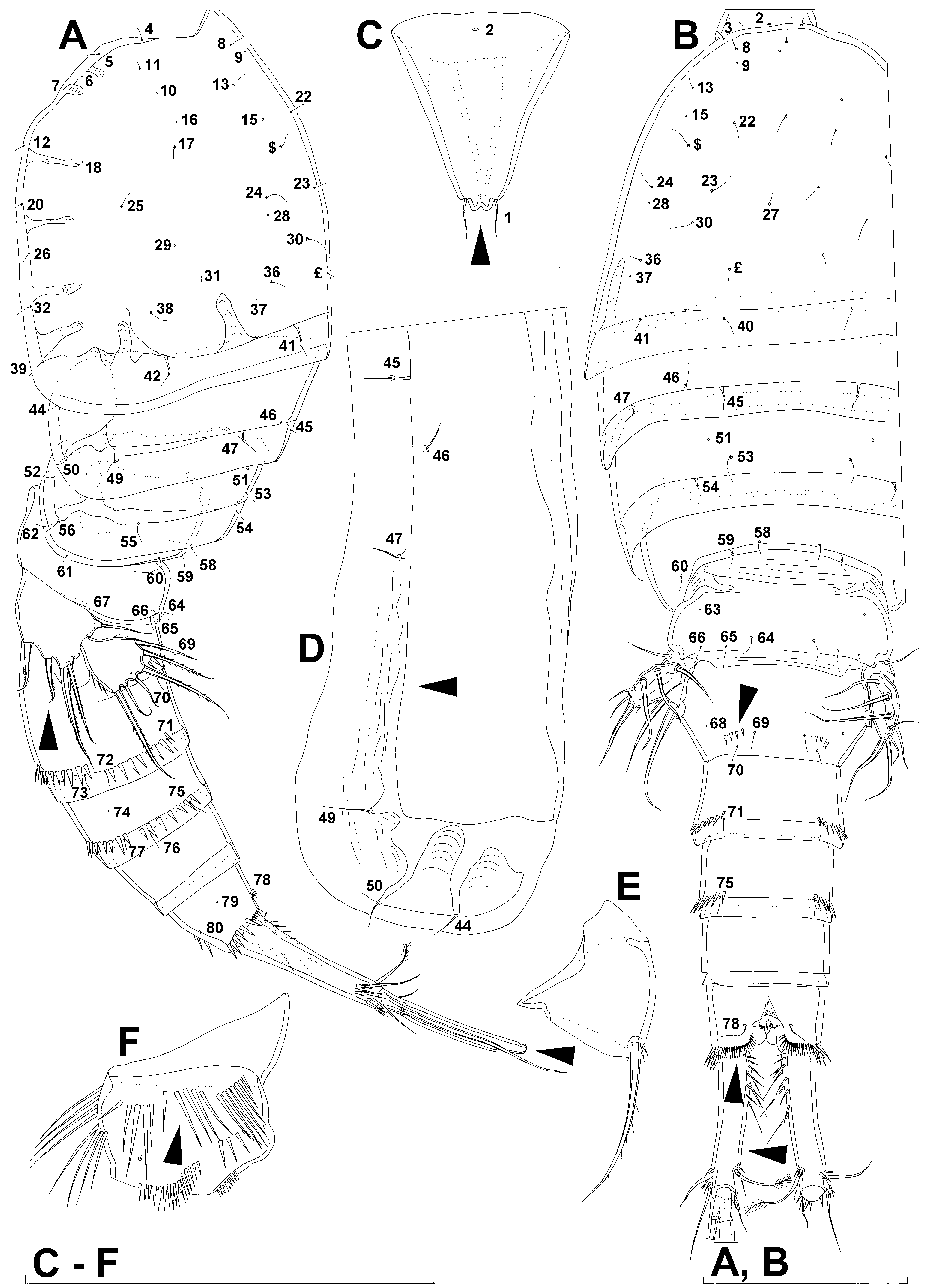
Rostrum ( Figs. 2C View FIGURE 2 , 10E View FIGURE 10 ) large, trapezoidal, clearly demarcated at base, reaching midlength of second antennular segment, with bilobate tip, about 1.1 times as long as wide; with two dorsal sensilla near tip (no. 1) and single central dorsal pore at base (no. 2); base of rostrum about 3.4 times as wide as its anterior margin; sensilla inserted into deep recesses.
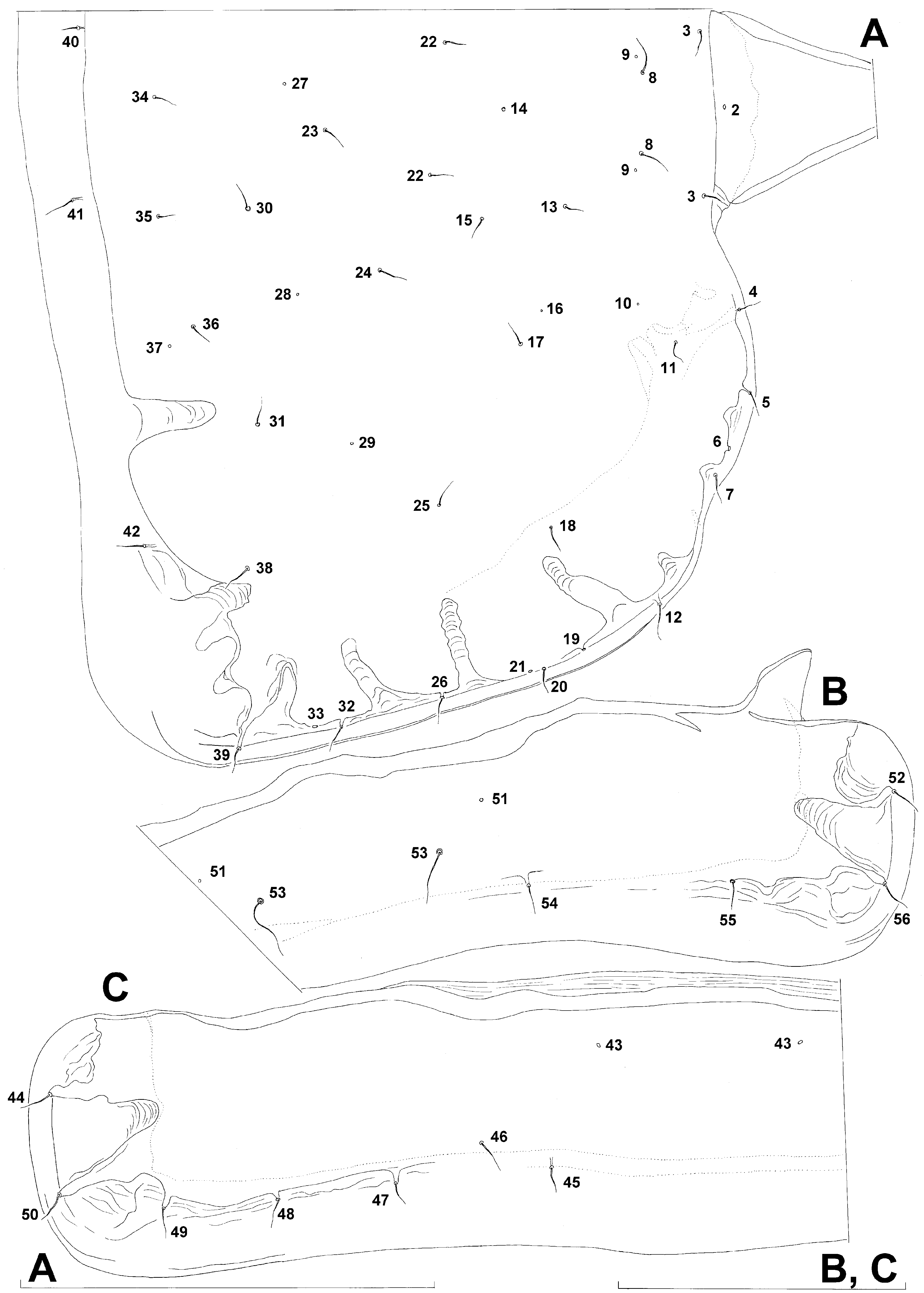
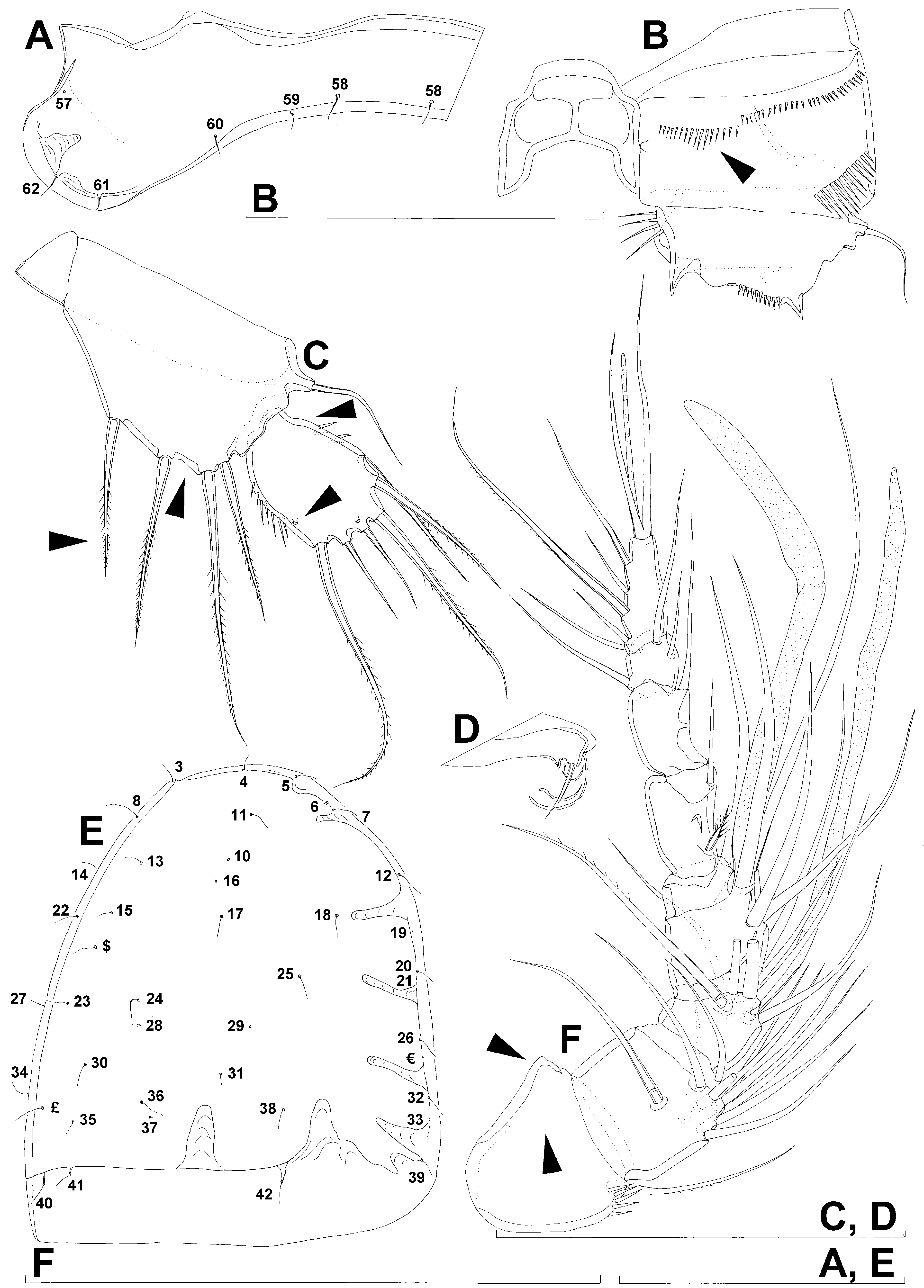
Cephalothorax ( Figs. 2A, B View FIGURE 2 , 3A View FIGURE 3 , 10A View FIGURE 10 ) tapering anteriorly in dorsal view, about 0.9 times as long as wide; comprising 30% of total body length. Surface of cephalothoracic shield with two unpaired dorsal pores (nos. 14, 27), two unpaired dorsal sensilla (nos. 34, 40), 10 pairs of pores (nos. 6, 9, 10, 16, 19, 21, 28, 29, 33, 37), and 28 pairs of long sensilla (nos. 3–5, 7, 8, 11–13, 15, 17, 18, 20, 22–26, 30–32, 35, 36, 38, 39, 41, 42); sensilla and pores 32–42 belonging to first pedigerous somite incorporated into cephalothorax.
Pleuron of second pedigerous somite (first free) ( Figs. 2A, B View FIGURE 2 , 3C View FIGURE 3 , 10B View FIGURE 10 ) with one pair of anterior dorsal pores (no. 43) and seven pairs of long sensilla (nos. 44–50); lateral pairs of sensilla nos. 44, 50, and 49 serially homologous to pairs nos. 32, 39, and 42 on first pedigerous somite respectively; other homologies difficult to define.
Third pedigerous somite ( Figs. 2A, B View FIGURE 2 , 3B View FIGURE 3 , 10B View FIGURE 10 ) slightly smaller than second pedigerous somite, pleuron with one pair of anterior dorsal pores (no. 51) but with only five pairs of sensilla (nos. 52–56); anterior pores more widely spaced than on second pedigerous somite; recognising serially homologous pairs easier with lateral (52=44, 56=50, 55=48) than with dorsal sensilla (possibly 53=45 and 54=46).
Fourth pedigerous somite ( Figs. 2A, B, D View FIGURE 2 , 10B, C View FIGURE 10 ) much smaller and shorter than previous two somites, especially in dorsal view, pleuron with antero-lateral pair of pores (no. 57) and five pairs of sensilla (nos. 58–62); pores not serially homologous to previous two somites, but all sensilla share homologues on third pedigerous somite (58=53, 59=54, 60=55, 61=56, and 62=52).
First urosomite ( Figs. 2A, B View FIGURE 2 , 10C View FIGURE 10 ) about as long as fourth pedigerous somite, with one pair of dorsal anterior pores (no. 63), one pair of lateral pores (no. 67), and three pairs of sensilla along distal margin (nos. 64–66); hyaline fringe much wider than in fourth pedigerous somite.
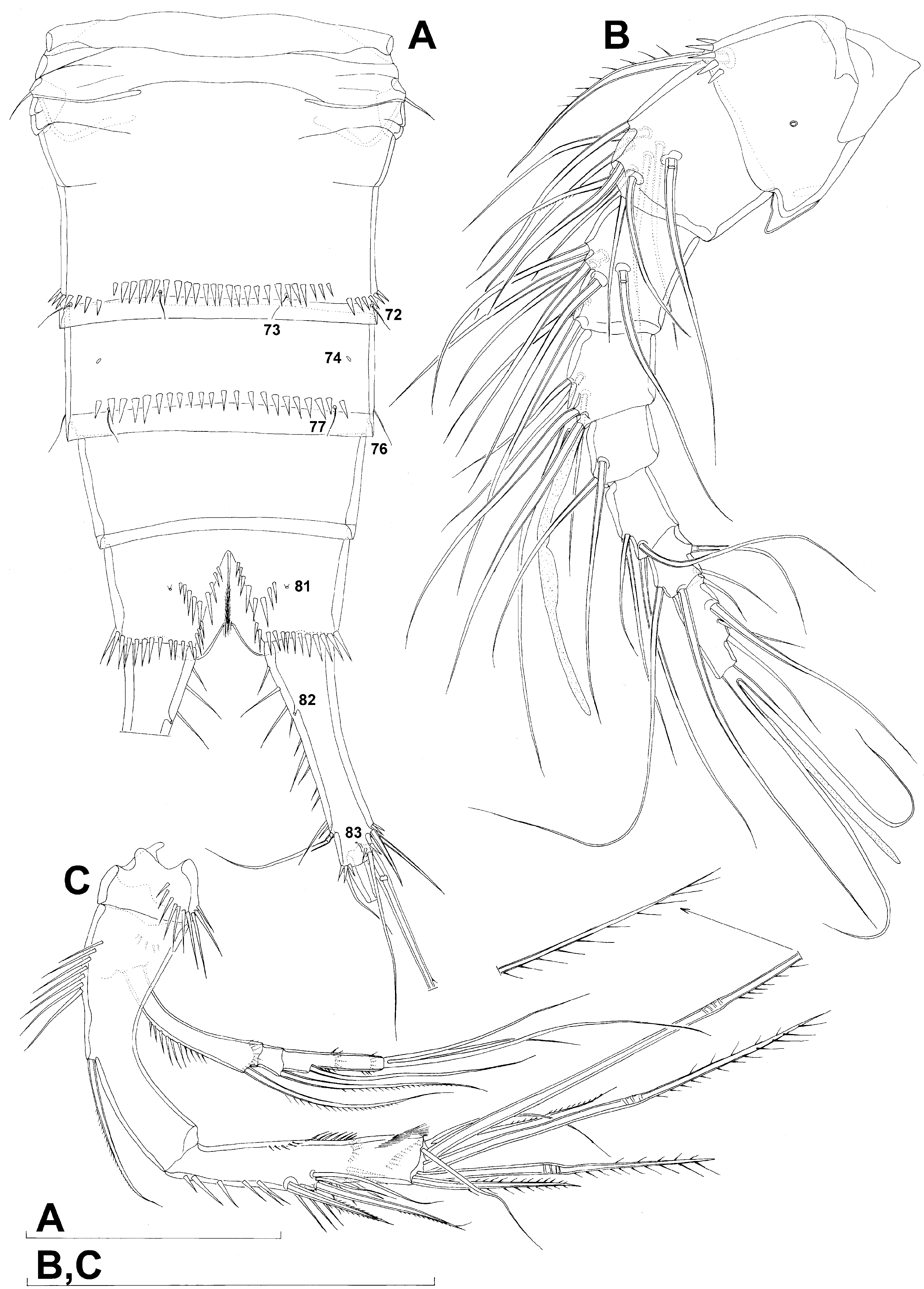
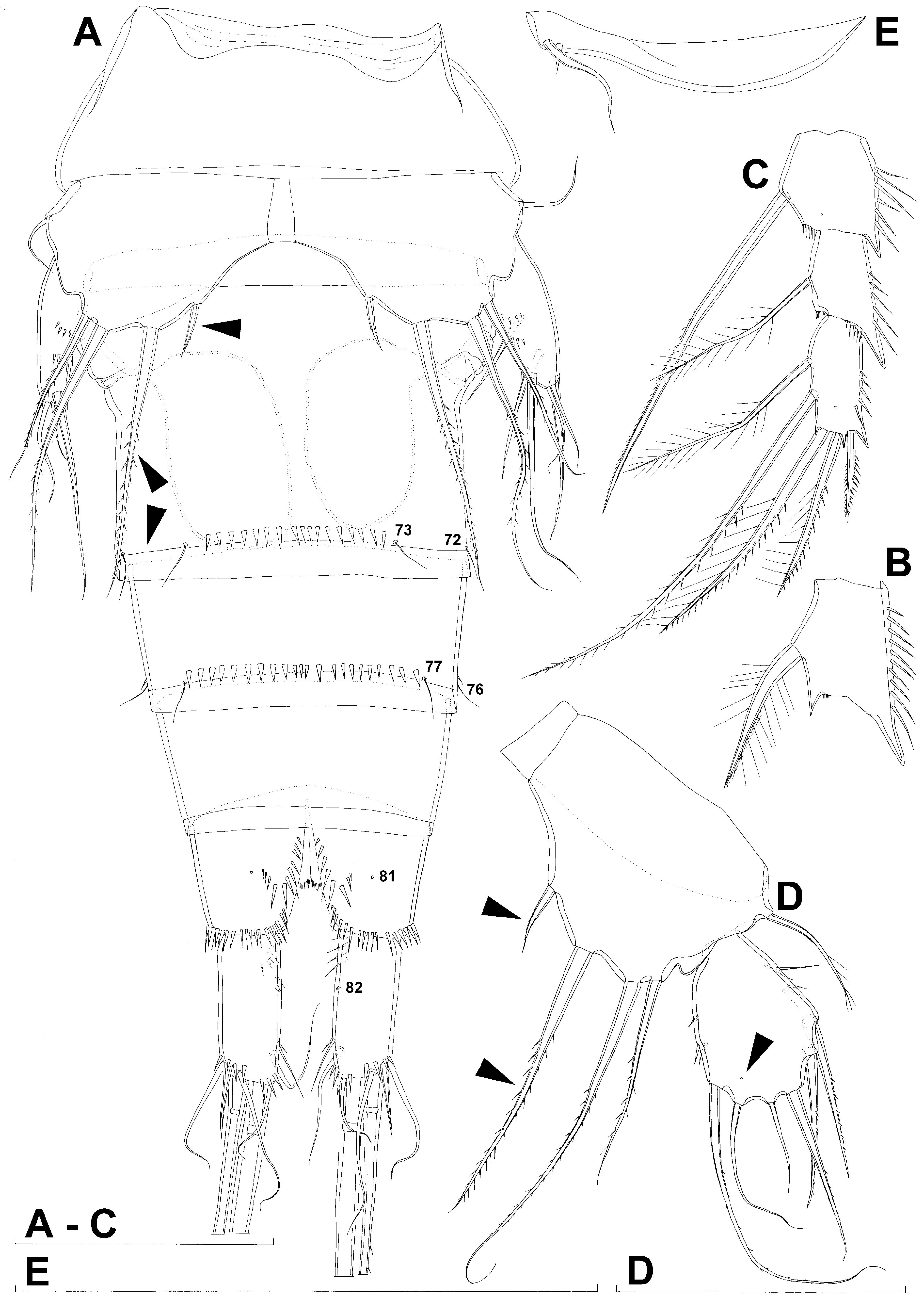
Genital double-somite ( Figs. 2A, B View FIGURE 2 , 4A View FIGURE 4 , 10C View FIGURE 10 ) 1.4 times as wide as long (ventral view); completely fused ventrally but with deep suture indicating original segmentation between genital and third urosomites dorsolaterally, thus dividing double-somite into equally long halves; anterior half of genital double-somite 1.2 times as wide as posterior, inflated laterally; anterior part with one pair of dorso-lateral pores (no. 68), two pairs of long dorsal sensilla (nos. 69 & 70), and two short rows of 6–8 strong spinules above sensilla no. 70; serially homologous pores and sensilla of anterior part of double-somite and those of first urosomite relatively easily established (i.e. 68=63, 69=64, and 70=65); posterior part with three pairs of posterior sensilla (nos. 71–73) and long row of strong spinules, interrupted dorsally between sensilla pair no. 71 and slightly ventro-laterally halfway between sensilla nos. 72 & 73; establishing serially homologous sensilla of posterior and anterior part of double-somite not easy (probably only 71=70); hyaline fringe wider than in first urosomite. Female genital complex ( Fig. 4A View FIGURE 4 ) weakly sclerotized and hardly distinguishable from internal sutures and soft tissue, copulatory pores not exposed on surface; paired genital apertures situated ventro-laterally, close to anterior margin and covered by reduced sixth legs.
Third urosomite ( Figs. 2A, B View FIGURE 2 , 4A View FIGURE 4 ) with one pair of anterior ventro-lateral pores (no. 74), three pairs of posterior sensilla (nos. 75–77), and posterior row of spinules interrupted dorsally between dorsal pair of sensilla (no. 75) and laterally on both sides of lateral sensilla (no. 76); lateral interruption of posterior row of spinules wider than in genital double-somite; all sensilla with homologous pairs on genital double-somite (i.e. 75=71, 76=72, 77=73) but ventral pair (no. 77) much more widely spaced; hyaline fringe as wide as in genital double-somite.
Fourth urosomite (preanal) ( Figs. 2A, B View FIGURE 2 , 4A View FIGURE 4 ) without ornamentation; hyaline fringe narrower than in third urosomite.
Anal somite ( Figs. 2A, B View FIGURE 2 , 4A View FIGURE 4 , 10D View FIGURE 10 ) clefted medially in posterior half, with one pair of large dorsal sensilla (no. 78), two pairs of lateral pores (nos. 79 & 80), one pair of ventral pores (no. 81), posterior row of spinules at base of each caudal ramus, and two curved ventral rows of spinules between median cleft and ventral pores; anal operculum short, reduced to narrow and thin membrane dorsally at end of medial cleft, concave and situated anterior to dorsal sensilla, representing less than 10% of somite's width, unornamented; anal sinus with several diagonal rows of hair-like spinules on both sides of median cleft, widely open, with weakly sclerotised walls, and without chitinous projections.
Caudal rami ( Figs. 2A, B, E, F View FIGURE 2 , 4A View FIGURE 4 , 10D View FIGURE 10 ) long and slender, about twice as long as anal somite, widest at base, about 3.5 times as long as wide (ventral view), slightly divergent and nearly cylindrical, with space between them about one ramus width; armature consisting of seven setae (three lateral, one dorsal and three apical), all in posterior sixth of ramus length; ornamentation consisting of anterior ventro-median pore (no. 82), posterior ventral pore (no. 83), two or three short spinules at base of each lateral seta, three short spinules at base of innermost apical seta, and two parallel rows of long inner spinules. Dorsal seta smooth and slender, inserted close to inner margin, about half as long as caudal ramus, triarticulate at base (i.e. inserted on two pseudojoints). Lateral setae all smooth; ventralmost one longest and most slender, inserted very close to distal margin, about as long as dorsal seta; dorsalmost one strongest, about 0.7 times as long as ventralmost one, inserted more anteriorly than ventralmost one but more posterior than dorsal seta; central one half as long as dorsalmost one, also strong, inserted at about same level as dorsal seta. Inner apical seta smooth and very slender, 0.6 times as long as dorsal seta. Principal apical setae fused basally, both with breaking planes and unipinnate distally along outer margin; middle apical seta much stronger and longer, about 1.7 times as long as outer apical one and 3.5 times as long as caudal rami.
Antennula ( Fig. 4B View FIGURE 4 ) eight-segmented, joined to cephalotholax with small triangular pseudosegment laterally, approximately 0.8 times as long as cephalothorax, with single short posterior row of spinules and single cuticular pore on first segment. Distal posterior corner of first segment produced into sharp process. Long aesthetasc on fourth segment slender, fused basally with adjacent large seta, and reaching tip of appendage; slender apical aesthetasc on eighth segment fused basally with two apical setae, forming apical acrothek. Setal formula: 1.11.8.6+ae.2.4.4.6+ae. Seta on first segment unipinnate, all others smooth. Dorsal setae on first and second segments with breaking planes. Length ratio of antennular segments, measured along caudal margin, 1: 0.8: 1: 0.7: 0.5: 0.6: 0.5: 1.
Antenna ( Fig. 4C View FIGURE 4 ) relatively short, composed of coxa, allobasis, one-segmented endopod and three-segmented exopod. Coxa short, with arched row of long posterior spinules. Allobasis longest and most robust segment of antenna, more than four times as long as coxa and about 1.1 times as long as endopod, widest at base and about three times as long as wide, with single unipinnate inner seta at about midlength and seven very long spinules along inner (convex) margin in proximal half. Endopod about as wide as distal part of allobasis, almost cylindrical, about five times as long as wide, with two surface frills subdistally, two lateral spines flanking two thin setae; apical armature consisting of seven setae, three strong, long, and geniculate, innermost one strong but short, and three short and slender; two slender apical setae fused basally; two lateral and two apical slender setae smooth; other armature pinnate; with row of long inner spinules. Exopod long and slender, almost cylindrical, about as long as allobasis but only half as wide; armature formula 1.1.4 and length ratio of segments 1: 0.15: 0.6; proximal segment with longitudinal row of strong inner spinules and transverse distal row of small anterior spinules, bearing a unipinnate seta at distomedial corner; second segment with a unipinnate setae at distomedial corner; distal segment with two arched transverse rows of small spinules anteriorly (one at midlength, the other close to distal margin), with one smooth inner seta, at about first third of its length, and three apical slender and smooth setae, which all fused basally.
Labrum large and complex tri-dimensional structure, trapezoidal in anterior view, rigidly sclerotized, with relatively wide and somewhat convex cutting edge, subapically with row of strong spinules and many rows of slender spinules apically and along posterior surface.
Paragnaths also forming complex tri-dimensional structure, trilobate, with two ellipsoid anterior lobes and one central posterior lobe, all fused at base, all lobes with numerous rows of slender anterior and apical spinules; posterior surface smooth.
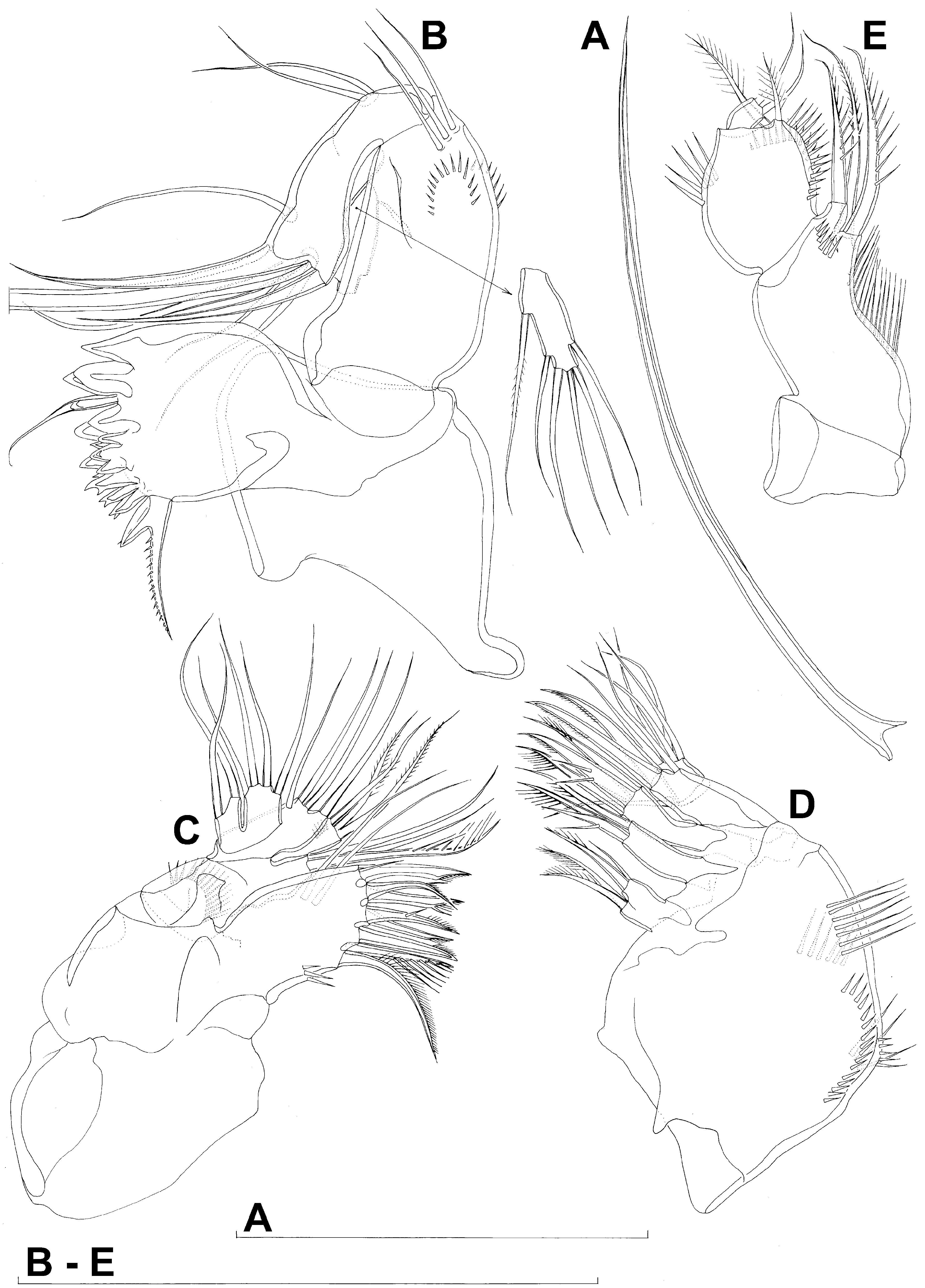
Mandibula ( Fig. 5A, B View FIGURE 5 ) with wide cutting edge on relatively short coxa, with two strong bicuspidate teeth ventrally, distally with long and slender central seta, eight bicuspidate teeth dorsally, and single dorsal unipinnate seta fused basally to tooth; teeth progressively decrease in size from ventral to dorsal side; dorsal seta only slightly longer but much stronger than central seta, and about twice as long as longest tooth; no ornamentation on coxa. Palp biramous, comprising basis, one-segmented exopod, and one-segmented endopod. Basis with somewhat inflated central part, about twice as long as wide, with three slender and smooth distal outer setae, with two arched rows of spinules in distal half. Exopod 0.75 times as long as basis and half as wide, narrowest medially, curved back towards coxa and parallel with basis, with three lateral and six apical smooth setae; all lateral and two apical setae slender, four apical setae strong and geniculate, one of them ( Fig. 5A View FIGURE 5 ) more than five times as long as exopod. Endopod only half as long as exopod, 2.7 times as long as wide, with one inner, three apical, and two outer slender and subapical setae; proximal outer seta bipinnate, other smooth.
Maxillula ( Fig. 5C View FIGURE 5 ) composed of praecoxa, coxa, basis, one-segmented endopod, and one-segmented exopod; endopod and exopod fused basally. Praecoxa large; arthrite rectangular, with three posterior spinules near dorsal margin and one spinule at base of ventralmost apical spine, apically and subapically with eight strong curved spines, each with a dense tuft of distal spinules along convex margin. Coxa with anterior arched row of long spinules, endite shorter than praecoxal arthrite, apically (on inner margin) with one curved and stout, bipinnate seta, and two smooth and slender setae. Basis smaller than coxa with two endites reaching further medially than coxal endite, almost in line with praecoxal arthrite, with five spinules and three setae on dorsal endite, and four setae on ventral endite; only two setae on dorsal endite bipinnate, others smooth. Endopod minute, rectangular, with four slender and smooth apical setae. Exopod smaller than endopod, with two slender and smooth apical setae.
Maxilla ( Fig. 5D View FIGURE 5 ) composed of large syncoxa, small basis and even smaller one-segmented endopod. Syncoxa with four rows of outer long spinules and with three endites; dorsal endite smallest, bilobate, with four setae, three of which strong and pinnate; central endite slender, with two pinnate setae, dorsal seta strong, with one spinule almost as strong as seta, giving it bifurcate appearance; ventral endite longest and strongest, with three strong, pinnate setae. Basis slightly larger than ventral endite of syncoxa, apically with two strong and geniculate, unipinnate spines, and two slender setae on ventral and posterior surfaces. Endopod only about 0.35 times as long as basis, 1.2 times as long as wide, with five slender and smooth apical setae, all equal in length.
Maxilliped ( Fig. 5E View FIGURE 5 ) not prehensile, four-segmented, composed of coxa, basis, and two-segmented endopod. Coxa short, almost triangular, unarmed and unornamented. Basis largest and longest segment, about 2.3 times as long as wide and 2.5 times as long as coxa, with longitudinal row of slender inner spinules and row of shorter anterior spinules at base of three inner distal spiniform setae; all setae close to each other, strong and of similar length, two unipinnate with large pinnules, one bipinnate with smaller pinnules. First endopodal segment 0.6 times as long as basis but slightly wider, almost ovoid in shape, with two parallel longitudinal rows of large inner spinules, one of them continuing as transverse distal row on posterior surface, and another row of five long outer spinules; with two slender distomedial plumose setae, shorter anterior seta reaching beyond second endopodal segment, longer seta situated posteriorly. Second endopodal segment minute, nearly rectangular, apically with two subequal smooth and slender setae.
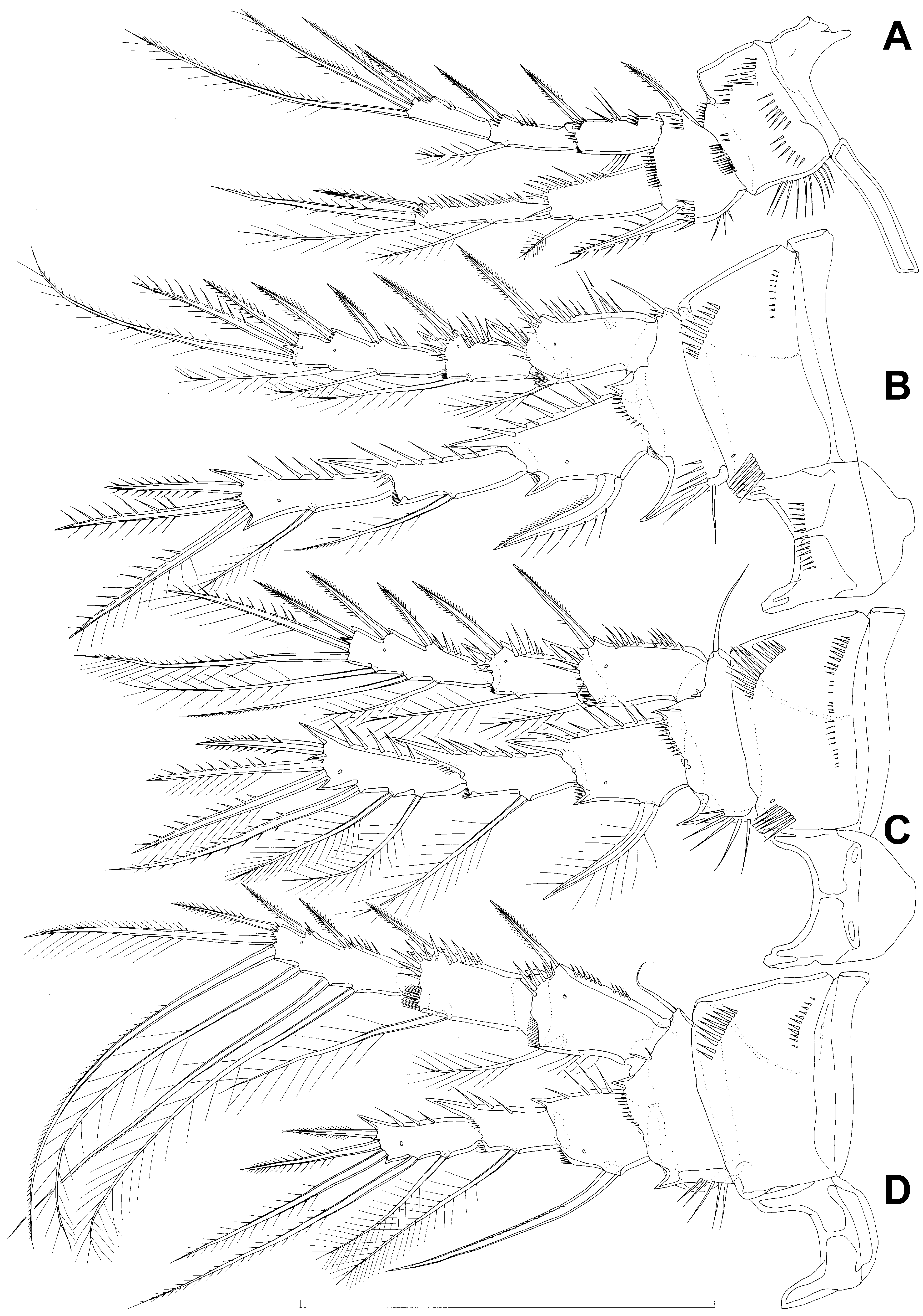
All swimming legs ( Fig. 6 View FIGURE 6 ) of similar size and long in comparison to body length, composed of small triangular and unarmed praecoxa, large rectangular and unarmed coxa, shorter and nearly pentagonal basis, slender three-segmented exopod, slender two- or three-segmented endopod; pair of legs joined by simple intercoxal sclerite.
First swimming leg ( Fig. 6A View FIGURE 6 ) with smooth and short intercoxal sclerite, its distal margin nearly straight. Praecoxa somewhat triangular, longer than intercoxal sclerite but shorter than coxa, unornamented. Coxa 1.5 times as wide as long, with longitudinal row of long inner spinules, four shorter transverse rows of smaller anterior spinules proximally, distal row of slender spinules and outer row of small spinules at base of basis. Basis with one short but strong and finely bipinnate outer spine and one longer and stronger inner spine; the latter 1.6 times as long as the former, with strong pinnules on both sides, and one long distomedial pinnule; ornamentation of basis consists of three strong inner spinules, an anterodistal row of slender spinules at base of endopod, and several strong spinules at base of both spines. Exopod with all segments of about same length, each about 2.5 times as long as wide and with outer spinules and subdistally on anterior surface; first segment with four inner slender spinules; first two segments with single strong and finely bipinnate distolateral spine; second segment with slender distomedial seta; third segment with two strong and pinnate outer spines and two setae apically; apical setae not prehensile, with short outer pinnules and long and sparse inner pinnules; length ratio of elements on third segment from outer to inner margin 1: 1.5: 2.1: 3.1. Endopod two-segmented, not prehensile, only slightly shorter than exopod; first endopodal segment 1.2 times as long as first exopodal segment and 2.3 times as long as wide, with strong inner and anterodistal spinules, with single bipinnate inner seta, the latter slender and about 0.6 times as long as segment; second segment slender, about 5.6 times as long as wide and 1.2 times as long as first segment, with continuous longitudinal row of strong outer spinules, with two slender inner seta, one strong and long apical spine, and another shorter spine distolaterally; apical spine about as long as inner distal seta, 1.9 times as long as outer distal spine, and 1.5 times as long as second segment but only about 0.7 times as long as longest exopodal seta.
Second swimming leg ( Fig. 6B View FIGURE 6 ), intercoxal sclerite with transverse distal row of small anterior spinules, distal margin deeply concave. Praecoxa short, unornamented. Coxa nearly 1.7 times as wide as long, anteriorly with pore and short row of long spinules near distomedial corner and two longer rows of spinules close to outer margin, proximal spinules smaller than distal ones. Basis with smooth, short and slender outer spine; inner distal corner produced into long and sharp process; anteriorly with distal row of small spinules and short row of long spinules close to inner margin; spine with two small spinules at base. First exopodal segment widest, third segment slender and about 3.6 times as long as wide, 1.5 times as long as second segment, and 1.4 times as long as first one; segments with single anterior pore, and outer and distal spinules (those on outer margin much stronger), and with inner distall frill on first two segments; first and second segments with single strong and finely bipinnate outer distal spine and slender bipinnate inner dista seta; third segment with three strong and finely bipinnate outer spines, two apical strong and bipinnate setae, and two slender and bipinnate inner setae; inner apical seta on third segment longest, about 1.7 times as long as outer apical one, twice as long as third segment, and 2.6 times as long as outer distal spine; outer distal corner of first and second segment produced into spiniform process. Endopod threesegmented, 1.1 times as long as exopod; all segments of about same length, but progressively narrower from proximal to distal end, each with outer distal corner produced into strong spiniform process and inner distal corner also spiniformly produced (though much less strongly than in exopod), each with row of strong outer spinules, first two segments additionally with small inner distal frill, and first and third segments with anterior cuticular pore; armature consisting of single bipinnate inner seta on first and second segments, and one inner and three apical elements on third segment (probably outermost spine and two strong setae); seta on first segment exceptionally strong and curved, other elements straight, inner seta on second segment slender and with distal inner row of minute pinnules in addition to long pinnules, inner seta on third segment also slender but just with long pinnules, apical setae with slender long inner pinnules and robust long outer pinnules; inner apical seta on third segment longest, 1.2 times as long as outer apical seta, 1.4 times as long as segment, and 1.6 times as long as outer apical spine.
Third swimming leg ( Fig. 6C View FIGURE 6 ) similar to second swimming leg, except for smooth intercoxal sclerite, longer proximal row of spinules on coxa, slender outer seta and shorter inner distal process on basis, and three inner setae on third endopodal and exopodal segments each; middle inner seta on third exopodal segment with only minute distal inner pinnules, distal inner seta on third endopodal segment more robust than other inner setae and with short but strong pinnules on both margins, all other inner setae on exopod and endopod bipinnate with long and slender pinnules; inner seta on first endopodal segment slightly less strong than serially homologous one on second leg, but also curved.
Fourth swimming leg ( Fig. 6D View FIGURE 6 ) relatively similar to third swimming leg, but with endopod only about 0.7 times as long as exopod, without pore or inner distal row of spinules on coxa, slightly shorter inner distal process on basis, much longer spiniform seta on first endopodal segment, only two inner setae on third endopodal segment, and longer and stronger inner setae on third exopodal segment.
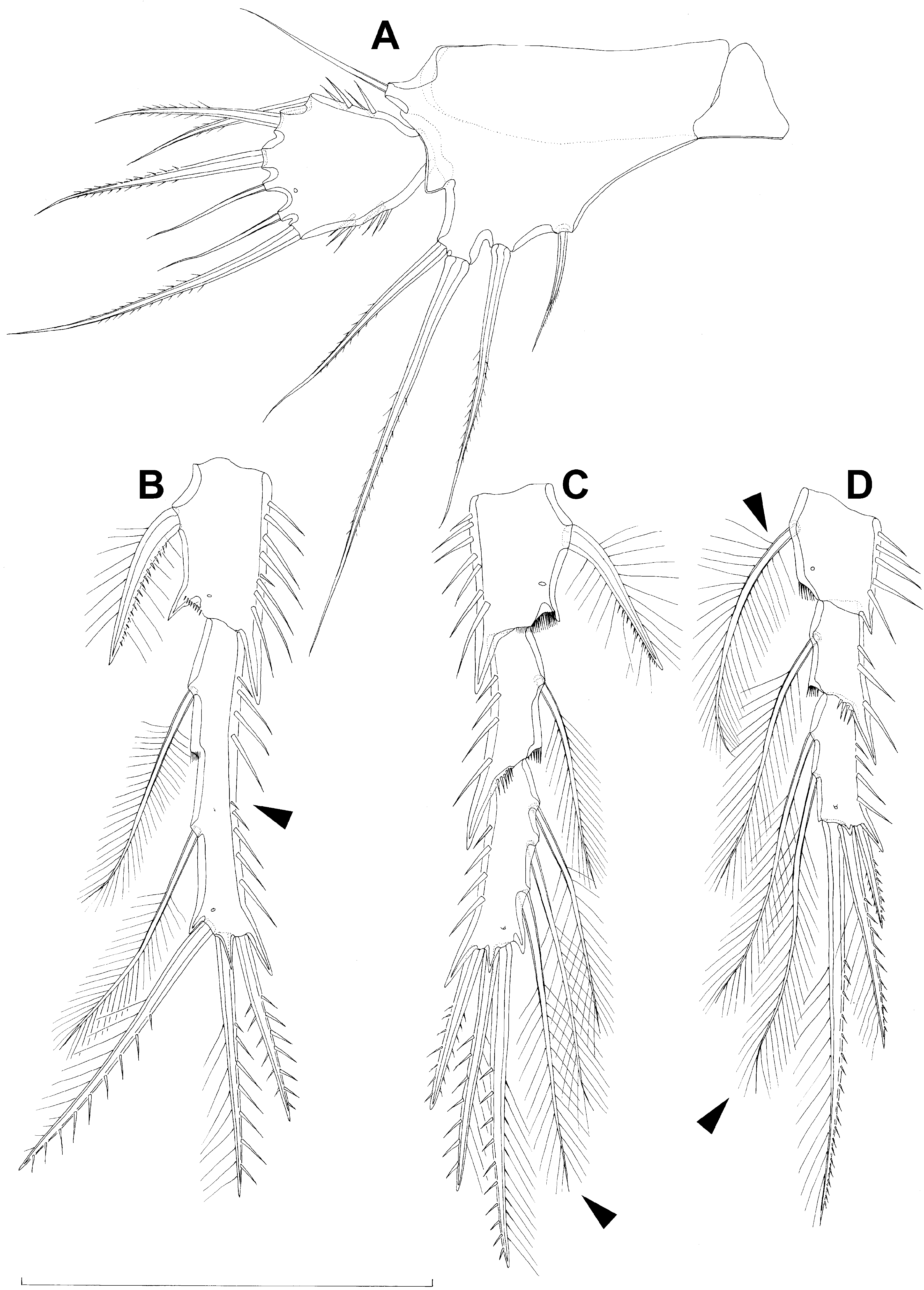
Fifth leg ( Fig. 7A View FIGURE 7 ) composed of wide baseoendopod (fused basis and endopod) and much smaller ovoid exopod, pair of legs joined by small triangular sclerite. Baseoendopod about 1.5 times as wide as long, more or less pentagonal, unornamented, with short spiniform process at base of exopod; outer basal seta slender and smooth, arising from short setophore, about 0.8 times as long as segment; endopodal lobe relatively narrow, trapezoidal, extending slightly beyond proximal third of exopod in length, with four stout, bipinnate setae, thier length ratio, starting from inner side, 1: 2.3: 3.4: 2. Exopod about 1.1 times as long as its maximum width, more or less trapezoidal, with narrow base, with two short rows of strong inner spinules, one row of strong outer spinules, and single anterior pore close to distal margin, with six setae; second and third seta from inner side short, slender, and smooth, other setae strong and bipinnate; length ratio of exopodal setae, starting from inner side, 1: 0.4: 0.4: 0.8: 0.6: 0.6.
Sixth leg ( Fig. 4A View FIGURE 4 ) minute flap covering ventro-lateral genital aperture, mostly fused to somite, unornamented, with single short and smooth seta near outer margin and two minute spines; inner minute spine fused basally to plate, forming small spiniform process. Sixth legs seemingly joined on ventral side by fold-like suture which hides copulatory pores.
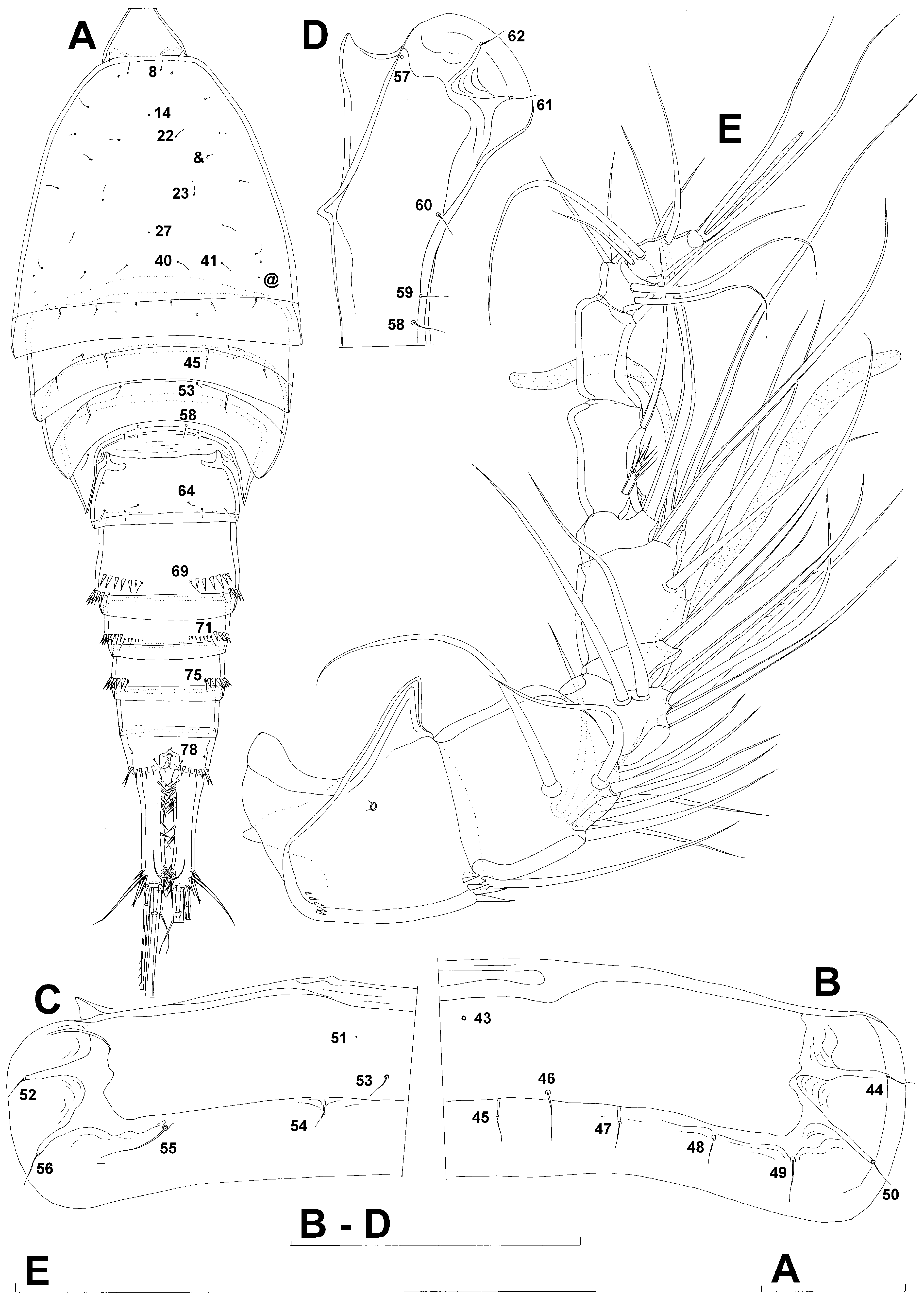
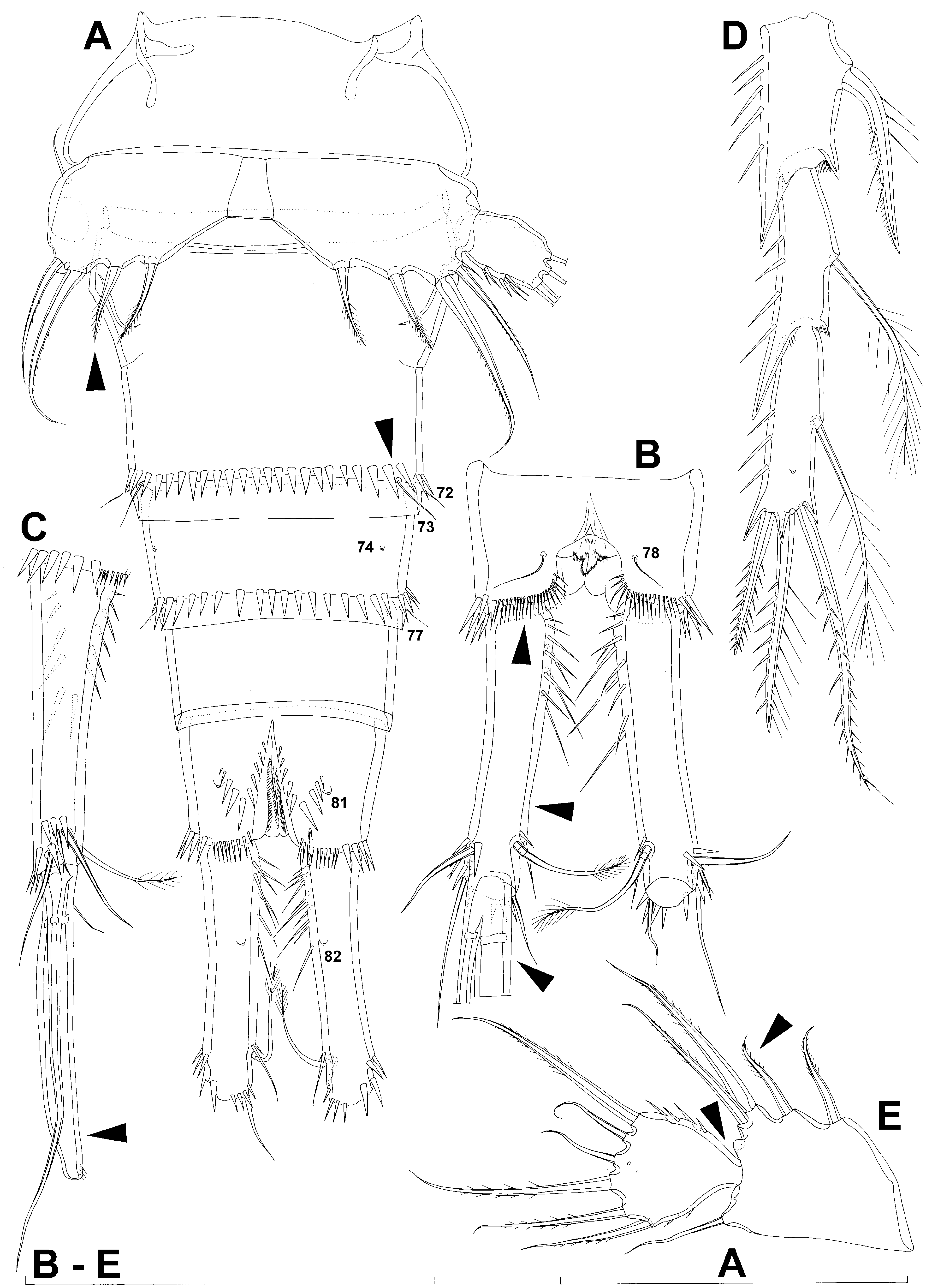
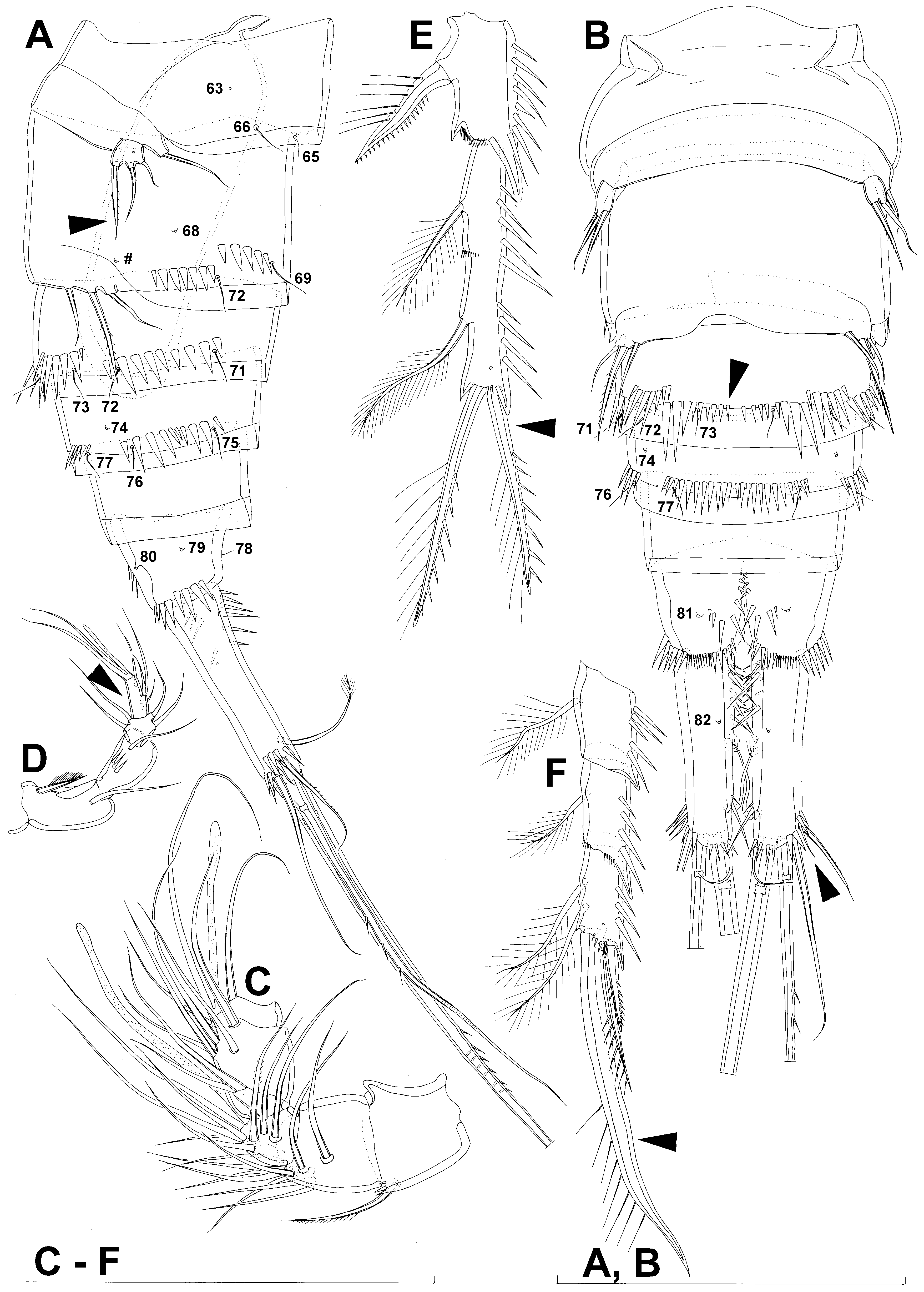
Male (based on allotype and five paratypes). Body length from 605 to 684 µm (610 µm in allotype). Habitus ( Figs. 8A View FIGURE 8 , 11C View FIGURE 11 ), colour, rostrum ( Figs. 8A View FIGURE 8 , 10F View FIGURE 10 ), shape and most ornamentation on cephalothorax ( Figs. 8A View FIGURE 8 , 10F View FIGURE 10 , 11D View FIGURE 11 ), shape and ornamentation of second pedigerous somite ( Figs. 8B View FIGURE 8 , 10F View FIGURE 10 , 11E View FIGURE 11 ), third pedigerous somite ( Figs. 8C View FIGURE 8 , 10F View FIGURE 10 , 11E View FIGURE 11 ), fourth pedigerous somite ( Figs. 8D View FIGURE 8 , 10F View FIGURE 10 , 11E View FIGURE 11 ), most ornamentation on first urosomite ( Fig. 9A, B, C View FIGURE 9 , 11E View FIGURE 11 ), ornamentation of last threeurosomites ( Figs. 9A, B, C View FIGURE 9 , 11F View FIGURE 11 ), caudal rami ( Fig. 9A, B, C View FIGURE 9 ), antenna, labrum, paragnaths, mandibula, maxillula, maxilla, maxilliped, first swimming leg ( Fig. 10F View FIGURE 10 ), and coxae, bases and exopods of second, third and fourth swimming legs as in female. Prosome/urosome ratio 1.05, greatest width at posterior end of cephalothorax, body length/width ratio about 3.1; cephalothorax twice as wide as genital somite in dorsal view. Genital somite and third urosomite not fused.
Cephalothorax ( Figs. 8A View FIGURE 8 , 10F View FIGURE 10 , 11D View FIGURE 11 ) in addition to all sensilla and pores present in female with one additional pair of dorsal anterior sensilla (no. &) and one additional pair of dorsal posterior pores (no. @).
First urosomite ( Figs. 9A, B, C View FIGURE 9 , 11E View FIGURE 11 ) slightly narrower and longer than in female, with two additional rows of minute spinules above sensilla pair no. 65 and without lateral pore pair no. 67.
Genital somite ( Figs. 9A, B, C View FIGURE 9 , 11F View FIGURE 11 ) homologous to anterior part of genital double-somite in female, 1.3 times as wide as long in dorsal view, with all sensilla, pores, and spinules homologous to those in female present, with additional lateral pair of pores (no. #) and additional lateral row of large spinule between sensilla no. 70 and pore no. #; large and longitudinally positioned spermatophore visible inside on right side, four times as long as wide, twice as long as genital somite, its posterior end reaching slightly beyond distal margin of genital somite, its anterior part protruding into first urosomite and even slightly into fourth pedigerous somite.
Third urosomite ( Figs. 9A, B, C View FIGURE 9 , 11F View FIGURE 11 ) only half as long as genital somite, with three posterior pairs of sensilla as in female, but pair no. 72 situated more ventrally and pair no. 73 very close to each other; ventral row of spinules interrupted between sensilla pair no. 73; additional minute spinules between sensilla pair no. 71 present in allotype ( Fig. 9A View FIGURE 9 ) but not in paratypes (11F).
Antennula ( Figs. 8E View FIGURE 8 , 11A View FIGURE 11 ) also as long as cephalothorax, but strongly geniculate and nine-segmented (basically female’s sixth segment subdivided), with geniculation between third and fourth and sixth and seventh segments. Segments that participate in geniculation strengthened with cuticular plates along anterior surface, largest ones on seventh segment. Aesthetascs as in female, on fourth and last segments, but additional large aesthetasc present on third segment. First two and last two segments similar to female, except for additional row of minute spinules on first segment; third segment much shorter and distal part of it fused with fourth segment (as can be judged from armature position); fourth segment accordingly longer; fifth segment shorter, while sixth female segment virtually unrecognisable. Setal formula 1.11.6+ae.7+ae.1.2.1.4.6+ae. All setae smooth, except for short proximal seta on sixth segment.
Second swimming leg ( Figs. 7B View FIGURE 7 , 10F View FIGURE 10 ) with second and third endopodal segments fused (arrowed in Fig. 7B View FIGURE 7 ) but armature and ornamentation as in female.
Third swimming leg ( Fig.7C View FIGURE 7 ) with distal inner seta on third endopodal segment slender and plumose (arrowed in Fig. 7C View FIGURE 7 ), other armature and all ornamentation as in female.
Fourth swimming leg ( Fig. 7D View FIGURE 7 ) with inner seta on first endopodal segment and distal inner seta on third endopodal segment slender and plumose (both arrowed in Fig. 7D View FIGURE 7 ), other armature and ornamentation as in female.
Fifth legs ( Figs. 9A, B, C View FIGURE 9 , 11B View FIGURE 11 ) much smaller than in female, without endopodal armature, and with baseoenopods fused medially into narrow plate. Exopod minute, ovoid, with single anterior proximal pore, and with two smooth setae and innermost bipinnate seta; length ratio of exopodal setae, starting from inner side, 1: 1.7: 0.5.
Sixth legs ( Fig. 9B, C View FIGURE 9 ) almost completely fused medially and to somite, forming simple flap, with concave hyaline fringe as only remnant of former subdivision; only functional genital aperture under right leg; each leg with three smooth setae, their length ratio from inner side, 1: 2.1: 1.7.
Variability. Most morphological features are conservative, including the sensilla and pores pattern of somites, and length ratio of different armature on appendages. The only significant form of morphological variability, except body length, was presence/absence of minute dorsal spinules on the third urosomite in males ( Figs. 9A View FIGURE 9 , 11F View FIGURE 11 ).
Morphological affinities. Wellstenhelia calliope sp. nov. differs from all congeners by the very narrow endopodal lobe on the female fifth leg ( Fig. 7A View FIGURE 7 ), where the reduced space between two central setae can be considered as a clear autapomorphy. This species has long caudal rami (l/w index of about 3.5), which distinguishes it at once from the sympatric Wellstenhelia clio sp. nov., Wellstenhelia erato sp. nov., and Wellstenhelia euterpe sp. nov., as well as from the Mediterranean Wellstenhelia bocqueti ( Soyer, 1971) comb. nov., and the Artcic Wellstenhelia melpomene sp. nov. The Swedish Wellstenhelia hanstromi ( Lang, 1948) comb. nov. also has somewhat shorter caudal rami than Wellstenhelia calliope , but differs additionally by its long innermost seta on the female fifth leg endopod, and short seta on the first endopodal segment of the fourth swimming leg (this character being a clear autapomorphy of Wellstenhelia hanstromi ). Only the sympatric Wellstenhelia qingdaoensis ( Ma & Li, 2011) comb. nov. has caudal rami as elongated as Wellstenhelia calliope , but the former differs by its short second seta from inner side on the female fifth leg endopod ( Fig. 25E View FIGURE 25 ), inflated inner principal caudal seta ( Fig. 25C View FIGURE 25 ), longer spinules on the first leg coxa ( Fig. 24F View FIGURE 24 ), narrower rostrum ( Fig. 24C View FIGURE 24 ), reduced armature on the male second leg endopod ( Fig. 26E View FIGURE 26 ), and transformed inner apical seta on the male fourth leg endopod ( Fig. 26F View FIGURE 26 ), as well as many details in the ornamentation of somites, including absent pores and sensilla nos. 14, 19, 21, 33, 34, 35, 43, 48 ( Fig. 24A, B, D View FIGURE 24 ), present sensilla nos. £, $, wider space between sensilla no. 69, only four spinules in the dorsal row on the anterior part of the genital double-somite ( Fig. 24B View FIGURE 24 ), much more slender and denser dorsal spinules along distal margin of the anal somite ( Fig. 25B View FIGURE 25 ), etc. In fact, so many morphological differences between Wellstenhelia calliope and Wellstenhelia qingdaoensis suggest that their elongated caudal rami probably originated convergently (see also Ma & Li 2011). In Wellstenhelia calliope the innermost endopodal seta on the female fifth leg is much shorter than the next one ( Fig. 7A View FIGURE 7 ), which distinguishes it at once from Wellstenhelia clio (with both setae long; Fig. 14C View FIGURE 14 ), Wellstenhelia qingdaoensis (both setae short; Fig. 25E View FIGURE 25 ), Wellstenhelia euterpe (innermost seta missing; Fig. 31G View FIGURE 31 ), Wellstenhelia melpomene (both setae long; Kornev & Chertoprud 2008), and Wellstenhelia hanstromi (both setae long; Lang 1948). Only Wellstenhelia erato and Wellstenhelia bocqueti have the innermost setae significantly shorter than the next one (similar to that in Wellstenhelia calliope ), but in Wellstenhelia erato this difference is much more pronounced ( Fig. 19D View FIGURE 19 ), while in Wellstenhelia bocqueti there is a deep notch between these two setae ( Soyer 1971). The proportion of different armature elements on the female fifth leg could be used alone to distinguish between different species of Wellstenhelia gen. nov., except perhaps between Wellstenhelia clio and Wellstenhelia hanstromi (their affinities are discussed below).
No known copyright restrictions apply. See Agosti, D., Egloff, W., 2009. Taxonomic information exchange and copyright: the Plazi approach. BMC Research Notes 2009, 2:53 for further explanation.
|
Kingdom |
|
|
Phylum |
|
|
Class |
|
|
Order |
|
|
Family |
|
|
Genus |