
Turbanella cuspidata, Yamauchi & Kajihara, 2018
|
publication ID |
https://doi.org/ 10.12782/specdiv.23.183 |
|
publication LSID |
lsid:zoobank.org:pub:3FA0A429-1676-4326-B8D3-FFBA8FE403C9 |
|
persistent identifier |
https://treatment.plazi.org/id/0397C909-FFE5-301E-FC30-D988FF5B2DBF |
|
treatment provided by |
Felipe |
|
scientific name |
Turbanella cuspidata |
| status |
sp. nov. |
Genus Turbanella Schultze, 1853 View in CoL Turbanella cuspidata sp. nov. ( Figs 5 View Fig , 6 View Fig )
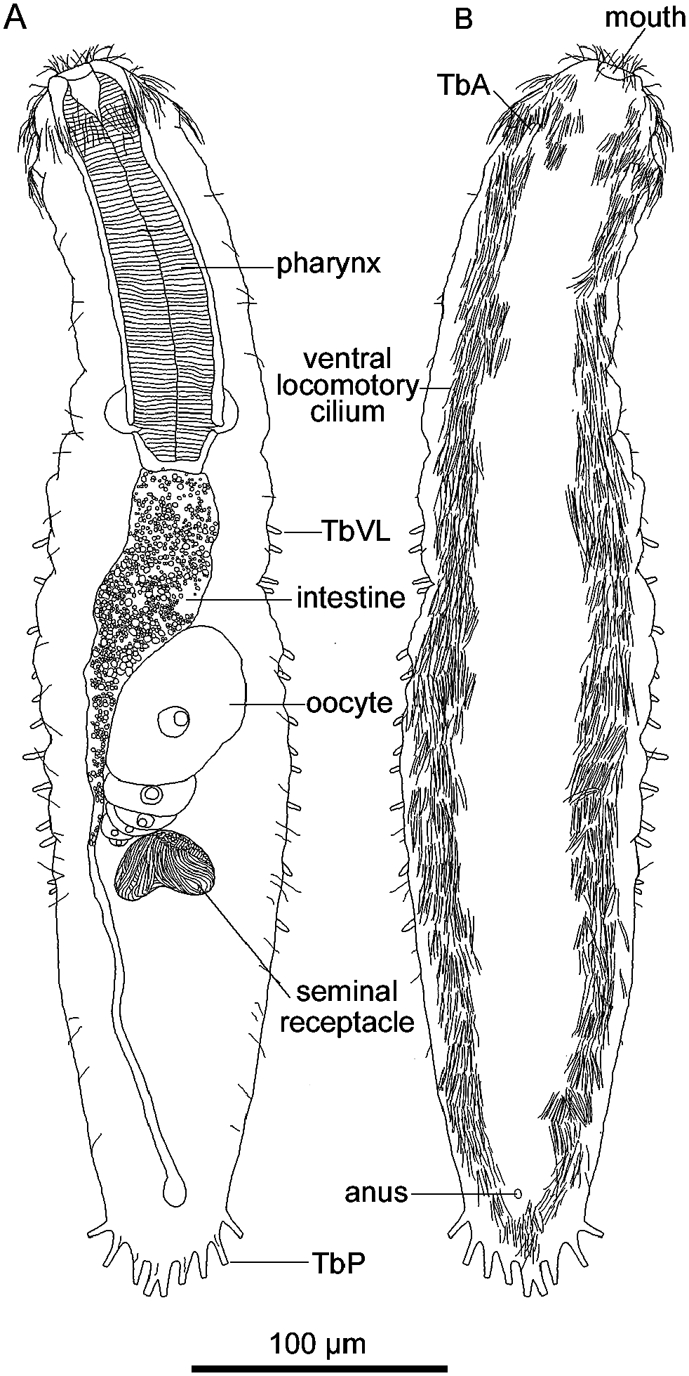
U07; sensory cilia (L 13 µm) in lateral and dorsal columns on trunk; ventral locomotory cilia (L 13 µm) running posteriorly from circum-cephalic ring in two longitudinal bands along lateral body margins, remaining separate throughout, but with post-anal ciliary patch.
Digestive tract. Mouth terminal, of medium width (18 µm); buccal cavity cup-shaped, shallow; walls lightly cuticular; pharynx medium throughout, with basal pharyngeal pores at U28–32; intestine divided into broad, anterior, granular region with refractive granules at anterior and posterior ends, and narrower, posterior, non-granular region; anus at U91.
Reproductive tract. Hermaphroditic, probably protogynous; testes not seen; oocytes lying along anterior part of intestine (U50–70), developing from posterior to anterior, largest (67×44 µm) anteriorly ( Figs 2A View Fig , 3C View Fig ); seminal receptacle present behind oocytes ( Figs 2A View Fig , 3C View Fig ).
Remarks. Among the specimens examined, only one had multiple eggs, thus indicating the direction of oocyte development, from posterior to anterior. This orientation,
Material examined. Twelve adults. Holotype, ICHUM 4980 View Materials , mounted on glass slide, Higashi-Shizunai, Hokkaido, Japan (42°17.333′N, 142°27.590′E), medium-grained sand, surface layer at water’s edge, 19 May 2012. Six paratypes, same collection data as for holotype: ICHUM 4981 View Materials , mount- ed on glass slide; ICHUM 4991–4995 View Materials , on SEM stubs. Four additional specimens destroyed after observation. GoogleMaps
Etymology. The new specific name is an adjective from the Latin cuspidatus (made pointed), indicating the pair of small, ventrolateral, projective organs.
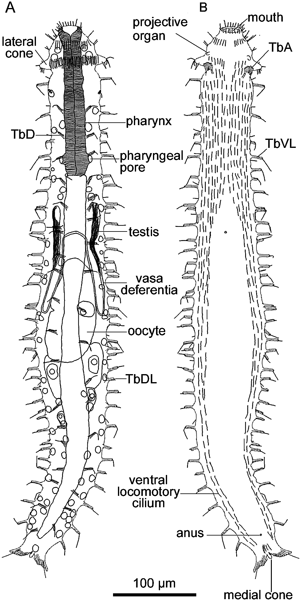
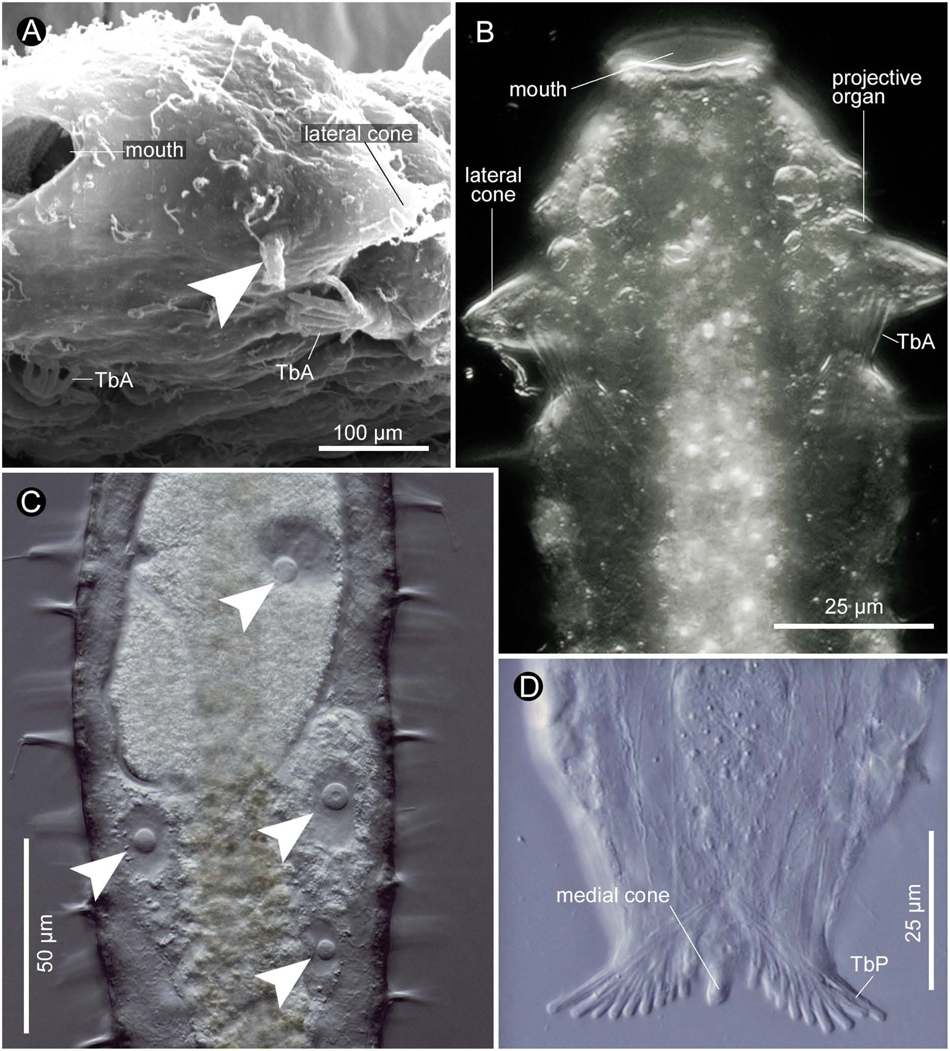
Description. Habitus. Adult Lt 570–670 µm (640 µm in holotype); L of anterior end to PhJIn (at U32–27) 160– 180 µm (177 µm in holotype). Body medium in length; head sculpted, with lateral cones at U07 ( Figs 5A View Fig , 6A, B View Fig ) and small ventrolateral projective organ at U06 on each side ( Figs 5B View Fig , 6A, B View Fig ); neck constriction at U09; trunk widest in mid-body region, tapering gradually to caudal base; caudum slightly cleft, incised from tips to U98, bearing a medial cone (L 5–10 µm) ( Figs 5B View Fig , 6D View Fig ). Glands 30–40 per side, medium size (6 µm in diameter), scattered along lateral and medial columns.
Adhesive tubes. TbA 7–8 per side (L 4–12 µm), occurring on lobe inserted at U09–11 ( Figs 5B View Fig , 6A, B View Fig ), most medial tube on hand always set lower than others; TbL/ TbVL 18–25 per side (L 10–16 µm, insertions difficult to distinguish), irregularly spaced and often asymmetrically arranged, with five in pharyngeal region, one at PhJIn, and others along intestine, but none behind anal opening, most bearing cilia; TbDL 8–13 per side, evenly spaced and sym- metrically arranged, with three in pharyngeal region and remainder along intestine, most bearing cilia; TbD 15–20 per side, with three in pharyngeal region and remainder along intestine, most bearing cilia; ‘cirrata’ [Seitenfüsschen] tubes occurring at U39; TbP 10–12 per side, arrayed along rear edge of each lobe, lengthening medial to lateral (L 4–13 µm).
Ciliation. Mouth surrounded by short sensory cilia (L 6 µm), with longer cilia (L 11 µm) inserted at points of head sculpting on each side; ciliary hairs (L 11 µm) forming circum-cephalic band at U07; sensory cilia (L 7 µm) occurring on trunk in lateral and dorsal columns; each Tb on trunk bearing cilium (L 13 µm) arising from rear apex of tube support; ventral locomotory cilia (L 15 µm) running in two longitudinal bands along lateral body margins to anus, separate except beneath (i.e., ventrally in) pharyngeal region ( Fig. 5B View Fig ).
Digestive tract. Mouth terminal, of medium width (18 µm); buccal cavity cup-shaped, shallow; walls lightly cuticular; pharynx of medium width throughout, with basal pharyngeal pores at U29–24; intestine narrows anterior to posterior; anus at U94.
Reproductive tract. Hermaphroditic; paired testes extending posteriorly from U32, their vasa deferentia recurving anteriorly and exiting at U38; bilateral oocytes developing in posterior to anterior direction, largest (125×57 µm) in anterior region of intestine ( Figs 5A View Fig , 6C View Fig ).
Remarks. Among approximately 30 species in the genus Turbanella , four species ( T. amphiatlantica Hummon and Kelly, 2011 ; T. bocqueti Kaplan, 1958 sensu Boaden (1974) ; T. varians Maguire, 1976 ; and T. wieseri Hummon, 2010 ) share many features with T. cuspidata sp. nov., but differ from the latter in the following ways. Turbanella amphiatlantica lacks the slight neck constriction; T. bocqueti has larger body size (Lt 800–1320) ( Hummon 2008); T. varians lacks lateral head cones; and T. wieseri bears nine TbD per side. In addition, the ventrolateral projective organ on each side the head is characteristic of T. cuspidata sp. nov.; the organ is a simple, cylindrical projection, about 45 µm in length and 20 µm in width, sticking out ventrally from a portion slightly anterior to the base of the lateral cone ( Fig. 6A, B View Fig ).

No known copyright restrictions apply. See Agosti, D., Egloff, W., 2009. Taxonomic information exchange and copyright: the Plazi approach. BMC Research Notes 2009, 2:53 for further explanation.