
Tromikosoma rugosum, Anderson, Owen F., 2016
|
publication ID |
https://doi.org/ 10.11646/zootaxa.4092.4.1 |
|
publication LSID |
lsid:zoobank.org:pub:EA66CAE5-F6CE-44BA-A5FF-67F2BEE6DEE8 |
|
DOI |
https://doi.org/10.5281/zenodo.6055446 |
|
persistent identifier |
https://treatment.plazi.org/id/03A4AB67-FFF3-FF9C-FF1D-FB50FDA960DF |
|
treatment provided by |
Plazi |
|
scientific name |
Tromikosoma rugosum |
| status |
sp. nov. |
Tromikosoma rugosum View in CoL sp. nov.
( Figures 14–17 View FIGURE 14 View FIGURE 15 View FIGURE 16 View FIGURE 17 )
Diagnosis. Small (adults possibly not seen), up to 69 mm TD; colour of test and appendages brownish-white; test puffed-up and wrinkled, the ambitus rounded and ill-defined; interambulacra wider than ambulacra (possibly juvenile condition); primary tubercles absent adradial to pore series in oral ambulacra; adapical secondary plates of aboral ambulacra elongate, occasionally reaching the perradius; madreporite indistinct; tridentate pedicellariae with minutely serrated edges.
Holotype. East coast North Island: 1 specimen (69 mm TD), 38° 59.8´S, 179° 15.8´E, 2756– 2478 m, NIWA6610. Stored in 80% ethanol.
Paratypes. East coast North Island: 1 specimen (43 mm TD), 35° 29.8´S, 178° 31.2´E, 3036– 3034 m, NIWA64937. 1 specimen (51 mm TD), 39° 0.0´S, 179° 19.2´E, 2748– 2605 m, NIWA6611. All stored in 80% ethanol.
Etymology. Named rugosum , because of its distinctive wrinkled appearance.
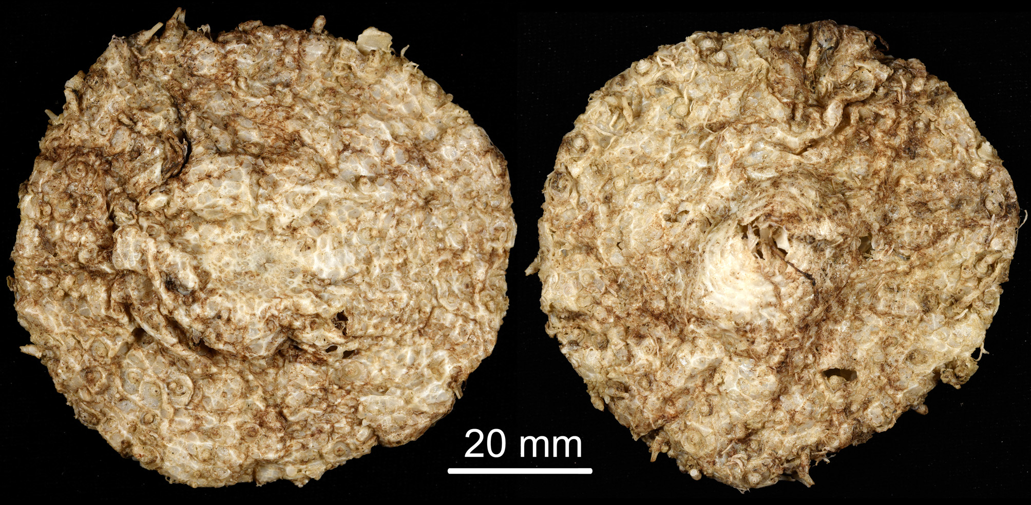
Description. Test of holotype ( Figure 14 View FIGURE 14 ) small (69 mm TD), circular, with rounded ambitus, the transition from the oral to the aboral surface not clearly defined. Test very flexible and thin and unable to maintain its form out of water, with the test surface much puffed-up and wrinkled, and very uneven. The test plates bear numerous fracture lines which blur delineation of the plate boundaries. Spines very fragile, only the basal sections of a few primary spines remaining. Colour of the test and spines of preserved specimens brownish-white, with little variation within and between specimens. Tubercles non-crenulate and perforate, circular except for a few of the primary tubercles near the ambitus which are slightly expanded laterally. Ratio of interambulacrum to ambulacrum at the ambitus about 1.3:1.
Interambulacral columns with about 15 plates, about 6 on the oral side and 9 on the aboral side. Ambulacral columns with about 20 plates, about 10 on each side; these number approximate as difficult to count due to the breaking up of the test plates. The specimen in NIWA 6611 (51 mm TD) has about 15–16 interambulacral plates and 18 ambulacral plates, divided evenly between the oral and aboral surfaces.
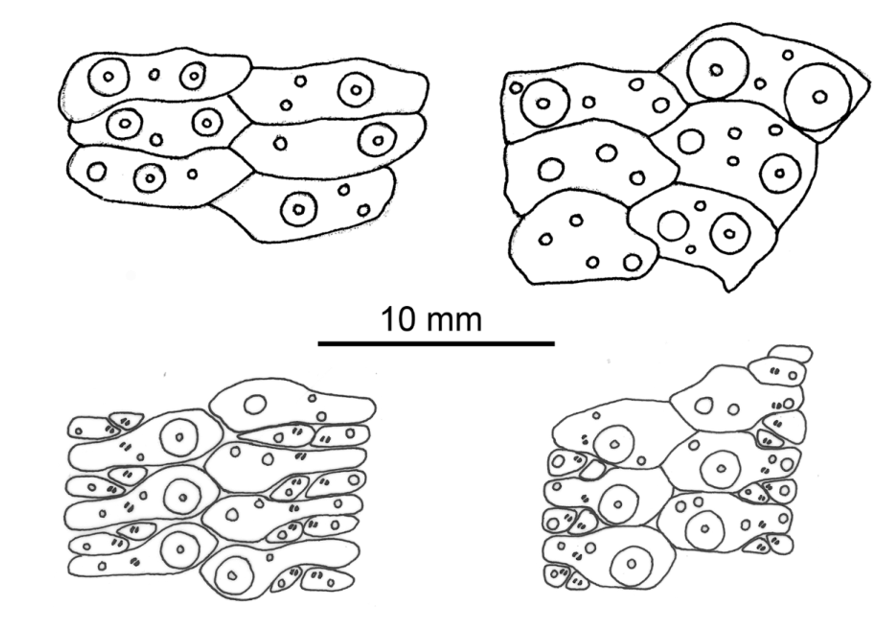
Oral test plating ( Figure 15 View FIGURE 15 ): An inner series of large primary tubercles near the perradius on 1–3 of the outer plates in the ambulacra; areoles circular. No primary tubercles adradial to the pore series. The plate configuration difficult to determine over much of the surface due the breaking up of the plates, but where visible the secondary plates small and limited to the adradial half of the main plate. The outer secondary plate shares the adradial boundary with the main plate; the inner plate slightly smaller and laterally abutting the outer plate. The podia are fairly well developed but lack obvious terminal disks (although these can be made out in NIWA 6611) and are arranged in a single, uneven line. One or two large primary tubercles per plate in the outermost plate of each interambulacral column, those adradially placed combining with a smaller primary tubercle on a few adoral plates to form a series down each column. Secondary tubercles scattered thinly over the remainder of the oral surface.
Peristome 21 mm in diameter, bearing six ambulacral plates (with podia) per column (five in NIWA 6611). Peristomial plates angled with up to seven small tubercles spread either side of the roughly central podium. These tubercles about half the height of the plate and located more towards the adoral edge—the podium located nearer the opposite edge. Buccal sacs not discernable, possibly absent.
Aboral test plating ( Figure 15 View FIGURE 15 ): Primary tubercles all smaller than those of the oral side, but fairly abundant. An inner series of primary tubercles near the perradius in the ambulacra on the outer 1–3 plates, and 1 or 2 smaller primary tubercles variably placed on plates further towards the apical system. Where visible, the arrangement of secondary plates with respect to the main plate is similar to the oral surface but differs in that the secondary plates are more elongate, the inner plate occasionally reaching the perradius, and the podia form into two parallel series. Plates of the interambulacra bear 1–2 primary tubercles, forming no obvious pattern, from the ambitus to the apical system (in NIWA 6611 a few of these plates have no tubercles and only one or two have two tubercles). Secondary tubercles scattered over the surface in a similar way to the oral surface.
Apical system not well defined; ocular and genital plates barely distinguishable from anal and coronal plates. The madreporite indistinct, the genital pore appearing to open into a membranous area distal to plate G2, but other pores not distinguishable. In NIWA 6611 the plates of the apical system are slightly more clearly defined; the madreporite and ocular plates roughly circular, the former more distinctive and prominent than that of the holotype.
Spines: The spines all broken, only the very base of a few remain on any of the specimens, and no hoofs could be seen. Primary spines circular and hollow, very delicate, with about 30 longitudinal striations; the largest spine about 0.9 mm diameter at the base.
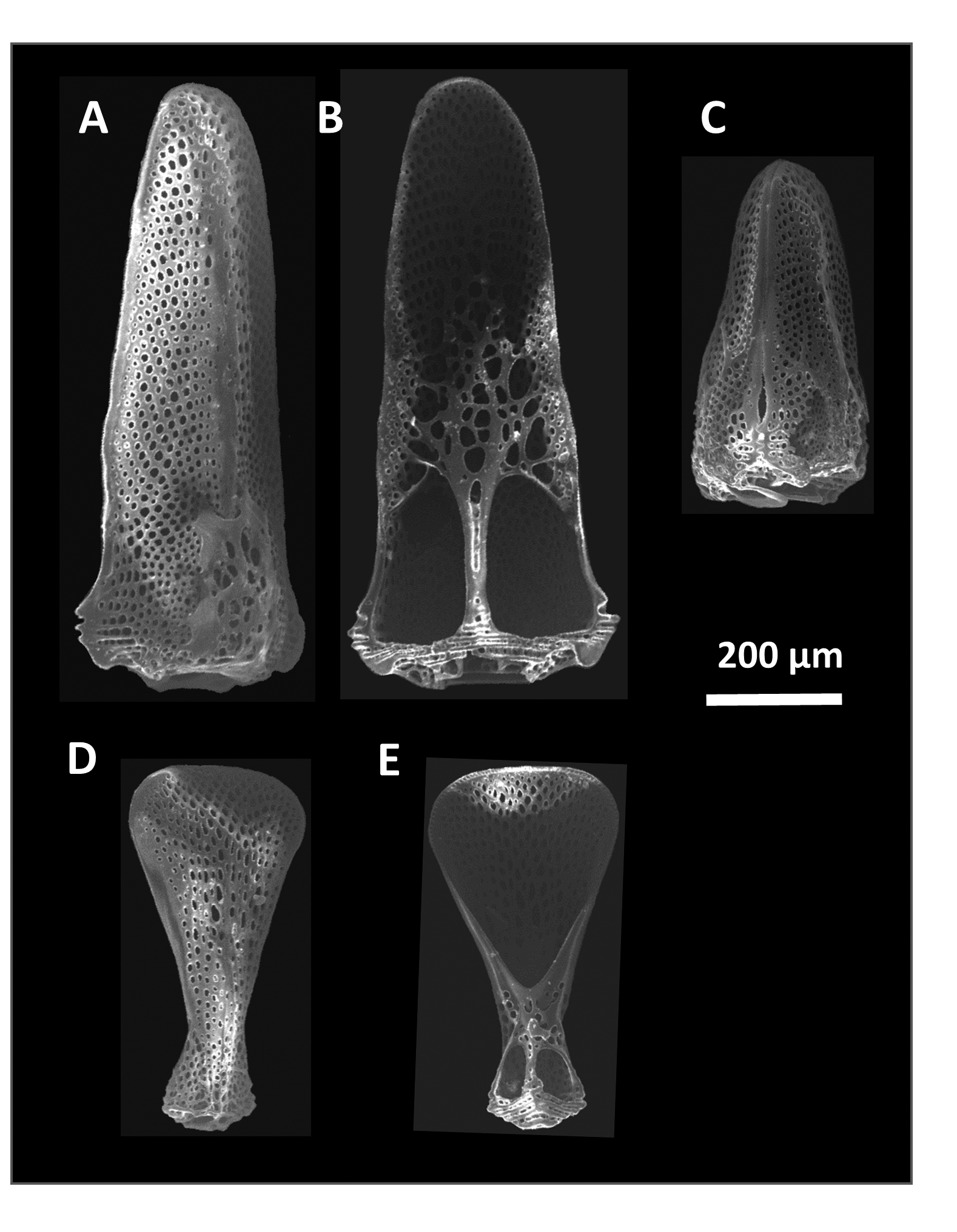
Pedicellariae ( Figure 16 View FIGURE 16 ): Only a few specimens were found on the holotype, and none at all on the paratypes. One type of rostrate tridentate pedicellariae was found, 0.5–0.9 mm long, with open blades evenly tapering to a rounded tip; blade edges minutely serrated and strengthened in the proximal part with a dense meshwork. The triphyllous pedicellariae are about twice as long as wide, with truncated blades 0.5 mm long and the cover-plate not well developed. Ophicephalous pedicellariae not found.
Sphaeridia: Oval-shaped (about 0.7 long by 0.4 wide), transparent with no obvious ornamentation, located adoral to the foot of the podium of the inner secondary ambulacral plates.
Size range —The three specimens range from 43–69 mm TD. It is uncertain as to whether these specimens represent juveniles or small adults of a species which reaches a maximum size more typical for the genus (170–200 mm TD). Although genital pores were not clearly distinguishable in these specimens, this does not necessarily indicate immaturity due to the difficulty in interpreting details of the apical system.
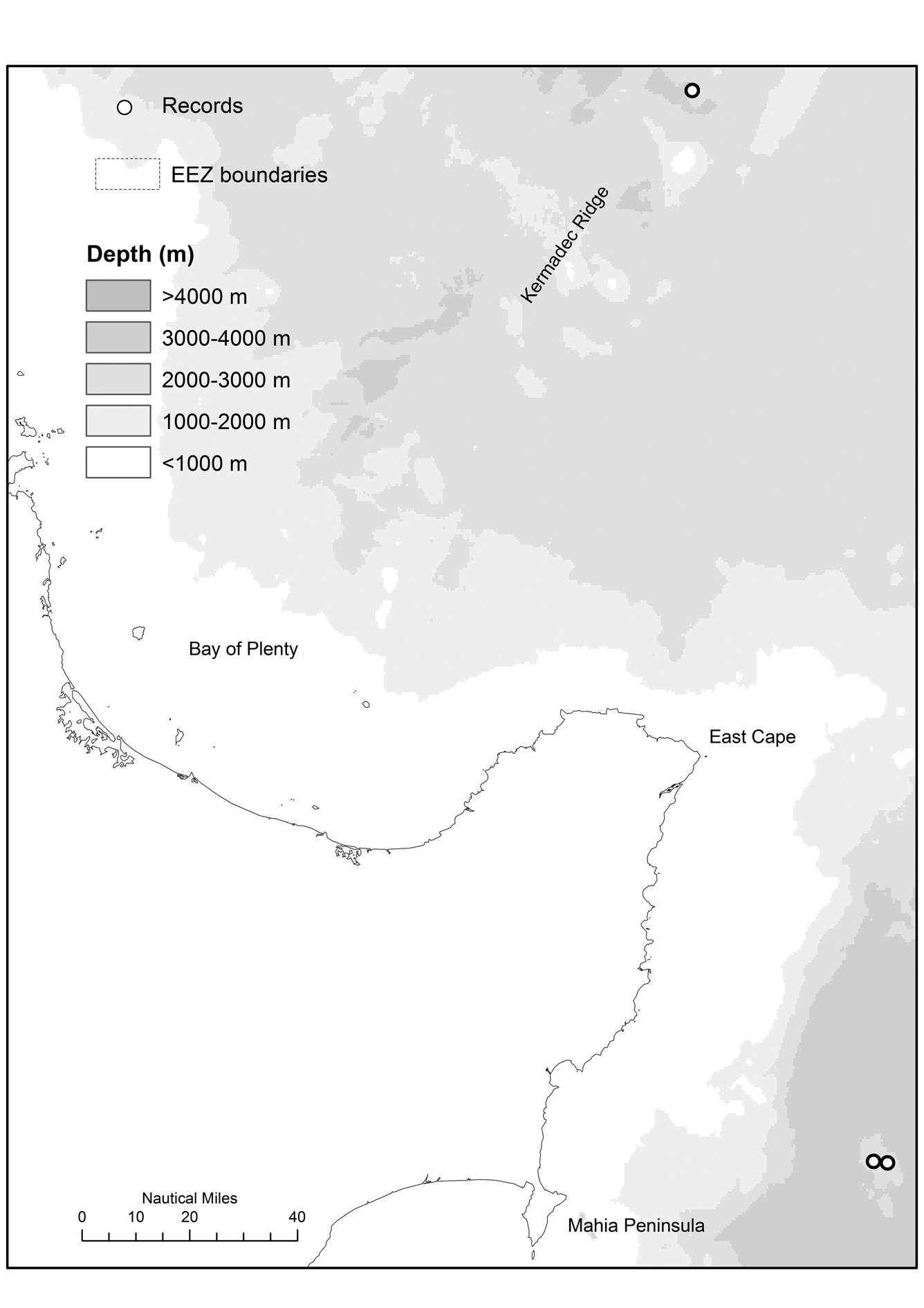
Occurrence. There are only three records (and three specimens) of T. rugosum sp. nov., all collected from deep water off the east coast of the North Island, between the Bay of Plenty and Mahia Peninsula ( Figure 17 View FIGURE 17 ). The potential depth range for the species is 2478–3036 m. There has been relatively little sampling at these depths in the New Zealand region (<200 research trawls/dredges) and the number of records is unlikely to be a reflection of their abundance in this habitat.
Remarks. This species is easily recognized and distinguished at a glance from other species of Tromikosoma , including similar sized specimens of T. australe , due to its exceedingly thin test, puffed-up and wrinkled appearance with a broadly rounded ambitus, and brownish-white colouration.
The small size of the specimens make it difficult to properly compare plating patterns with those of other species of Tromikosoma and so it is difficult to say with which it is most closely aligned. However from what can be seen of these patterns and of the arrangement of the primary tubercles it seems fairly typical of the genus. The lack of a primary tubercle outside the pore series in the oral ambulacra may separate it from T. australe and T. uranus (although the lack of such tubercles in smaller specimens of T. australe examined suggests this may be a size-related character), and the separation of the primary plates of the aboral ambulacra by the elongated secondary plates has also been reported for T. tenue and larger specimens of T. australe . Where this arrangement was observed in T. rugosum , it appeared to be the exception rather than the rule, and it may be that this character is also sizerelated.
The shape of the large tridentate pedicellariae is most like that of T. panamense , a species it otherwise differs from in the lesser abundance small tubercles and non-exclusion of the primary ambulacral plate from the adradial boundary by the outer secondary plate—a marked feature of T. panamense . The lack of an obvious madreporite in the apical system of the holotype is unusual, this feature usually being fairly obvious in echinothurioids in general, although a similar characteristic was found in T. koehleri , with Mortensen (1935) also unable to distinguish the madreporite in the type specimen.
No known copyright restrictions apply. See Agosti, D., Egloff, W., 2009. Taxonomic information exchange and copyright: the Plazi approach. BMC Research Notes 2009, 2:53 for further explanation.