
Trigoniophthalmus alternatus (Silvestri, 1904)
|
publication ID |
https://doi.org/ 10.5531/sd.sp.55 |
|
DOI |
https://doi.org/10.5281/zenodo.7730559 |
|
persistent identifier |
https://treatment.plazi.org/id/038D8781-FFFF-205A-FF2D-FB7CA232FD64 |
|
treatment provided by |
Felipe |
|
scientific name |
Trigoniophthalmus alternatus |
| status |
|
“Jumping bristletail”
Figures 8 View FIGURE 8 (lateral), 9 View FIGURE 9 (dorsal, ventral)
Plates 1 (lateral), 2 (dorsal), 3 (ventral)
Trigoniophthalmus , as the representative of the most basal order in Class Insecta, is notable in its tracheal architecture by a complete lack of longitudinal connections between spiracles. This taxon is critical to understanding the apparent insect ground-plan tracheal structure.
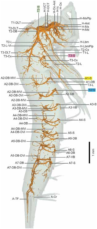
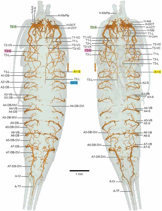
The thoracic tracheae of most taxa are supplied by both T2-S and T3-S, with T2-DLT connecting longitudinally. In Trigoniophthalmus , however, T3-S appears to only supply T3-L and its associated coxa. Branches from T2-S extend posteriorly into the metathorax, nominally supplied by T3-S, including an apparent T3-DLT. The naming of T3-DLT here is an instance of using the positional criterion in homology (sensu Remane, 1952), in that the connectivity of this trachea is not consistent with other taxa; however the position suggests its identification as T3-DLT ( de Pinna, 1991). T2-VB extends posterior, almost in the opposite direction of the anterior-reaching cephalic branches. While the position of T2-VB may not appear to be “ventral” in nature, a comparison of its relative position in the hump-backed Trigoniophthalmus and in particular its connection to T2-L with apterygote taxa from Zygentoma and Dermaptera demonstrates its homology as a ventral branch. Several tracheae range anteriorly or posteriorly beyond segment boundaries, most prominently A5-DB-DVi, which extends posteriorly past A6-S, reaching A7-S; and A7-Cr, which likewise extends posteriorly from A7-S into both the cerci and terminal filament. However, tracheae for most segments, particularly in the abdomen, remain restricted to their individual segment, placing Trigoniophthalmus among the simplest tracheal body plans in this study and unique in its lack of longitudinal connections. This corroborates observations made by Palmén (1877) and reviewed by Dittrich and Wipfler (2021).
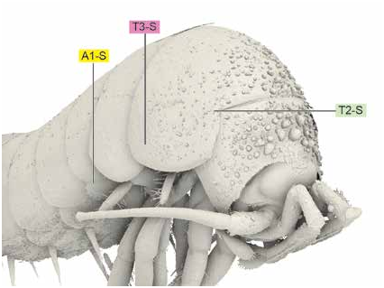
DESCRIPTION: HEAD: Characteristically arched thorax, pronotum covering much of head, making boundary between thoracic tracheae and head tracheae rather indistinct. T2-S at anteriormost margin of mesothorax, covered by overhanging tergum, shown in figure 10 View FIGURE 10 . H-DCT very thick, with three branches: H-Ic, extending anteriorly and dorsad; H-Oc-Ant anterior and medially before dividing into H-Oc toward midline and H-Ant laterally; H-Mx proceeds anteriad with H-MxPlp branching ventrally and anteriad. H-VCT very thick, with two branches: H-Lbm ventrad, extending into H-LbmPlp; H-Md anteriad, crossing over H-Md from opposite side of head but not connecting.
THORAX: T2-S present, with five tracheae: H-DCT and H-VCT leading anteriad directly into head, beginning at T2-S and running parallel for length of prothorax; T2-DB leading dorsad; T2-VB and T2-L directly posteriad. H-DCT with no thoracic branches; H-VCT with T1-L branching ventrally; T1-VC present, extending from T1-L. T2-DB-Vi branches just dorsal of T2-S, extending anteriorly; T2-DB continuing dorsad, dividing into what appear to be anterior T1-DLT and posterior T2-DLT near midline; T1-DLT and T2-DLT extend into viscera with no connections to neighboring spiracles. T2-L running posteriad and ventrally, into midleg; large T2-L-Vi and smaller T2-VC divide from T2-L posterior from T2-S. T2-VC joins with anterior-arching T3-VC to form T2-VX intersection. T2-L-Vi extends posteriad into metathorax, with apparent dorsal T3-DLT, T3-Cx ventral, and numerous small visceral tracheae. T3-S ventral to and much smaller than T2-S, with two tracheae: T3-L and T3-VB. T3-L extending dorsad with small T3-L-Vi before arcing ventrad into hind leg; T3-VB directly toward midline with small T3-VC branch that arcs anteriorly to join T2-VC at T2-VX.
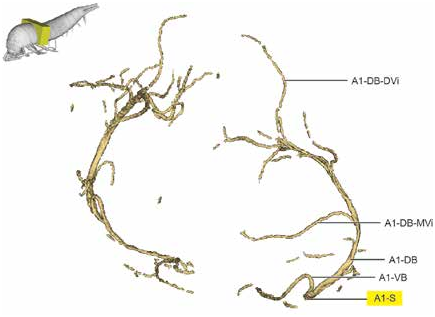
ABDOMEN: A1..7-S present, all located ventrally. No longitudinal connections present; figure 11 View FIGURE 11 with representative abdominal segment. A n -S with A n -DB running dorsad along arc of body wall, turning inward toward midline of body but not forming DC; A n -VB branching dorsally for a short distance before arcing ventrad toward midline. A n -VC absent. A n -DB with visceral tracheae A n -DB-MVi, branching anterior and medially halfway up body; A n -DB-DVi branches dorsally toward tergal wall. A5-DB-DVi extends posteriorly to 8th abdominal segment; A7-DB-DVi extends posteriorly past 8th abdominal segment, dividing into A-TF and A-Cr; neither A5-DB-DVi nor A7-DB-DVi connect to spiracles of other segments. A-TF with two tracheae per side (4 total); A-Cr single trachea per cercus.
No known copyright restrictions apply. See Agosti, D., Egloff, W., 2009. Taxonomic information exchange and copyright: the Plazi approach. BMC Research Notes 2009, 2:53 for further explanation.