
Trichocateres fasciculifer, JIŘÍ KOLIBÁČ, 2010
|
publication ID |
https://doi.org/ 10.5281/zenodo.275634 |
|
DOI |
https://doi.org/10.5281/zenodo.6196977 |
|
persistent identifier |
https://treatment.plazi.org/id/425A8793-076D-FFE8-9DD5-FC1FFB82FE8D |
|
treatment provided by |
Plazi |
|
scientific name |
Trichocateres fasciculifer |
| status |
sp. nov. |
Trichocateres fasciculifer n. sp.
Type specimens: Holotype (not dissected): “NE INDIA; ASSAM; 1999 / 5km N of Umrongso; 700m / 25°27’N 92°43’E; 17.–25.v / Dembický & Pacholátko leg.”. Two paratypes (males): same data as holotype; “ LAOS north, 13.–24.V.1997 / 15 km NW Louang Namtha / N 21°07.5, S 101°21.0 / at. 750± 100 m / E. Jendek & O. Šauša leg.” (all three type specimens deposited in the Moravian Museum, Brno, Czechia; J. Kolibáč coll.).
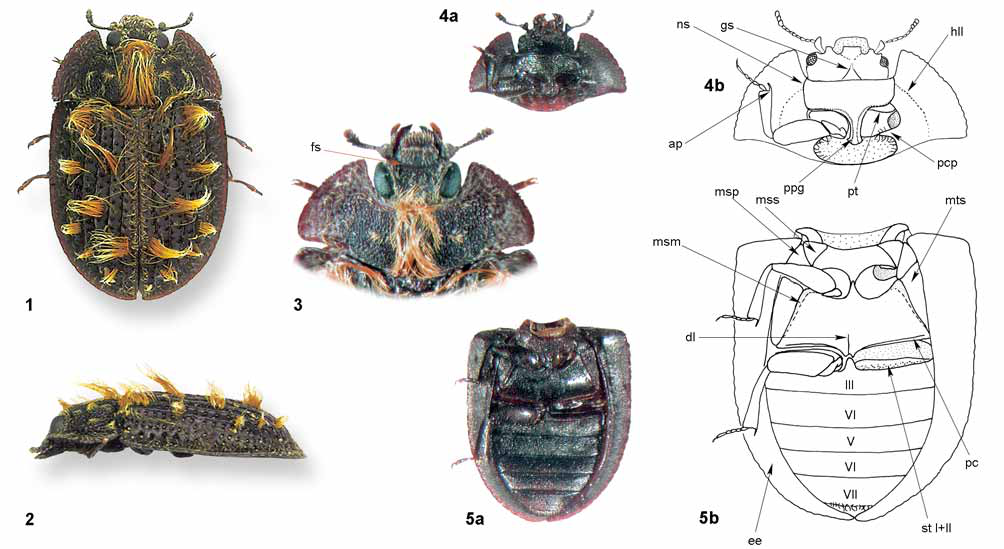
Description: Measurements: Holotype – body length (from elytral apex to clypeus) 5.2 mm, pronotum length 1.3 mm, elytra length 3.5 mm, pronotum width 3.1 mm, elytra width (together) 3.3 mm, antenna length c. 0.8 mm. Paratype from Laos – body length 5.5 mm, body width 3.5 mm.
Coloration and sculpture ( Figs 1, 2, 3, 4 View FIGURES 1 – 5 a, 5a): Body oval, conspicuously convex; dorsal surface dark brown to black in Assam specimens, brown in Laos specimen; margins of pronotum and elytra red-brown, colour lighter in Laos specimen. Head finely, densely granulate, with short, decumbent, yellowish setae. Pronotum granulate at centre, sparsely punctate along explanate lateral margins, with semi-erect yellowish pubescence; longitudinal tuft of long, erect, yellow-orange hairs situated at centre, small patches of decumbent hairs occurring along it. Each elytron with eight tufts of yellow-orange hairs (same pattern of tufts in all three specimens), a number of other long, erect hairs situated along mesonotum and elytral suture; explanate lateral part of elytra with semi-erect yellowish pubescence, finely and sparsely punctate; five elytral carinae present; one regular row of deep punctures running along each side of carina, the last two rows of punctures (in lateral direction) not accompanied by carina. Ventral surface of body dark brown to black; sparsely punctate, punctures minute; pubescence formed by short, sparse, decumbent pale hairs. Legs without sculpture, sparsely pubescent in the same way as ventral surface of body.
Head ( Figs 3–4 View FIGURES 1 – 5 ): gular sutures widely separated, extending to approximately midpoint of cranium, strongly convergent; frontoclypeal suture well-developed, deep, arcuate; antennal grooves in ventral side of cranium short, shallow; antennal sockets partly visible from above; two eyes present, relatively large (space between them twice as wide as eye diameter), distinctly elevated, not emarginate, elliptic, greater part situated dorsally; epicranial acumination conspicuous but not deep.
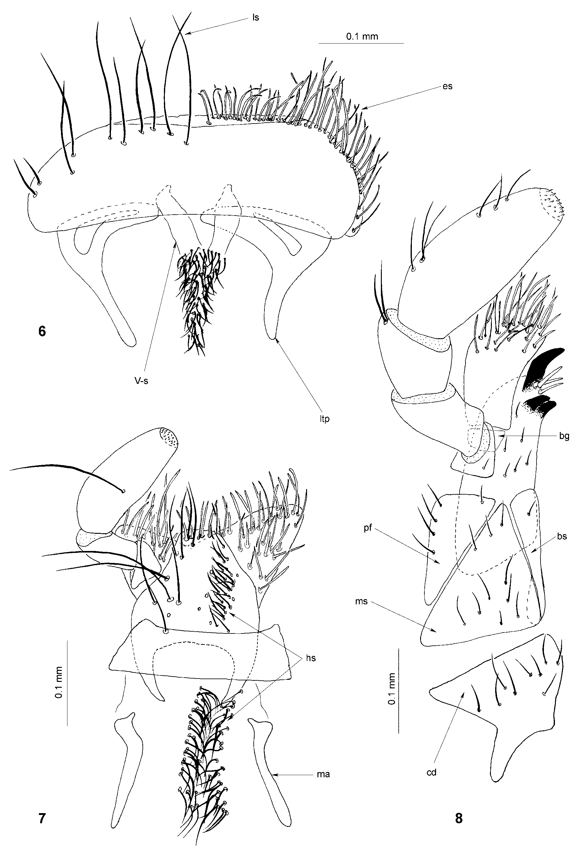
Maxilla ( Fig. 8 View FIGURES 6 – 8 ): lacinia relatively robust, with three dark spines at apex in pattern 1+2 and c. 3 pale setae between them, apical spine hook-like and two spines beneath it shorter; basigalea well-developed, pigmented; galea very weakly clavate, with numerous elongate setae, without spines and ciliate setae; mediostipes perfectly free, not fused with lacinia; basistipes elongate, rectangular; palpifer large, not denticulate along outer margin; maxillary palps 4-segmented, terminal joint conical, elongate.
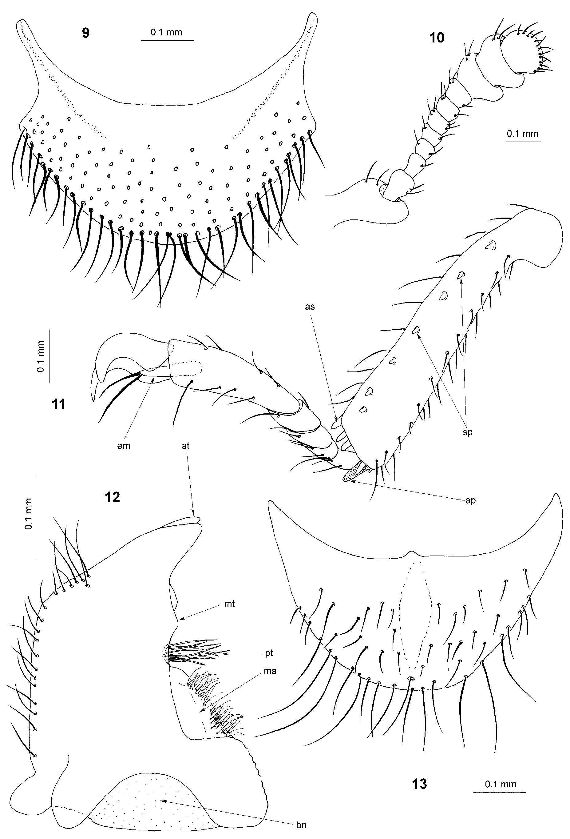
Mandible ( Fig. 12 View FIGURES 9 – 13 ): with two apical teeth situated side by side (in horizontal axis); medial tooth very small, double; mola present, denticulate along margin; membranous, finely but densely pubescent appendage situated above mola (penicillus?); prostheca situated below medial tooth, formed by tuft of long, robust setae; ventral ciliate furrow absent; basal notch deep.
Labrum ( Fig. 6 View FIGURES 6 – 8 ): free (not fused with cranium); oval, not emarginate in apical part; epipharynx without distinct sclerite; lateral tormal processes not connected in middle; V-shaped sclerite with dense pubescence situated between tormal processes.
Labium ( Fig. 7 View FIGURES 6 – 8 ): submentum without tuft of setae; mentum trapezoidal, anterior corners extended; prementum without notch and not divided into two parts; ligula membranous, deeply emarginate, without ciliate setae, with long pale setae; mental apodeme composed of two elongate sclerites; hypopharyngeal bar absent; hypopharyngeal sclerite absent; hypopharynx with two strips of short setae along ligula, strips fused into one in mental area; terminal joint of labial palps shorter than the labial ones, conical.
Antennae ( Fig. 10 View FIGURES 9 – 13 ): 11-segmented; antennal club 3-segmented, relatively compact; antennal joints symmetrical or very weakly asymmetrical (joints 9–10), without conspicuous sensorial fields, with short pubescence; scapus robust, pedicellus smaller, but larger than joint 3; antennae relatively short, extending backwards to midway along pronotum.
Prothorax ( Figs 3, 4 View FIGURES 1 – 5 ): pronotum transverse; anterior margin deeply emarginate (arcuate), anterior corners projecting; lateral margins excavate; lateral edge present, finely undulating but not denticulate; prosternal process dilated at apex, with two sharp grooves running along margins of process and procoxal cavities; procoxal cavities externally open to halfway, internally closed; postcoxal projections pubescent along margins; trochantin well-developed; inner part of hypomeron with fine longitudinal line.
Mesothorax ( Figs 3, 5 View FIGURES 1 – 5 ): prepectus present in anterior part of mesepisternum only, absent above mesosternum; mesocoxal cavities externally widely open, internally closed; trochantin minute; mesonotum wide, scutum with extended basal corners, scutellum oval.
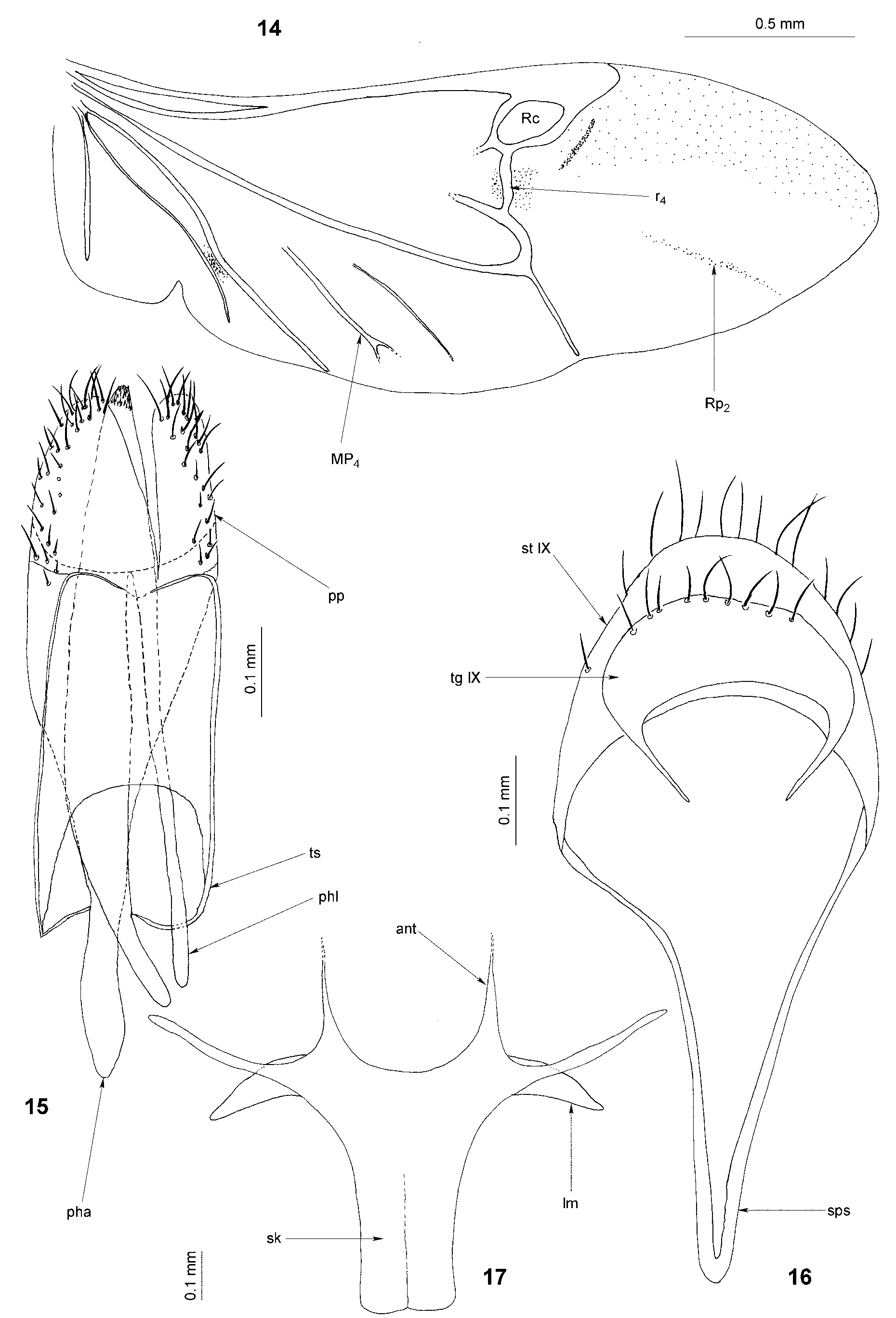
Wing ( Fig. 14 View FIGURES 14 – 17 ): radial cell displaced downwards; r4 relatively short; pigmented fleck (below Rc) small, pale, not complete; medial field with all four veins, cross-veins absent (wedge cell absent); MP4 shortly bifurcate at apex; wing pigmented above vein Rp2.
Metathorax ( Figs 5 View FIGURES 1 – 5 , 17 View FIGURES 14 – 17 ): metasternum flat and wide, distinctly narrowed towards anterior portion, widely bordered along lateral sides; discriminal line (discrimen) short; paracoxal sutures well-demarcated, parallel with coxae; metepisternum distinctly triangular; metanotum rectangular, extremely wide; metendosternite with broad stalk, laminae narrowed at apex, anterior tendons long and widely separated.
Elytra ( Figs 1, 2, 5 View FIGURES 1 – 5 ): regularly punctate, with extremely wide epipleura; elytral locking mechanism absent; five elytral carinae present; lateral sides of elytra explanate.
Legs ( Figs 4, 5 View FIGURES 1 – 5 , 11 View FIGURES 9 – 13 ): procoxae not projecting, transverse; mesocoxae oval, metacoxae extended to lateral surface of metathorax; trochanters relatively small, triangular; femora very weakly clavate; tibiae with row of small spines along outer margin; tibial apical spur pattern 1–1–1; protibial spur hooked; apices of all tibiae with row of c. 5 spines; tarsomere 1 in all pairs of legs conspicuous, approximately as long as tarsomere 2; tarsomere 5 as long as, or slightly longer than, 1–4 combined; tarsal lobes absent; claws without denticles; empodium large, projecting, bisetose; tarsal formula 5–5–5.
Abdomen ( Figs 5 View FIGURES 1 – 5 , 9, 13 View FIGURES 9 – 13 , 15, 16 View FIGURES 14 – 17 ): five sternites visible, the last one (sternite VII) with row of short hairs; male sternite VIII without spiculum gastrale, with longitudinal unpigmented area in centre (sternite VIII in the second paratype shorter); male segment IX complete; tegmen composed of two parts (parameres separated from phallobase, dorsal and ventral pieces fused), inverted (phallobase ventrally open); phallobasic apodeme well-developed; tegminal struts coalescent with base of phallobasic apodeme; parameres densely pubescent; phallus stout, slightly shorter than phallobase, with several minute spines at apex.
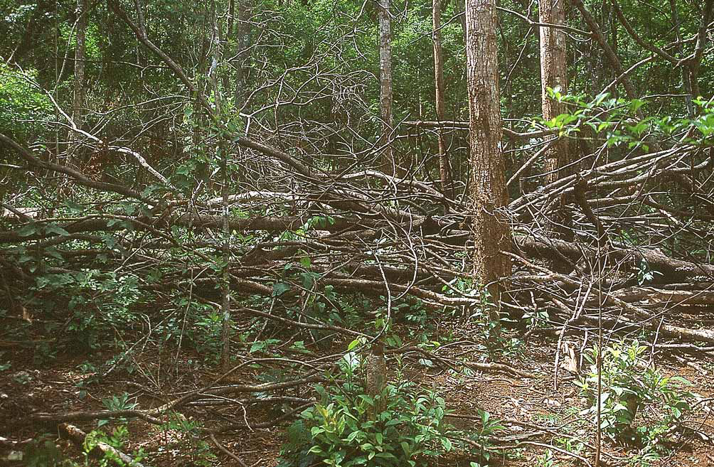
Note on distribution and biology: Two specimens were knocked down from fallen logs in a forest clearing ( India: Assam) ( Fig. 18 View FIGURE 18 ); one specimen from northern Laos was probably caught in the same manner. The species may also be present in Myanmar ( Burma) between the two known localities but is perhaps relatively rare (numerous coleopterists, including myself, have collected beetles in the surroundings of Louang Namtha in recent years but only the single specimen is known from Laos to date). Gut content of the both dissected paratypes with small remnants of insect cuticle, paratype from Laos with the remains of a small insect larva. The species is predatory.
Etymology: Fascicula = small beam, bundle, sheaf; ferre = to carry, bear, hold.
| ASSAM |
Botanical Survey of India, Eastern Regional Centre |
No known copyright restrictions apply. See Agosti, D., Egloff, W., 2009. Taxonomic information exchange and copyright: the Plazi approach. BMC Research Notes 2009, 2:53 for further explanation.