
Capsaloides magnaspinosus Price, 1939
|
publication ID |
https://doi.org/ 10.5281/zenodo.172308 |
|
DOI |
https://doi.org/10.5281/zenodo.6255193 |
|
persistent identifier |
https://treatment.plazi.org/id/03FC8787-E162-FFC9-FED3-FB929EF1FA29 |
|
treatment provided by |
Plazi |
|
scientific name |
Capsaloides magnaspinosus Price, 1939 |
| status |
|
Capsaloides magnaspinosus Price, 1939 View in CoL ( Figs 1 View FIGURE 1 D, 2D, 5–7)
Typehost: Tetrapterus imperator (Bloch & Schneider, 1801) (Istiophoridae) .
Typelocality: Woods Hole, Massachusetts, USA [Atlantic Ocean].
Additional records: Xiphias gladius Linneaus, 1758 , Aguadilla, Puerto Rico [Atlantic Ocean] (see Williams & BunkleyWilliams 1996); T. albidus Poey, 1860 , Woods Hole [Atlantic Ocean] (see Williams & BunkleyWilliams 1996); Tetrapturus audax (Philippi, 1887) , Nelson Bay, New South Wales, Australia [Pacific Ocean] (present study).
Site: Nares.
Infection details: Two of 5 specimens of T. audax from Nelson Bay, New South Wales, Australia infected with a total of 6 specimens (present study).
Specimens examined: Four mounted specimens collected in present study (vouchers SAMA AHC 28913–16); 1 paratype (USNPC 35648).
Redescription
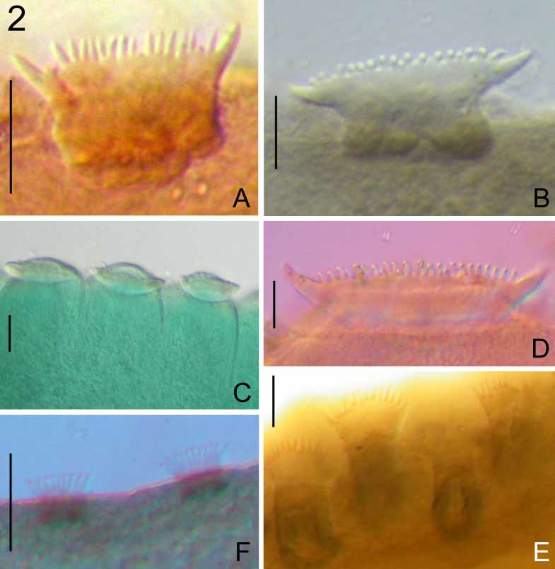
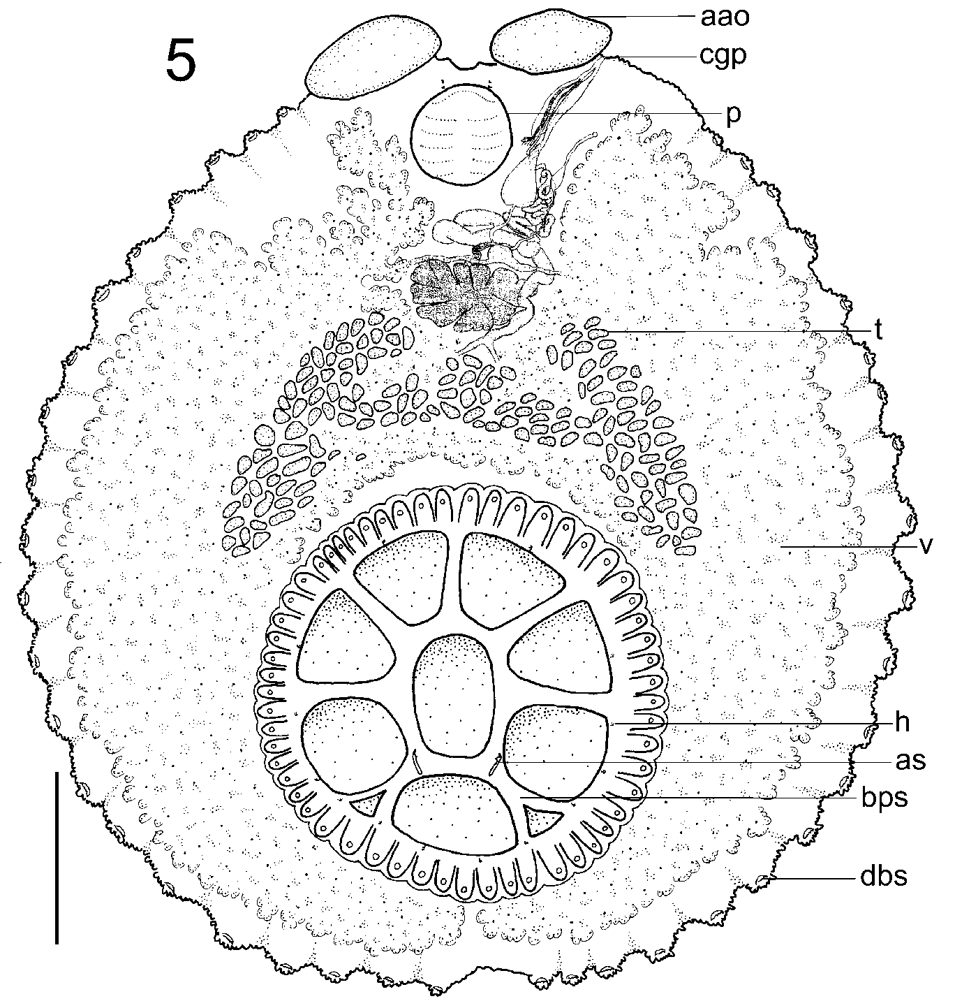
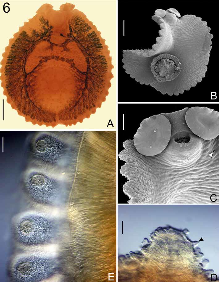
Capsalinae (sensu Egorova 1989). Redescription and measurements based on 4 flattened specimens and 1 specimen examined using SEM. Specimens a pink colour in life. Total length, including haptor 10,520 (9,000–11,600, n = 4); maximum width 9,017 (7,933–10,667, n = 4) at level of posterior region of testes. Haptor 4,067 (3,467–4,867, n = 4) in diameter; located within posterior body margin ( Figs 5 View FIGURE 5 , 6 View FIGURE 6 A, 6B). Haptor divided by septa into 1 central and 7 peripheral loculi; posterior pair of septa bifid ( Fig. 5 View FIGURE 5 ). Haptoral accessory sclerites 217 (163–282, n = 6) long, with distinct distal point ( Fig. 1 View FIGURE 1 D); associated with anterior region of posterior septal pair. Fourteen hooklets 16 (15–18, n = 14) long, distributed as illustrated ( Fig. 5 View FIGURE 5 ). Haptor surrounded by muscular scalloped marginal valve ( Figs 5 View FIGURE 5 , 6 View FIGURE 6 B, 6E); dorsal surface of each scallop with single distinct papilla ( Figs 5 View FIGURE 5 , 6 View FIGURE 6 E).
Dorsal body surface with large conspicuous papillae ( Fig. 6 View FIGURE 6 B), capable of independent ripplinglike movement in live worms. Ventral body surface apapillate ( Fig. 6 View FIGURE 6 B). Margin of body sinuous; each sinuation surmounted by numerous minute tegumental papillae ( Figs 6 View FIGURE 6 C, 6D) and single crownshaped sclerite comprising 20–30 cusps ( Figs 2 View FIGURE 2 D, 5). Left anterior isolated group of dorsomarginal body sclerites just posterior to common genital pore ( Fig. 5 View FIGURE 5 ); comprises 4–5 sclerites each with 3–8 cusps. Right anterior isolated group of dorsomarginal body sclerites not seen. Anterior attachment organs oval 1,378 (1,267– 1,466, n = 3) long, 971 (954–1,001, n = 3) wide. Two pair of eyespots anterodorsal to pharynx. Pharynx oval without distinct constriction 1,283 (905–1,425, n = 4) long, 1,072 (970–1,141, n = 4) wide.
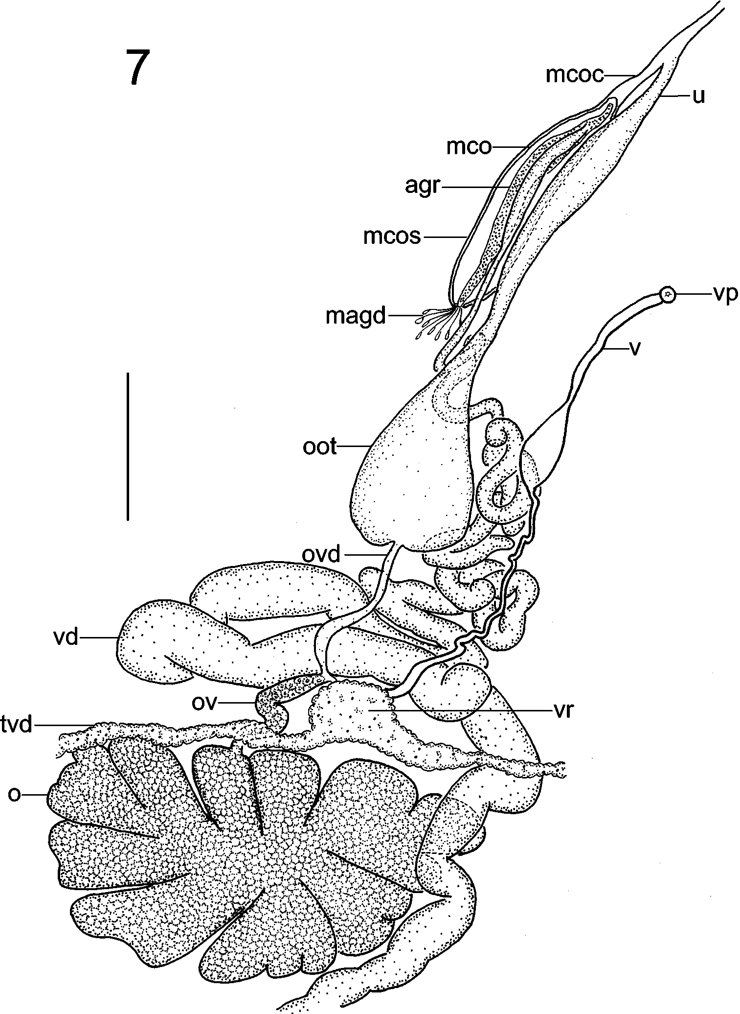
Testes numerous, arranged in “M” shape ( Fig. 5 View FIGURE 5 ). Vas deferens passes leftside of ovary and loops medially immediately anterior to vitelline reservoir; convoluted then straightening, passing dorsal to vagina and oötype ( Fig. 7 View FIGURE 7 ). Vas deferens enters male copulatory organ sac posteriorly and joins accessory gland reservoir at distal end of male copulatory organ ( Fig. 7 View FIGURE 7 ). Male copulatory organ simple with no spines or protuberances ( Fig. 7 View FIGURE 7 ).
Ovary lobed 710 (511–977, n = 4) long, 1,119 (781–1,345, n = 4) wide. Oviduct passes dorsal to transverse vitelline duct; presumably receives duct (not seen) from vitelline reservoir to form ovovitelline duct ( Fig. 7 View FIGURE 7 ). Ovovitelline duct passing ventral to vas deferens before entering oötype. Mehlis’ glands not seen. Oötype opens into short uterus which joins male copulatory organ canal just prior to common genital pore ( Fig. 7 View FIGURE 7 ). Common genital pore opens at lateral body margin just posterior to margin of left anterior attachment organ ( Fig. 5 View FIGURE 5 ). Vaginal pore opens midway between midline and left lateral body margin at level of midpoint of pharynx ( Fig. 5 View FIGURE 5 , 6 View FIGURE 6 C); vagina mostly narrow with slight swelling midway along duct at level of oötype proximally, joining vitelline reservoir ( Figs 5 View FIGURE 5 , 7 View FIGURE 7 ). Vitelline follicles extensive filling most of body proper; transverse vitelline duct just anterior to ovary; transverse ducts join to form swollen vitelline reservoir ( Figs 5 View FIGURE 5 , 7 View FIGURE 7 ). Eggs not observed.
Remarks
The original description of C. magnaspinosus by Price (1939) was based on 3 specimens collected from the nasal tissue of T. imperator off Woods Hole, USA by MacCallum in 1924 and deposited in the USNPC. The original illustration was small and it is difficult to see the details of the male and female reproductive system. Price (1939) distinguished C. magnaspinosus from other species in the genus by the large size of the dorsomarginal body sclerites and the relatively large haptor. The presence of a single papilla associated with the dorsal surface of each haptoral scallop ( Figs 5 View FIGURE 5 , 6 View FIGURE 6 E) in the marginal valve was not described by Price (1939) but it also serves to easily distinguish this species since it was not a character seen in any other species from the material examined here.

No known copyright restrictions apply. See Agosti, D., Egloff, W., 2009. Taxonomic information exchange and copyright: the Plazi approach. BMC Research Notes 2009, 2:53 for further explanation.