
Tenedos piedecuesta, Martínez & Brescovit & Quijano, 2022
|
publication ID |
https://doi.org/ 10.11646/zootaxa.5130.1.1 |
|
publication LSID |
lsid:zoobank.org:pub:ABF61117-DD64-4A32-BD61-20E577F80C3D |
|
DOI |
https://doi.org/10.5281/zenodo.6520669 |
|
persistent identifier |
https://treatment.plazi.org/id/03C787B1-FF80-FF17-D49C-FB010EB4F99A |
|
treatment provided by |
Plazi |
|
scientific name |
Tenedos piedecuesta |
| status |
sp. nov. |
Tenedos piedecuesta View in CoL sp. n.
Figs 90–92 View FIGURE 90 View FIGURE 91 View FIGURE 92 ; 107 View FIGURE 107 .
Type material. Holotype: COLOMBIA. Santander: Santa Bárbara, Vereda Salinas, Finca San Francisco , Pristine andean forest fragment, Manual , 2429m [6°59′54.0′′N, 72°52′55.5′′W], M. Castro, J. Neita, J. Park & E. Torres leg., 9-11.IX.2018, 1 ♂ (IAvH-I-603) GoogleMaps . Paratypes: COLOMBIA. Santander: Piedecuesta, El Rasgón, Secondary forest fragment, Pitfall trap, 2150m [7°3′N, 72°57′W], I. Quintero & E. González leg., 21-23.IX.2004, 1 ♂ (IAvH-I-580), 1 ♀ (IAvH-I-649), 2 ♀ (IAvH-I-650) GoogleMaps .
Etymology. The specific name is a noun in apposition taken from the type locality of the paratype.
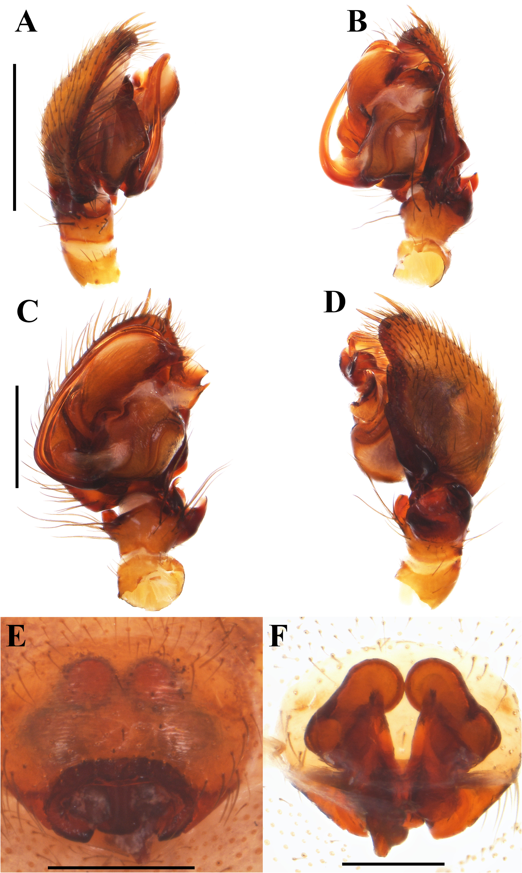
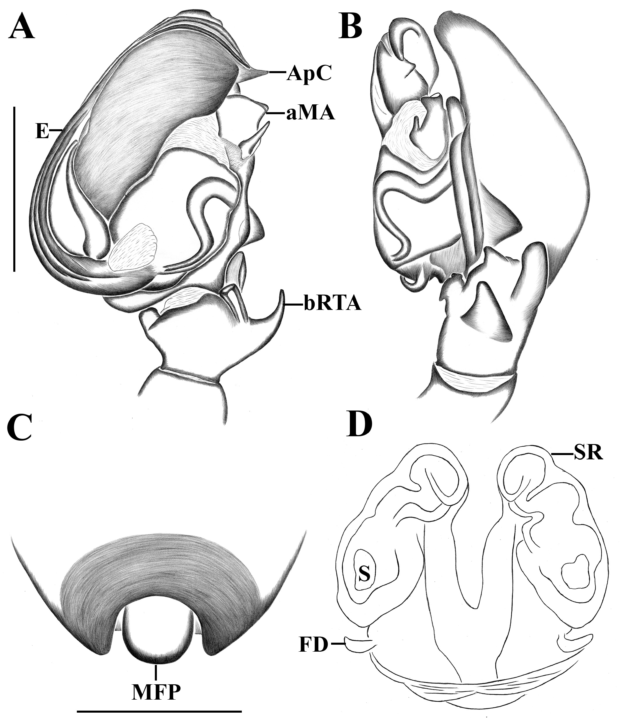
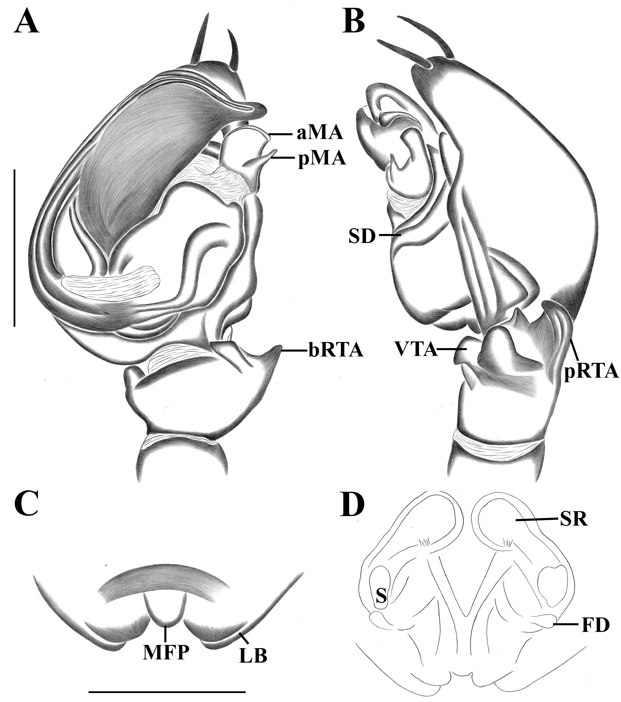
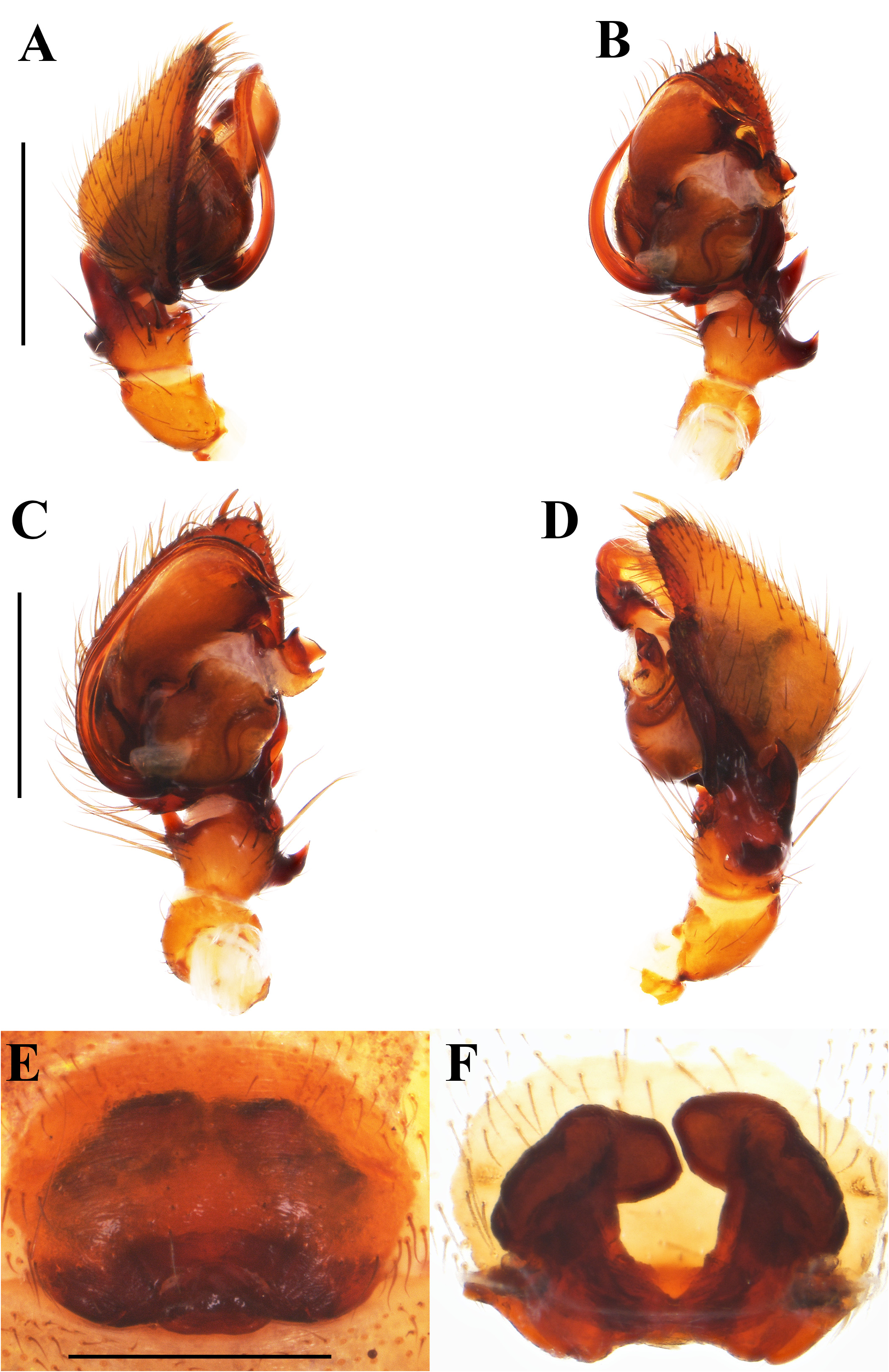
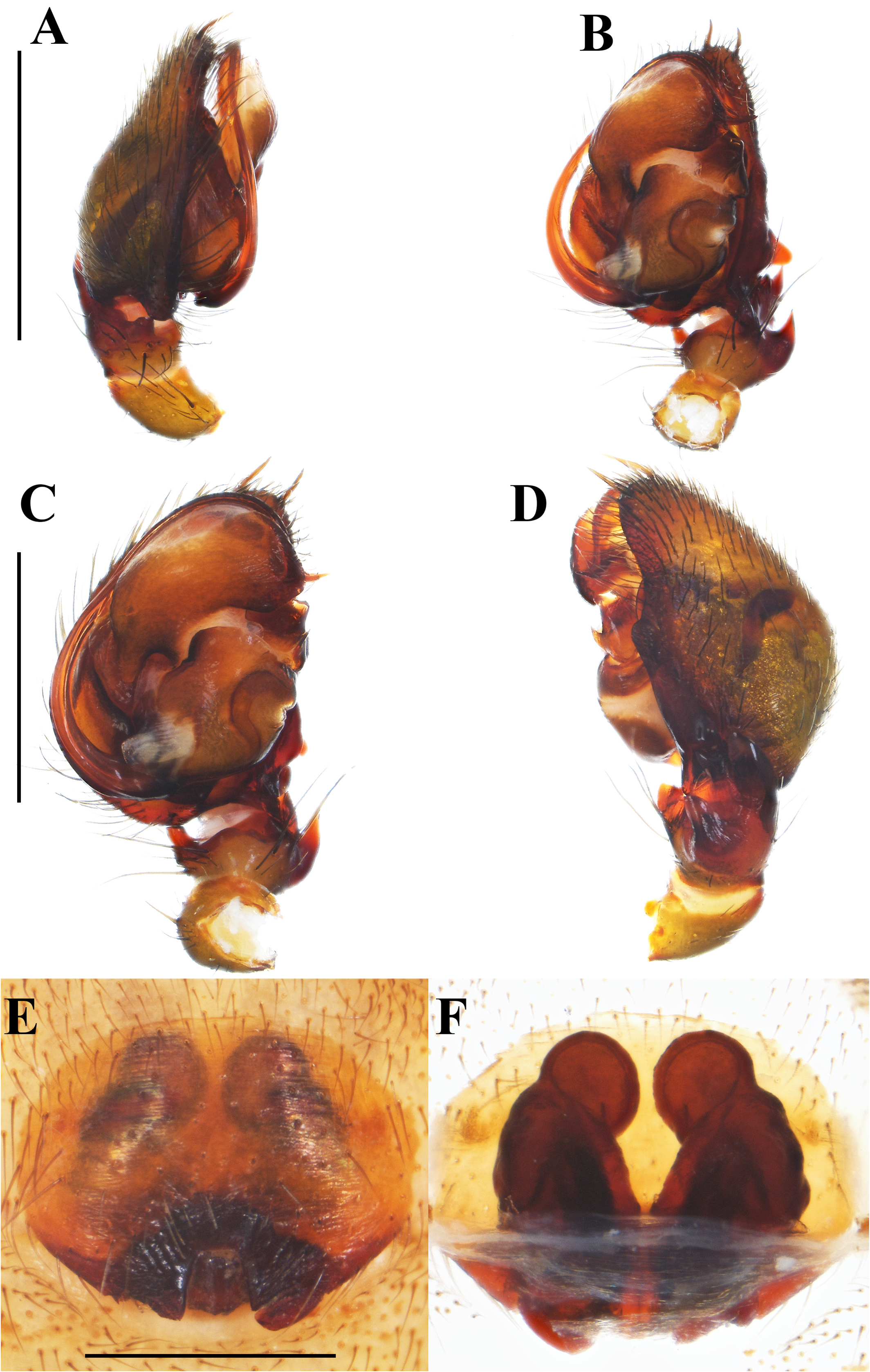
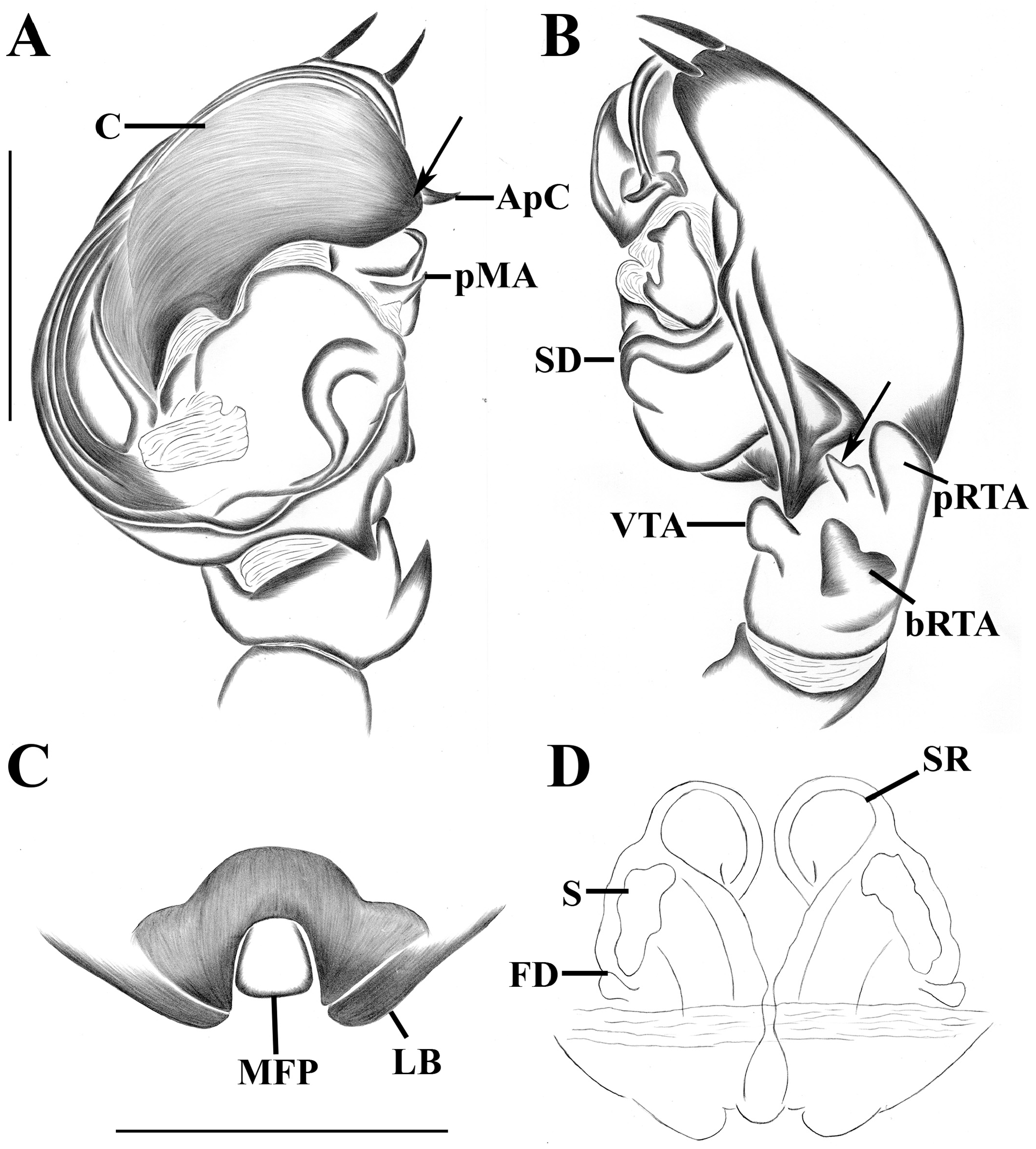
Diagnosis. Males of Tenedos piedecuesta sp. n. resemble T. dankittipakuli sp. n., T. griswoldi sp. n., T. tama sp. n., and T. humboldti sp. n. by similar shape of median apophysis (MA); triangular projection at medial side of retrolateral process of cymbium (RPC); basal retrolateral tibial apophysis (RTA) ( Figs 77A–D View FIGURE 77 ; 78A–B View FIGURE 78 ; 80A–D View FIGURE 80 ; 82A–D View FIGURE 82 ; 83A–B View FIGURE 83 ; 85A–D View FIGURE 85 ; 86A–B View FIGURE 86 ; 88A–D View FIGURE 88 ; 89A–B View FIGURE 89 ; 91A–D View FIGURE 91 ; 92A–B View FIGURE 92 ), but are distinguished by rounded sclerotized process at anterior side of conductor (C); very thin appendix of conductor (ApC); wider, laminar, apically bifid anterior branch of the retrolateral tibial apophysis (aRTA); wider basal retrolateral tibial apophysis (bRTA), bifid in tip lateral borders ( Figs 91A–D View FIGURE 91 ; 92A–B View FIGURE 92 ). Females are similar those of dankittipakuli sp. n., T. tama sp. n., and T. humboldti sp. n., by very wide at base seminal receptacles (SR), slightly curved towards median septum ( Figs 77E–F View FIGURE 77 ; 78C–D View FIGURE 78 ; 80E–F View FIGURE 80 ; 85E–F View FIGURE 85 ; 86C–D View FIGURE 86 ; 88E–F View FIGURE 88 ; 89C–D View FIGURE 89 ; 91E–F View FIGURE 91 ; 92C–D View FIGURE 92 ), but are characterized by large median field plate (MFP) with squared basal edge; longer than wide, basally divided lateral borders (LB); large spermathecae (S) ( Figs 91A–D View FIGURE 91 ; 92A–B View FIGURE 92 ).
Description. Male (Holotype, IAvH-I-603). Coloration ( Fig. 90A–B View FIGURE 90 ): carapace uniformly dark brown. Chelicerae with paturon brown, fangs brown-reddish. Endites dark brown, white on anterior region. Labium and sternum dark brown. Legs: Coxae I–IV light yellow. Femora I–IV light brown from basal to medial region, dark brown the rest of their extension. Patellae I–IV dark brown. Tibia I light yellow, dark brown on basal and distal sides, II–IV dark brown. Metatarsi-tarsi brown. Abdomen: dorsally dark gray with nine white guanine spots organized as follows: two large and oval spots, anteriorly positioned; two lateral and rounded spots larger than previous one, medially positioned; four small and rounded spots, in posteromedial position; a wide and transversal band, posteriorly positioned. Laterally dark gray three obliques stripes, posteriorly positioned. Ventrally dark gray covered by two large longitudinal spots. Spinnerets brown. Measurements: total length 5.95, carapace length 3.28, width 2.23, height 1.33. Clypeus height 0.55. Eye diameters and interdistances: AME 0.10, ALE 0.12, PME 0.10, PLE 0.13; AME–AME 0.24, AME–ALE 0.23, AME–PME 0.27, PME–PME 0.26, PME–PLE 0.43, ALE–PLE 0.31. Chelicerae 0.62 length. Sternum length 1.15, width 1.07. Legs: I—femur 1.60/ patella 0.72/ tibia 2.04/ metatarsus 1.86/ tarsus 1.21/ total 7.43; II—1.72/ 0.78/ 1.53/ 1.51/ 0.63/ 6.17; III—1.71/ 0.84/ 1.30/ 1.45/ 0.62/ 5.92; IV—2.25/ 0.89/ 1.87/ 2.24/ 1.08/ 8.33. Abdomen length 2.28. Legs spines pattern (only the differences from the general pattern): II—tibia v1r-1r-2. Palp: retrolateral process of cymbium (RPC) long, wide, widening slightly towards base; tegulum (T) large, rounded, almost as long as wide, retrolateral excavation poorly accentuated; subtegulum (St) longer than wide; conductor (C) developed, wide with sclerotized process on distal side; appendix (ApC) long, very thin; embolus (E) long laminar at base, filiform towards apex; base of embolus (EB) with small projections, approximately two times as long as basal tegular membrane; basal tegular membrane (BTM) originated basally on tegulum, very wide, ending as a very short, apically rounded; spermatic ducts (SD) S-shaped with anterior fold more open, wider than posterior; ventral tibial apophysis (VTA) very small, tubular, strongly sclerotized; median apophysis (MA) large, bifid with squared anterior branch (aMA) with folded edges, posterior branch (pMA) very short, thin; retrolateral tibial apophysis (RTA) bifid shorter than palpal tibia with posterior branch (pRTA) wider than anterior branch (aRTA) ( Figs 91A–D View FIGURE 91 ; 92A–B View FIGURE 92 ).
Female (Paratype, IAvH-I-649). Coloration and abdominal pattern of spots as male, except legs uniformly brown ( Fig. 90C–D View FIGURE 90 ). Measurements: total length 8.01, carapace length 3.47, width 2.44, height 1.59. Clypeus height 0.66. Eye diameters and interdistances: AME 0.11, ALE 0.13, PME 0.16, PLE 0.15; AME–AME 0.23, AME–ALE 0.27, AME–PME 0.38, PME–PME 0.32, PME–PLE 0.54, ALE–PLE 0.39. Chelicerae 1.12 length. Sternum length 1.32, width 1.27. Legs: I—femur 1.85/ patella 0.74/ tibia 1.77/ metatarsus 1.56/ tarsus 1.16/ total 7.08; II—1.67/ 0.81/ 1.39/ 1.05/ 0.73/ 5.65; III—1.58/ 0.73/ 1.02/ 1.36/ 0.79/ 5.48; IV—1.82/ 0.81/ 1.50/ 2.17/ 1.06/ 7.36. Abdomen length 4.27. Legs spines pattern (only the differences from the general pattern): I—tibia v2-1r-1p, metatarsus v1r-0-2; II—tibia v1r-1r-2, metatarsus v1r-0-2. Epigyne: lateral borders (LB) short, not projected, curving towards posterior region of epigyne, forming a posterior, small atrium (A), apically bifid; median field plate (MFP) very small, longer than wide; copulatory ducts (CD) short, wide, almost undistinguished from spermathecae; seminal receptacles (SR) long, very wide at base, distally thin, lightly curved towards median septum; spermathecae (S) large, irregulars, posteriorly positioned; fertilization ducts (FD) almost as long as spermathecae length ( Figs 91A–D View FIGURE 91 ; 92A–B View FIGURE 92 ).
Variation. Males (n=2): total length: 5.95–7.32; carapace length: 3.28–3.53; femur I length: 1.09–1.60. Females (n=3): total length: 6.58–8.01; carapace length: 3.15–3.47; femur I length: 1.21–1.85.
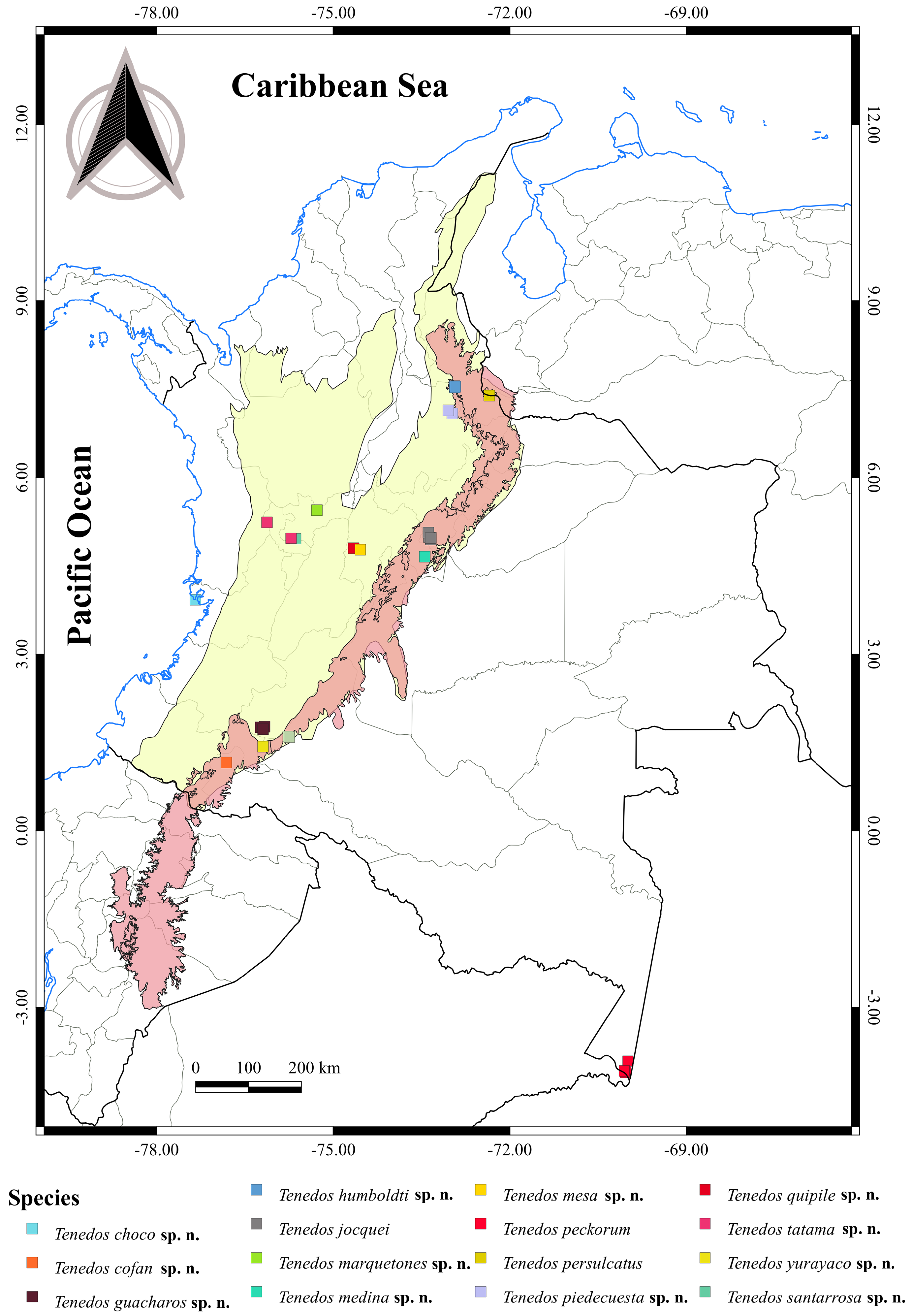
Distribution. Known from Santander department ( Fig. 107 View FIGURE 107 ).
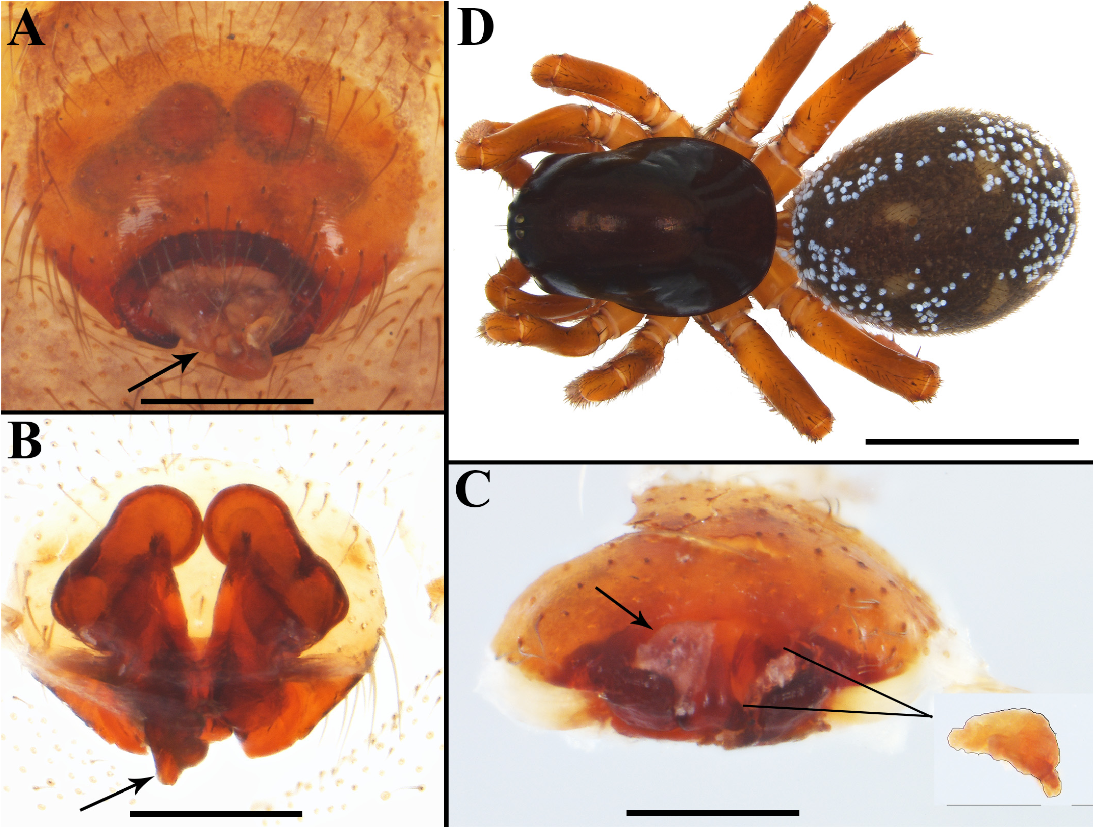
Natural history. The specimens were collected with Winkler extractor traps. We observed that this species was being attacked by entomopathogenic fungus, mainly the females ( Fig. 10B View FIGURE 10 ).


No known copyright restrictions apply. See Agosti, D., Egloff, W., 2009. Taxonomic information exchange and copyright: the Plazi approach. BMC Research Notes 2009, 2:53 for further explanation.
|
Kingdom |
|
|
Phylum |
|
|
Class |
|
|
Order |
|
|
Family |
|
|
Genus |