
Tenedos andes Jocqué & Baert, 2002
|
publication ID |
https://doi.org/ 10.11646/zootaxa.5130.1.1 |
|
publication LSID |
lsid:zoobank.org:pub:ABF61117-DD64-4A32-BD61-20E577F80C3D |
|
DOI |
https://doi.org/10.5281/zenodo.7625313 |
|
persistent identifier |
https://treatment.plazi.org/id/03C787B1-FF7C-FF14-D49C-F9F10979F8B9 |
|
treatment provided by |
Plazi |
|
scientific name |
Tenedos andes Jocqué & Baert, 2002 |
| status |
|
Tenedos andes Jocqué & Baert, 2002 View in CoL
Figs 93–95 View FIGURE 93 View FIGURE 94 View FIGURE 95 ; 106 View FIGURE 106 .
Tenedos andes Jocqué & Baert, 2002: 83–84 View in CoL , figs 7A–C. ( Male holotype from Finca San Pablo, 3km de N Alban, Cundinamarca, Colombia, 1800m, P. & B. Wygodzinsky leg., deposited in AMNH_ IZC 00217585 View Materials , examined).
Other material examined. COLOMBIA. Cundinamarca: Topaipí, Finca El Alirio, Corn crop, Manual , 1377m [5°24,055’N, 74°17.64’W], M. Medrano & A. García leg., 5.I_ 18-23.IX.2012, 3 ♂ (ICN-Ar-8020), 2 ♀ (ICN-Ar-8021) GoogleMaps .
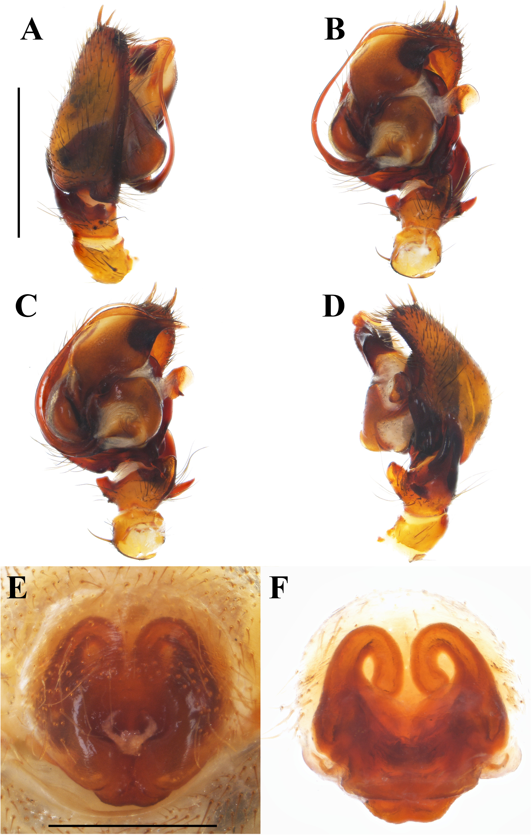
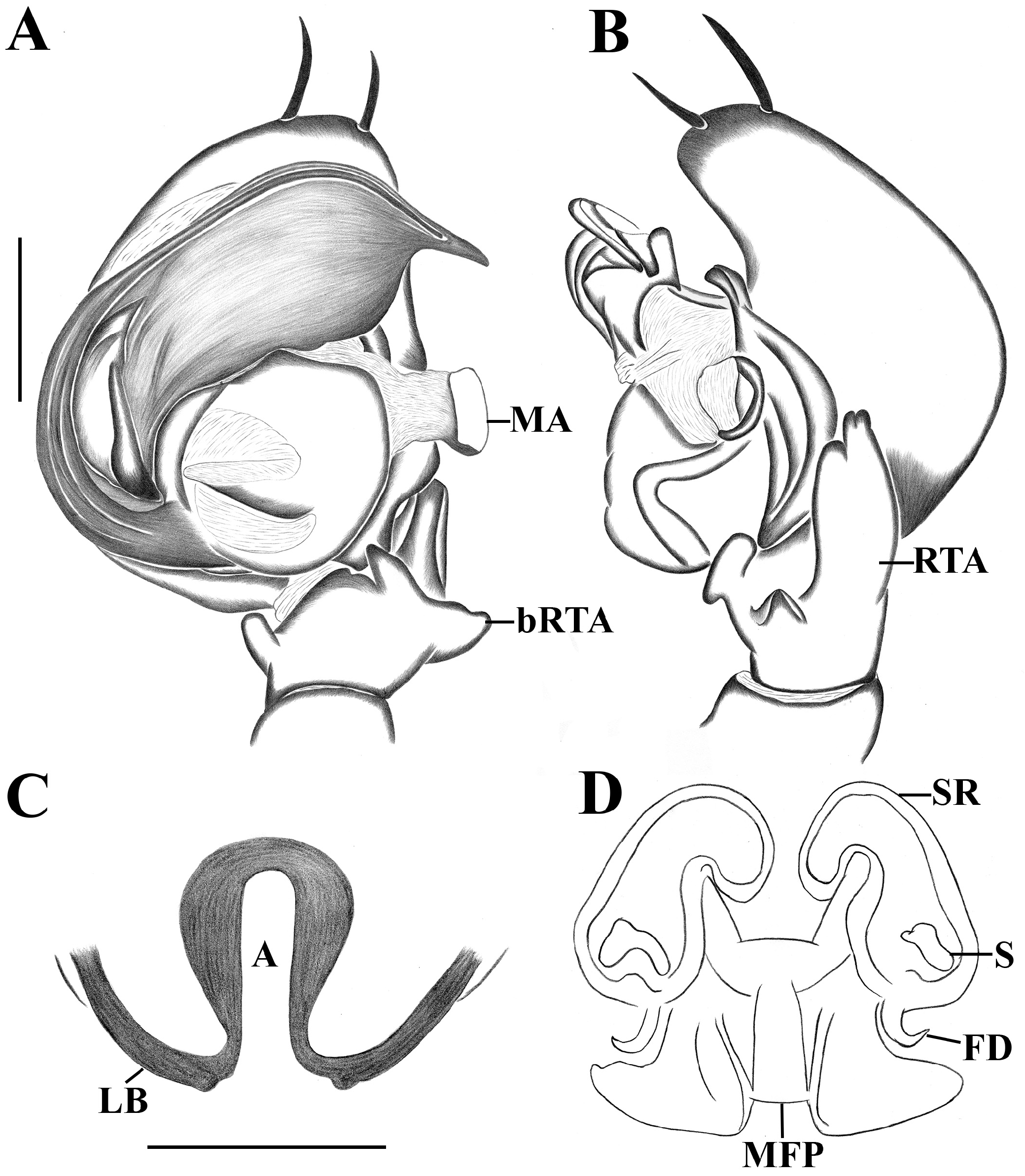
Diagnosis. Males of Tenedos andes Jocqué & Baert, 2002 , resemble those of T. medina sp. n. and T. carlosprietoi sp. n., by the presence of basal retrolateral tibial apophysis (bRTA); rounded concave median apophysis (MA) (see Jocqué & Baert, 2002: 83, fig. 7A–B; figs 94A–D; 95A–B; 97A–D, 98A–B; 99A–F; 101A–D; 102A–B; 104A–D), but can be distinguished by median apophysis with slight notch at external edge, long, wide; apically rounded retrolateral tibial apophysis (RTA); short and quadrangular basal retrolateral tibial apophysis close to retrolateral tibial apophysis ( Figs 94A–D View FIGURE 94 ; 95A–B View FIGURE 95 ). Females are similar to those of Tenedos medina sp. n. and T. carlosprietoi sp. n., by curved lateral borders, forming large atrium (A) ( Figs 94E–F View FIGURE 94 ; 95C–D View FIGURE 95 ; 97E–F View FIGURE 97 , 98C–D View FIGURE 98 ; 101E–F View FIGURE 101 ; 102C–D View FIGURE 102 ; 104E–F View FIGURE 104 ), but can be recognized by very large median field plate (MFP), occupying almost 50% of dorsal region of epigyne, short lateral borders (LB) thin at basal side; long and thin seminal receptacles (SR) ( Figs 94E–F View FIGURE 94 ; 95C–D View FIGURE 95 ).
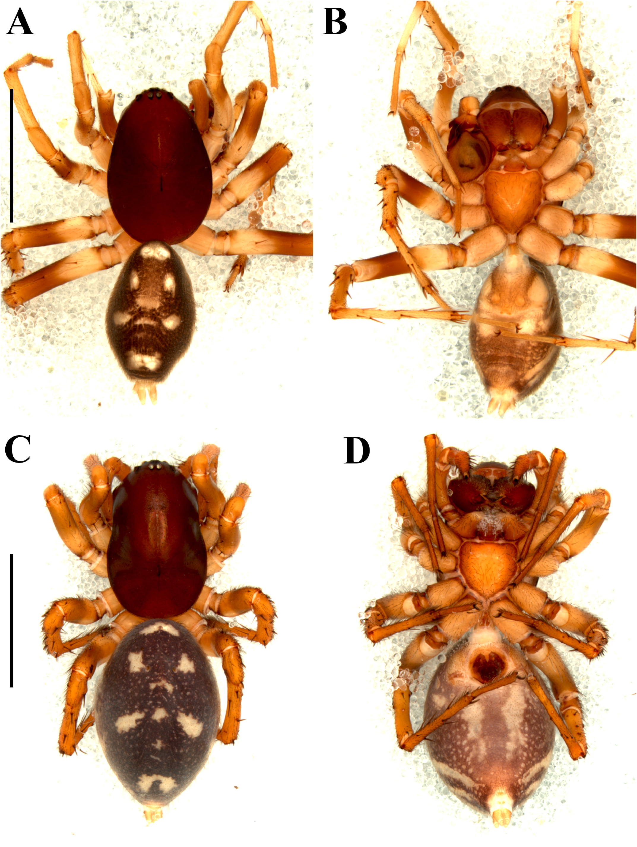
Redescription. Male (Holotype, AMNH_IZC 00217585). Coloration ( Fig. 93A–B View FIGURE 93 ): carapace brown-reddish. Chelicerae with paturon brown, fangs brown-reddish. Endites light brown, white on anterior region. Labium and sternum light brown. Legs: Coxae I –IV pale beige. Femora I –IV light yellow from base to medial region, brown the rest of their extension. Patella I yellow, II–IV light brown. Tibia I pale yellow with dark spots on the base, II–IV yellow with dark lateral spots. Metatarsus I brown, II–IV yellow with dark spots on the base. Tarsi I –IV yellow. Abdomen: dorsally dark gray with ten white guanine spots organized as follows: two irregular spots very close to each other partially fused in medial region, anteriorly positioned; two longitudinal spots larger than previous ones, anteromedially positioned; four irregular spots, forming a cross, medially positioned; two large and irregular spots, posteriorly positioned. Laterally dark gray with a wide and oblique longitudinal stripe, extending to posterior side. Ventrally light gray with two large anterior spots and several small light spots uniformly distributed on abdomen. Spinnerets light yellow. Measurements: total length 5.61, carapace length 2.82, width 1.84, height 1.21. Clypeus height 0.55. Eye diameters and interdistances: AME 0.09, ALE 0.10, PME 0.09, PLE 0.11; AME–AME 0.22, AME–ALE 0.23, AME–PME 0.21, PME–PME 0.24, PME–PLE 0.37, ALE–PLE 0.23. Chelicerae 0.90 length. Sternum length 1.12, width 0.97. Legs: I—femur 1.64/ patella 0.64/ tibia 1.70/ metatarsus 1.41/ tarsus 0.92/ total 6.31; II—1.36/ 0.64/ 1.25/ 1.28/ 0.81/ 5.34; III—1.45/ 0.45/ 1.25/ 1.58/ 0.78/ 5.51; IV—2.01/ 0.63/ 1.48/ 2.12/ 1.14/ 7.38. Abdomen length 2.61. Legs spines pattern (only the differences from the general pattern): II—metatarsus p0- 1d-1; III—femur d0-0-1, metatarsus p1d-1-2; IV—metatarsus d1-1-0. Palp: retrolateral process of cymbium (RPC) long, wide with small rounded projection on anterior side; tegulum (T) large, rounded, almost as long as wide; subtegulum (St) large, longer than wide; conductor (C) developed, wide with granulate cuticle; appendix (ApC) short, apically sharp; embolus (E) long, filiform towards apex; base of embolus (EB) approximately as long as three times as basal tegular membrane width; basal tegular membrane (BTM) originated proximally on tegulum, proximally flattened, ending as short, sharp appendix; spermatic ducts (SD) S-shaped, both folds full open, thin; ventral tibial apophysis (VTA) small, triangular-shaped in ventral view; median apophysis (MA) large, rounded, slight notch on external edge; retrolateral tibial apophysis (RTA) large, longer than palpal tibia, shovel-shaped; basal retrolateral tibial apophysis (bRTA) short, square-shaped ( Figs 94A–D View FIGURE 94 ; 95A–B View FIGURE 95 ).
Female (ICN-Ar-8021). Coloration and abdominal pattern of spots as male ( Fig. 93C–D View FIGURE 93 ). Measurements: total length 7.23, carapace length 3.71, width 2.47, height 1.59. Clypeus height 0.71. Eye diameters and interdistances: AME 0.10, ALE 0.11, PME 0.13, PLE 0.14; AME–AME 0.24, AME–ALE 0.32, AME–PME 0.36, PME–PME 0.32, PME–PLE 0.51, ALE–PLE 0.35. Chelicerae 1.51 length. Sternum length 1.31, width 1.30. Legs: I—femur 2.18/ patella 0.73/ tibia 1.78/ metatarsus 1.49/ tarsus 1.14/ total 7.32; II—1.85/ 0.63/ 1.48/ 1.41/ 0.92/ 6.29; III—1.71/ 0.62/ 1.25/ 1.66/ 0.94/ 6.18; IV—2.02/ 0.85/ 1.67/ 2.51/ 1.18/ 8.23. Abdomen length 3.72. Legs spines pattern (only the differences from the general pattern): IV—femur d0-0-1d, tibia v1p-1p-2, metatarsus d1p-1p-0. Epigyne: lateral borders (LB) short, curved towards posteromedial region of epigyne, forming medial large atrium (A); median field plate (MFP) very large and quadrangular-shaped; copulatory ducts (CD) very short and wide, almost undistinguished from spermathecae; seminal receptacles (SR) long, thin, curved towards median septum; spermathecae (S) large, irregulars, posteriorly positioned; fertilization ducts (FD) almost as long as spermathecae length ( Figs 94E–F View FIGURE 94 ; 95C–D View FIGURE 95 ).
Variation. Males (n=4): total length: 5.61–6.11; carapace length: 2.82–2.99; femur I length: 2.11–2.23. Females (n=2): total length: 6.99–7.23; carapace length: 2.91–3.71; femur I length: 2.18–2.31.
Distribution. Known from Cundinamarca department, Colombia ( Fig. 106 View FIGURE 106 ).


No known copyright restrictions apply. See Agosti, D., Egloff, W., 2009. Taxonomic information exchange and copyright: the Plazi approach. BMC Research Notes 2009, 2:53 for further explanation.
|
Kingdom |
|
|
Phylum |
|
|
Class |
|
|
Order |
|
|
Family |
|
|
Genus |
Tenedos andes Jocqué & Baert, 2002
| Martínez, Leonel, Brescovit, Antonio D. & Quijano, Luis G. 2022 |
Tenedos andes Jocqué & Baert, 2002: 83–84
| Jocque, R. & Baert, L. 2002: 84 |