
Tegenaria incognita, Bolzern, Angelo, Crespo, Luís & Cardoso, Pedro, 2009
|
publication ID |
https://doi.org/ 10.5281/zenodo.187030 |
|
publication LSID |
lsid:zoobank.org:pub:5970CC87-D3F4-4C1F-92AA-AEC122AE7724 |
|
DOI |
https://doi.org/10.5281/zenodo.5627590 |
|
persistent identifier |
https://treatment.plazi.org/id/14F2A432-FC4F-43CF-B043-2206A7CB16AF |
|
taxon LSID |
lsid:zoobank.org:act:14F2A432-FC4F-43CF-B043-2206A7CB16AF |
|
treatment provided by |
Plazi |
|
scientific name |
Tegenaria incognita |
| status |
sp. nov. |
Tegenaria incognita sp. n.
( Figs. 6–10 View FIGURES 6 – 10 , 15–18 View FIGURES 11 – 18 )
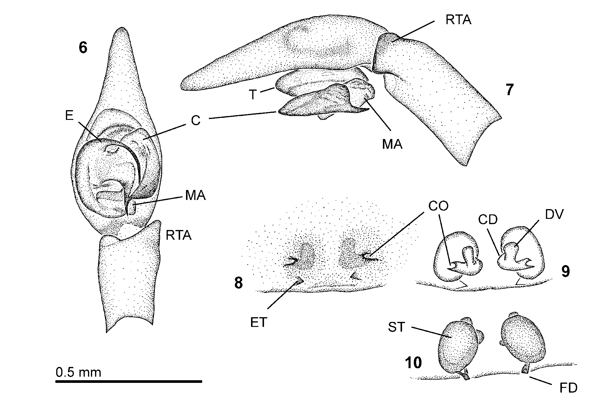
Type material. Holotype 3 (deposited at the NMB, Nr.: 2805a, Figs 6–7 View FIGURES 6 – 10 , collected by pitfall trap); Portugal, Lisbon, “Parque Florestal de Monsanto” (38°43’N, 9°11’W); leg. A. Rebelo, 9. November 2004. 1 3, paratype (deposited at the NMB, Nr.: 2805b); same locality and collecting details as holotype. 2 Ƥ, paratypes (deposited at the NMB, Nr.: 2805c, Figs 8–10 View FIGURES 6 – 10 , 15–18 View FIGURES 11 – 18 , collected by hand), same locality as holotype; leg. L. Crespo, 13. April 2008.
Further material examined. 101 3, 5 Ƥ same locality as holotype (deposited at the NMB, Nr.: 2805d and 2805e, collected by pitfall traps); leg. L. Crespo, 16. October - 1. November 2008. 2 3, 2 Ƥ (deposited in the personal collection of Luìs Crespo, Portugal); same locality and collecting details as previous.
Diagnosis. T. incognita sp. n. is closely related to T. barrientosi sp. n., T. bucculenta (L. Koch, 1868) , T. feminea (Simon, 1870) and T. montigena ( Simon, 1937) . It can be separated from the later three species by the absence of a patellar apophysis (present in T. feminea, Barrientos 1980 : fig. 1A), the broad and flat lateral tibial apophysis (fig. 7; absent in T. feminea ; much smaller in T. bucculenta, Barrientos 1991 : fig. 3; protruding ventrodistally in T. montigena, Simon 1937 : fig. 1541) and the presence of small diverticulae at the copulatory ducts (fig. 9). T. incognita sp. n. can be separated from T. barrientosi sp. n. by the characters mentioned in the diagnosis section of the description of T. barrientosi sp. n (see above).
Etymology. The name refers to the fact that this species has not been described until now even though the only known finding site lies in a forest among the largest Portuguese city. The name is derived from the Latin adjective “incognitus” and is female in gender.
Description. Prosoma: carapace: 1.82–3.95 (2.04) mm long, 1.39–3.00 (1.46) mm wide in males (n=4); 2.83–3.16 mm long, 1.99–2.21 mm wide in females (n=2). Fovea-length to carapace-length: 0.147–0.209 (0.181). Yellowish brown coloured with two longitudinal bands of darkened triangular dots (in the male specimens pigmentation very weakly visible, probably due to alcohol preservation or pitfall solution). Border of carapace continuously darkened ( Fig. 15 View FIGURES 11 – 18 ). Plumose hairs present. Head-region: 0.86–1.95 mm wide in males, 1.43–1.52 mm wide in females. Head-region only somewhat darker. PER 0.47–0.88 (0.48) mm wide in males, 0.72–0.73 mm wide in females. Diameter of PME: 0.06–0.10 mm; PLE: 0.08–0.14 mm; AME: 0.05–0.10 mm; ALE: 0.08–0.15 mm. Eye-formula ( Fig. 17 View FIGURES 11 – 18 ): ALE>PLE>PME=AME in males, ALE= PLE>PME>AME in females. PME separated by 1.5 times their diameter. PME and AME separated by the diameter of PME. PME and PLE separated by 1.5 times the diameter of PME. PME and ALE separated by 1–1.5 times the diameter of PME. AME separated by slightly less or about their diameter. AME and ALE separated by 0.5 time the diameter of AME or slightly more. Clypeus height (measured under AME): about 1.5–2 times the diameter of AME; clypeus height (measured under ALE): about 0.5–1 time the diameter of ALE. AER slightly recurved, PER straight in dorsal view ( Fig. 15 View FIGURES 11 – 18 ). Both eye-rows slightly procurved or AER straight in frontal view ( Fig. 17 View FIGURES 11 – 18 ). Chelicerae: 0.85–2.21 (0.97) mm long, 0.41–1.06 (0.48) mm wide in males; 1.46–1.56 mm long, 0.75–0.80 mm wide in females. Chelicerae protruding, visible in dorsal view. 3 teeth at the pro-margin, median tooth biggest. 8–10 teeth at the retro-margin, most proximal tooth very small. Gnathocoxa width to length: 0.510–0.543 (0.510). Labium width to length: 0.857–1.009 (1.009). Distal margin of labium concave. Sternum ( Fig. 16 View FIGURES 11 – 18 ): 1.12–2.18 (1.17) mm long, 0.92–1.67 (0.97) mm wide in males; 1.57–1.68 mm long, 1.23–1.35 mm wide in females. Somewhat lighter median band, but no other colourpattern.
Legs and palps: plumose hairs present. Yellowish-brown without pattern. Trochanter straight or slightly curved, not notched ( Fig. 16 View FIGURES 11 – 18 ). Number of dorsal trichobothria at tarsus I and IV: 6–7; at tarsus II and III: 5–6. Leg measurements are listed in tables 4–5. For spine formulae see table 3.
Opisthosoma: 1.83–3.69 (1.98) mm long, 0.97–2.42 (0.97) mm wide in males; 3.70–4.59 mm long, 2.21–2.95 mm wide in females. Colour dark gray-green, dorsally in the anterior half a narrow bright median band, not very distinct; more posteriorly shading into 3–5 chevrons (in male specimens these patterns are not visible due to preservation or capture liquid) ( Fig. 15 View FIGURES 11 – 18 ). Plumose hairs present. Spinnerets ( Fig. 18 View FIGURES 11 – 18 ): colulus is a rectangular plate, pale (distally slightly more sclerotized), distal margin strongly concave. PLS longer than all others. PMS as long as ALS. ALS not darkened. Both segments of PLS pale or distal segment dorsally somewhat darkened. Distal segment of PLS as long as basal segment. PLS separated by more than 2 times their diameter. Distance between ALS smaller than 0.5 their diameter.
Male palp ( Figs 6–7 View FIGURES 6 – 10 ): femoral and patellar apophyses absent. RTA with 2 branches: lateral branch simple, very broad, lamella-like; lateroventral branch almost completely hidden by the lateral branch, a long drawnout lobe. Dorsally at palpal-tibia short spine absent. Bulb length to cymbium length: 0.41–0.47. Embolus filiform, becoming more slender distally, shorter than cymbium width, curved through 90°. Embolus origin (free apex) on the left palp at 11½ o'clock position, terminal end at 4½ o'clock position. Tegulum anteriorlaterally constantly curved. Median apophysis pocket-like, consisting of a membranous basal part and terminally of a thin and broad sclerotized plate. Origin of median apophysis proximal at tegulum (between 5 and 7 o’clock position). Connection of median apophysis to tegulum membranous. Conductor droplet - shaped, parallel to cymbium margin and shorter than the alveolus. Conductor folded only at the terminal half. Terminal end is a strongly sclerotized and pointed peak, pointing towards posterior. Membranous ridge present from terminal end of conductor towards connection of the conductor to the tegulum (ventral branch of conductor). Connection of conductor to tegulum is membranous.
range (in mm) of all examined specimens (n=4). The measurements of the holotype are given in parenthesis. Epigynum and vulva ( Figs 8–10 View FIGURES 6 – 10 ): whole epigynal plate 0.34 mm long, 0.58–0.67 mm wide, sclerotized. Atrium 0.22 mm long, 0.34–0.38 mm wide. Atrium only an indistinct, strongly sclerotized trapezoidal depression. Anterior margin of atrium indistinct. Posteriorly the atrium reaches the epigastric furrow. Pair of posterior teeth present, pointing inwards. Ducts and spermathecae barely visible through the plate. Copulatory openings visible as gaps, outward directed ( Figs 8–9 View FIGURES 6 – 10 ). Copulatory ducts short. Small anteriorly originating diverticulae at the copulatory duct present ( Fig. 9 View FIGURES 6 – 10 ). Spermathecae expressed as smooth and strongly sclerotized globular structures, separated by less than their diameter ( Fig. 10 View FIGURES 6 – 10 ). Fertilisation ducts small.
Natural history. The specimens were captured in mixed woodland, with both pine and cork oak trees. Adult specimens were caught in spring and autumn.
Distribution. Only known from the mentioned forest in Lisbon, Portugal ( Fig. 19 View FIGURE 19 ).
femur patella tibia metatarsus tarsus total
Pedipalp 1.15–1.28 0.53–0.55 0.70–0.74 - 1.00–1.06 3.39–3.61 Leg I 2.18–2.29 1.00–1.02 1.82–1.99 1.74–1.83 1.10–1.17 7.85–8.29 Leg II 1.99–2.12 0.99–1.03 1.47–1.58 1.59–1.71 1.00–1.04 7.04–7.48 Leg III 1.91–2.05 0.90–0.96 1.29–1.37 1.74–1.87 0.93–1.00 6.77–7.24 Leg IV 2.52 1.05 2.21 2.56 1.12 9.46

| NMB |
Naturhistorishes Museum |
No known copyright restrictions apply. See Agosti, D., Egloff, W., 2009. Taxonomic information exchange and copyright: the Plazi approach. BMC Research Notes 2009, 2:53 for further explanation.