
Systelloderes loebli, Štys & Baňař, 2007
|
publication ID |
https://doi.org/ 10.5281/zenodo.4503564 |
|
DOI |
https://doi.org/10.5281/zenodo.4594260 |
|
persistent identifier |
https://treatment.plazi.org/id/03BC87A6-B34E-5A1B-FE4E-3E47DF1FFAD9 |
|
treatment provided by |
Felipe |
|
scientific name |
Systelloderes loebli |
| status |
sp. nov. |
Systelloderes loebli View in CoL sp. nov.
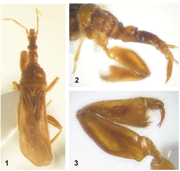
( Figs. 1-16 View Figs View Figs View Figs View Figs )
Type locality. New Caledonia, Mount Koghi, 400-500 m a.s.l.
Type material. HOLOTYPE: ♀, ‘ New Caledonia / Mt. Koghi, prim. for. / 400-500m, 18.-19.x / 1998, I. Löbl, litter’. The specimen bears the following red label: ‘ HOLOTYPE / Systelloderes / loebli sp. nov. / Štys & Baňař det. 2007’; collection of Muséum d’Histoire Naturelle, Genève ( Switzerland).
Diagnosis. Large species, over 5 mm long, macropterous, brownish. Antennal segment 2 longer than segment 3, segments 2-4 isomorphic, terete. Fore leg: coxa with a long, narrow, free, erect scale; femur with a prominent basidorsal extension rising high above the level of tarsus; femoro-tibial membrane with dorsal neopatellar sclerites; tibial process absent; tibial and tarsal armatures as illustrated.
Description. Body elongate, moderately robust, extremities short, coloration and vestiture uniform and inconspicuous ( Fig. 1 View Figs ).
Measurements. Total body length – 5.75-5.85 (abdomen deformed). Head. Anterior lobe, L – 0.71; posterior lobe, L – 0.31, W – 0.38; distance of eye to apex of antennifer – 0.49; diatone (max W across eyes) – 0.36; min interocular distance, dorsal – 0.24; min interocular distance, ventral – 0.18; eye, L – 0.15. Labium. Total, L – 0.95; segment 1, L – 0.12; segment 2, L – 0.21; segment 3, L – 0.48; segment 4, L – 0.16. Antenna. Segment 1, L – 0.24; segment 2, L – 0.62; segment 3, L – 0.56; segment 4, L – 0.42. Pronotum. Total L (max) – 0.98; collum: L (median) – 0.18, max W – 0.49; midlobe: L (max) – 0.44, W (max) – 0.89; hindlobe: L (max) – 0.36, L (median) – 0.31, W (max) – 0.98. Forewing. Max L – 2.95. Fore leg. Total L – 1.07, max W – 0.42; Ti 1: L – 0.87, max W – 0.41;. Ta 1: L – 0.29, max W – 0.17; anterior claw, L (basis – apex) – 0.31; posterior claw, L (basis – apex) – 0.26. Middle leg. F 2: L – 0.84, max W – 0.18; Ti 2: L – 0.69, max W – 0.13; Ta 2: L (without claw) – 0.24, max W – 0.09. Hind leg. F 3: L – 1.02, max W – 0.29; Ti 3: L – 1.16, max W – 0.14; Ta 3: L (without claw) – 0.38, max W – 0.09.
Coloration ( Fig.1 View Figs ). Body nearly unicolorous, between light-brown (antennae, middle and hind legs) and brown to dark-brown (head and midlobe of pronotum).
Texture. Moderately shiny (including extremities), head lustrous. Dorsum of head with irregularly distributed, rather sparse, small setigerous tubercles, lateral and ventral sides of head, prothoracic collum, and dorsum of pronotal midlobe with a continuous cover of regularly distributed, minute setigerous tubercles (the latter particularly distinct in preocular area, on posterior cephalic lobe and on midlobe of pronotum, creating their shagreened appearance). Setigerous tubercles replaced by indistinct, large and shallow alveoles laterally on parts of prothorax and dorsum of pronotal hindlobe, the latter slightly rugulose. Mesoscutellum, wings and abdomen without particular structures. Antennae, labium, Cx 1, Tr 1, Ta 1, middle legs, and hind legs smooth. Anterior and posterior faces of F 1 and Ti 1 with scattered minute setigerous tubercles; longitudinal depression of anterior (mesal) face of Ti 1 with transverse wrinkles.
Vestiture. Macrotrichia golden, ‘soft’, mostly short, straight, oblique, on some body parts (particularly posterior lobe of head, pronotum, and forewings) moderately to strongly curved. Macrotrichia mixed with much longer, diagonal, semierect to erect, conspicuous, mostly straight to only slightly curved trl setae with bilaterally symmetrical position; some of these may represent true trichobothria although the bothrium itself was not observed. Scales absent, except on Cx 1 (see below). Head, labrum, and labium. Distribution of trl setae (longitudinally arranged if more than 1+1) as follows. Head: several trl setae on and alongside anteclypeus and on labrum; 2+2 preocular (proximad to antennifers); 1+1 close to mesal eye margins, 2+2 postocellar; labial segment 1: dorsum/venter 1+1/0; 2: 3+3/0; 3: 3+3/2+2; 4: 3+3/3+3 (all strongly oblique and curved, similar to normal, elongate macrotrichia). Setation of ventral surface of head erect, short; longer and curved hairs on buccular part, gradually more curved and longer on proximal part close to neck, none trichobothrium-like. Antennae with uniform, short, straight, oblique setae; trl setae 1+1 on segment 1, then occurring from base of segment 2 to apex of segment 4, more elongate, denser and more erect distally. Prothorax. Dense, ‘soft’, short, mostly curved setae; trl setae: collum 1+1 anterolaterally, 1+1 posteromedially; midlobe: 1+1 posteromedially. Mesoscutellum. trl setae 2+2, in proximal part. Forewings with sparse, short, curved microtrichia only on veins, none on wing membrane. Fore leg. Cx 1 with curved, nearly adpressed pubescence and two long, distiventral, diagonally pointing trl setae; a long, narrow, moderately ectally curved scale (L 0.12, W 0.02) subapically on ventral surface, nearer mesal face ( Figs. 3 View Figs , 6 View Figs ). Tr 1 with curved, long setae; on ventral face, with 3-4 long, curved trl setae: the most distal one inserted at identical point as coxal scale; a few setae directed proximad, the other distad. F 1 with short, oblique setae on dorsal and ventral faces, anterior and posterior faces nearly bare; erect trl setae as follows: about 5+ 5 in double-row in distal half of dorsal face and multiple terminal dorsal cluster, and about 5+5 trl setae regularly distributed on ventral face. Ti 1 anterior face nearly bare, posterior and ventral faces with long, straight, oblique setae (particularly in distal half), dorsal and ventral faces and distal edge with numerous, irregularly distributed trl setae; other conspicuous macrotrichia: a single subpatellar trl seta, and three very long, curly setae at distiventral angle. Ta 1 with long, straight, diagonal setae, especially dense and mostly curly on ventral face; conspicuous trl setae: 1+1 dorsal, subterminal, strikingly long, and 1+1 apicilateral claw-guarding setae (anterior one, close to the shorter claw, conspicuously shorter). Middle and hind legs densely covered with short oblique to semierect setae on all faces; tibial and tarsal setae radiating relative to tibial and tarsal axes. Tr 2 and Tr 3 with two conspicuous erect ventral trl setae each. Ventral faces of F 2 and F 3 each with one basal (adtrochanteral), one postmedial, and several distal suberect to erect trl setae, dorsal faces of F 2 and F 3 each with 1+1 erect subterminal (adtibial) trl setae. Ti 2 and Ti 3 with long, oblique to erect trl setae developed along entire length,>15 and> 20 in number, respectively. Ta 2 and Ta 3 (second ’segments’) with about 10 strongly oblique trl setae on Ta 2, over 12 on Ta 3; setae on the latter undoubtedly homologous but not thin and trl like, and nearly spiniform instead. The only visually undoubtedly trl seta on Ta 3 is a ventral erect seta on its first ‘segment’. Abdomen. Dorsum with short setae, without scales. Venter with golden short to moderately long, straight to slightly curved semierect macrotrichia (length and density increasing distally) intermixed with short, blackish, adpressed hairs. Some trl setae occurring marginally; distribution of long trl setae on posterior segments as follows: ventrite 7 – 1+1 at posterolateral angles, 1+1 submedial near posterior margin; subgenital plate: 1+1 submedial (extremely long) in basal part, 1+1 (very long) at posterior margin.
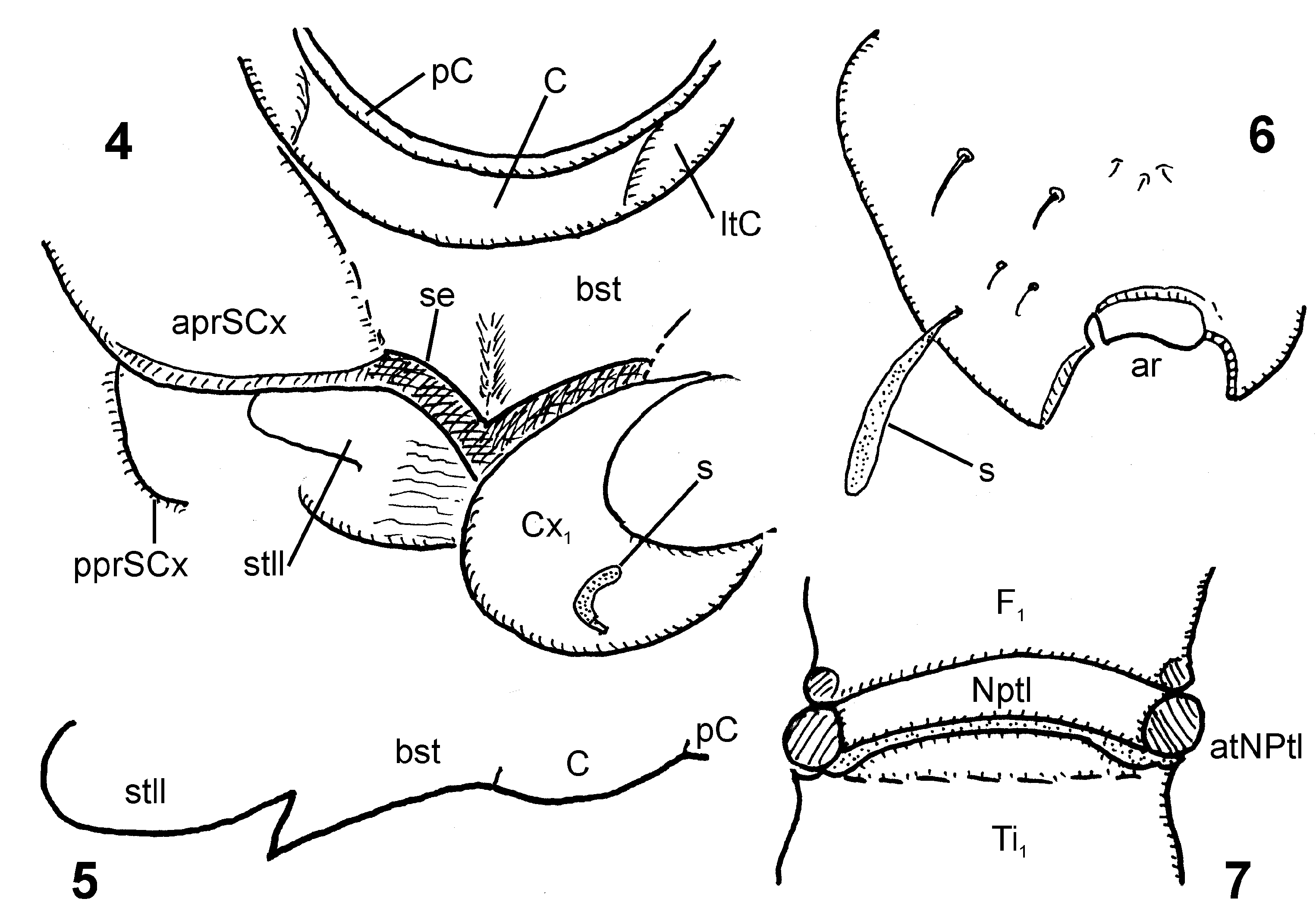
Structure. Head ( Figs. 1-2 View Figs ) strikingly narrow and long, slightly longer than pronotum (1.04 times as long as pronotum). Anterior lobe markedly longer than posterior lobe, 2.3 times as long. Lateral margin of preocular region parallel-sided proximally, its long distal part slightly convex, diverging towards antennifers; anteclypeus very long, narrow. Eye 0.31 times as long as distance between eye and antennifer. Postocular impression broad, shallow; lateral margin of postocular part of anterior cephalic lobe straight, diverging towards convex side of posterior lobe – postocular impression not marked in lateral outline. Posterior lobe transverse, 0.82 times as long as wide, its dorsum strongly convex, lateral sides moderately so. Eyes small, in lateral view not exceeding dorsal or ventral outlines of head; facets individually convex. Ocelli large, ocellar tubercles low. Ventral outline of head continuous, very slightly concave, only apex (fused bucculae) and basis (association with neck) outstanding. Dorsal ocular index 6.0, ventral ocular index 4.0. Antennae moderately long, thin; first segment strikingly long, cylindrical; segments 2-4 terete (not flagelliform, neither segment 4 subfusiform); antennal formula (longest segment first) 2, 3, 4, 1; segment 2 being 1.11 times as long as segment 3. Labium ( Fig. 2 View Figs ) moderately long, rather thin, directed anterad (segments 1, 2) and ventrad (segments 3, 4), without particular structures, labial formula (longest segment first) 3, 2, 4, 1. Segment 3: 2.2 times as long as segment 2. Ventral outline of segment 2 emarginate near base and at midlength. Labrum reaching to middle of segment 2.
Pronotum ( Figs. 1-2 View Figs ). Collum short, 2.45 times as wide as long, with narrow precollum, dorsum with a linear impressed median area and pair of low and broad elevations, lateral area with low ‘pleural’ tubercle not visible in dorsal view. Collar constriction sharply delimited. Midlobe (dorsal side) with a linear, nearly percurrent median impression terminating just in front of posterior margin; disc with markedly plastic relief, with i) an inversely triangular anteromedial depression, ii) broad, not distinctly delimited, subcircular posteromedial depression, and iii) paired deep lateral pits emitting a lateral depression each; lateral margins broadly convex, interrupted, slightly notched because of lateral pits; posterior margin entire, trisinuate, sublateral shallowly concave parts slightly depressed, medial convex part broadly rounded, without edge. No traces of Y-shaped impressions. Constriction between midlobe and hindlobe broad, sharply demarcated. Midlobe 2.5 times as long as collum, 1.2 and 1.45 times as long as hindlobe maximum and median length, respectively. Midlobe 2.0 times as long as wide. Hindlobe ample, its median twice as long as collum, indicated neither by ridge nor impression, lateral margins rounded, posterolateral angles (in strictly dorsal view) subrectangular; posterior margin bisinuate (tetrasinuate, if moderately protruding posterolateral angles are counted), medially broadly and shallowly concave, moderately convex sublaterally. Hindlobe 3.2 times as wide as medially long. ‘ Proepimeral lobe ’ (see ŠTYS & BAŇAŘ 2006) extensive, distinctly exceeding posterior prosupracoxale posteroventrad, but not enclosing fore acetabula. Mesoscutellum. Concave central part equilaterally triangular, produced in long, apically rounded mucro and included into a larger triangle due to lateral association with forewing grooves. Prothoracic coxa, prosupracoxale, proacetabula, and prosternum. For details see Figs. 4 and 6 View Figs . Cx 1 situated within proacetabulum, the latter open anteriorly and closed elsewhere. Proximal parts of lateral, anterior, and most of mesal faces of Cx 1 fully enclosed and externally delimited by prosupracoxalia. Anterior prosupracoxale excessively developed, anteromesally extended, embracing Cx 1 anterolaterally, anteriorly, and anteromesally. Transverse anterior part of anterior prosupracoxale fused with ventral part of collum (forming a not fully understood system of anteroventral prothoracic evaginations); anteromesal part of anterior prosupracoxale directed mesocaudad, fusing with triangular probasisternum, and taking part in formation of its strengthened, horizontal, sharp-edged lateral sides, here termed ‘ prosternal strigilatory edge(s) ’ (see Discussion). This composite triangular probasisternum horizontal, clearly delimited by its sharp edges; prosternellum adhering to probasisternum, sinuate in sagittal plane, more dorsal than probasisternum, with only the posterior, rounded, tongue-shaped part posteriad to coxae visible in ventral view.
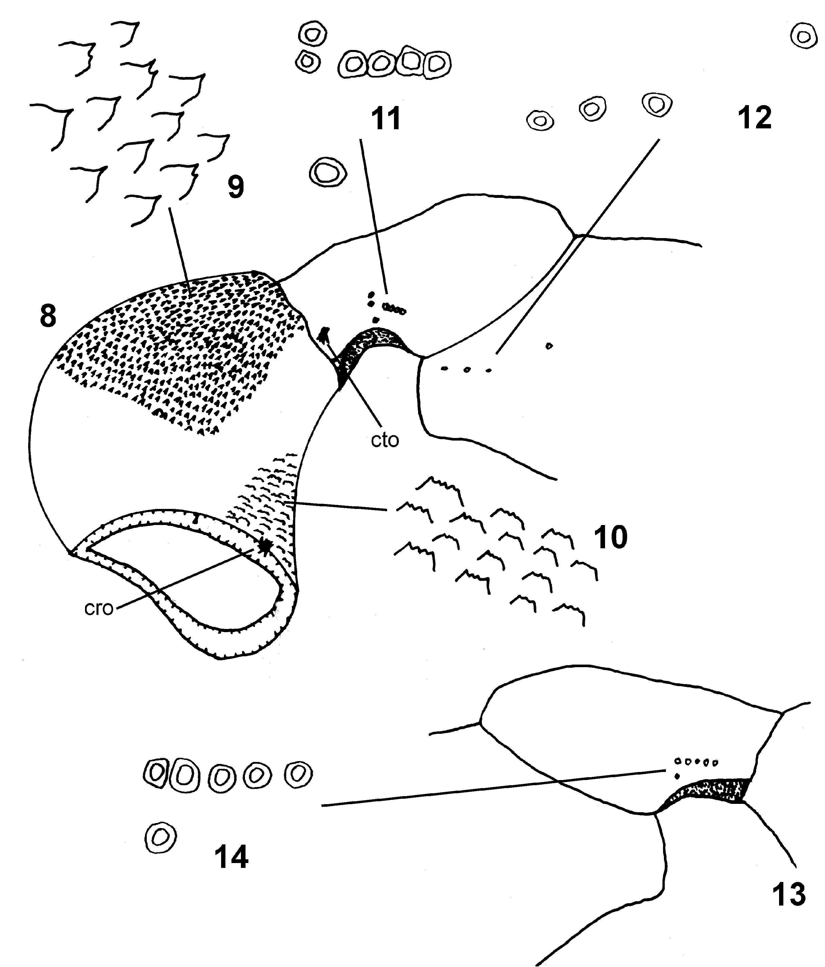
Legs rather short. Fore leg ( Figs. 3 View Figs , 6-16 View Figs View Figs View Figs ) extremely stout, femur and tibia incrassate. Cx 1 ( Fig. 8 View Figs ) conical, anteroventral face with dense, prominent, drop-like cuticular thorns ( Fig. 9 View Figs ) resembling and arranged like rasping files. Antero- and especially posterodorsal proximal parts of fore coxa with dense rows of different cuticular processes ( Fig. 10 View Figs ), resembling serrate rasping files, each of those with several apical teeth, thus very similar to Pseudohenschiella hauseri Baňař & Štys 2006 from Madagascar (cf. BAŇAŘ & ŠTYS 2006). Proximal region of Tr 1 very narrow, more so than robust distal part, both regions separated by concave impression; entire adcoxal (dorsal) surface deeply concave (accommodating distal part of Cx 1 during flexion of Tr 1) and delimited by sharp, lateral, free anterodorsal and posterodorsal edges (about as long as one third of tr-f junction). F 1 2.5 times as long as wide, with basidorsal angle forming subrectangularly produced, apically rounded dorsal extension, the latter rising strikingly above dorsal surface of Tr 1, and as high as its proximal diameter. Knee. Distinct remnant of a tripartite neopatella * (new term) visible dorsally in intersegmental F 1 -Ti 1 membrane when tibia maximally bent towards femur ( Fig. 7 View Figs ). Ti 1 broadly triangular, 2.1 times long as wide, compressed in anteroposterior plane, both anterior and posterior faces each with vaguely delimited longitudinal depression. Cleaning comb short, formed by tightly packed short setae; three ventralmost spiniform setae longer and stouter. Intersegmental tibiotarsal membrane forming an evaginated pocket stretching far ventrad beneath tarsus itself (enabling apparently its close appression towards distal margin of tibia). Distiventral, armature-bearing process absent. Apicitibial armature ( Fig. 15 View Figs ) consisting of seven spiniform setae: two short ventral (straight), three long subventral (all slightly oblique towards tarsus), and two short dorsal (strongly oblique towards tarsus). Ta 1 cylindrical, 1.7 times as long as wide, ventral surface slightly concave, tarsal armature ( Fig. 16 View Figs ) of 1+1 proximal, curved spiniform setae and 1+1 distal setae (anterior one semicircular, posterior one broadly spiniform, shorter than proximal setae). Claws all of same shape, regularly curved, posterior one shorter and narrower.
Fore leg sensilla on coxa, femur, and trochanter. Basal rim of Cx 1 anteromesally with coxal rim organ ( Fig. 8 View Figs ), consisting of cluster of several (5-7) differently directed, straight setae and one distant short seta. Condylar trochanteral organ ( Fig. 8 View Figs ) consisting of several (six?) poorly visible short setae. Anterior trochanteral organ ( Figs. 8, 11 View Figs ) consisting of 6+1 campaniform sensilla (group of six, one isolated; posterior trochanteral organ ( Figs. 13-14 View Figs ) consisting of six campaniform sensilla (five in straight row, one isolated). Anterior femoral organ ( Figs. 8, 12 View Figs ) consisting of 3+1 campaniform sensilla (group of three and one isolated) very close to base of F 1.
Neopatella (new term) = sclerite(s) situated within the dorsal section of the F-Ti intersegmental membrane in a position where the original patellar limb segment or its remnants would be situated (see Discussion). In fore leg of female S. loebli sp. nov., the dorsal apex of F 1 is concavely excised and the small dorsolateral projections of its posterior margin provide articulation with the neopatellar articulatory tubercles.The area between these projections is filled with a tripartite neopatella, its medial part forming a narrow arcuate strip adhering to the concavity of the apex of F 1, and its lateral parts being formed by strongly sclerotized, prominent, dorsally projecting articulatory tubercles associated with the medial strip of neopatella and articular projections of F 1. The distal margin of the neopatella is associated with a short intersegmental F-Ti membrane; the latter separates the neopatella from a produced and feebly delimited process of basidorsal margin of Ti 1.
Middle leg and hind leg. Lateral (dorsal, adfemoral) face of Cx 3 flat, largely covered by sandpaper-like file similar to that on Cx 1. F 3 moderately incrassate, its basidorsal margin arcuate, clearly extending above the level of dorsal face of Tr 3. Proximal segments otherwise without particular structures, anterior and posterior faces of Ti 2 and Ti 3 sulcate. Ta 2 and Ta 3 two-segmented, segment 1 extremely short, without dorsal surface in lateral view (dorsal part of segment 1 visible as dorsal part of basitarsal ring filling up apex of tibia in anterodorsal view). Apices of both middle and hind tibiae each with strikingly short posteroventral and anteroventral setal combs; every comb terminating ventrally with a long spiniform seta. Claws and parempodial setae isomorphic.
Forewing as usual for Systelloderes . Pterostigma strikingly well formed, long and wide, RP arising from its middle; rp-mp (= the anterior crossvein) entering open discal cell strikingly more distad than CuA3+4 (= the posterior ‘crossvein’); AP in claval area not developed.
Ventral side of abdomen (distorted in the holotype) with series of 1+1+1 large sclerites on ventrites 3-8. Terminalia (distorted in the holotype). Posteromedial part of ventrite 7 thickened. Ventral laterotergite 8 distinct from subgenital plate, the latter strongly sclerotized, elongate, basal margin convex, distal margin concave in front of proctiger.
Etymology. Dedicated to Ivan Löbl (Genève), an eminent coleopterist, our friend, and collector of the species.
Bionomics. The holotype was collected by sieving leaf litter in a patch of a primary tropical New Caledonian rainforest surrounded by a secondary forest (I. Löbl, in epist.).
Distribution. Known only from the type locality at Mount Koghi on the Grande Terre of New Caledonia.

No known copyright restrictions apply. See Agosti, D., Egloff, W., 2009. Taxonomic information exchange and copyright: the Plazi approach. BMC Research Notes 2009, 2:53 for further explanation.