
Systaria hainanensis, Zhang, Feng, Fu, Jianying & Zhu, Mingsheng, 2009
|
publication ID |
https://doi.org/ 10.5281/zenodo.275382 |
|
DOI |
https://doi.org/10.5281/zenodo.6222085 |
|
persistent identifier |
https://treatment.plazi.org/id/B50B87B1-1E75-015E-98F3-FC413B122E9C |
|
treatment provided by |
Plazi |
|
scientific name |
Systaria hainanensis |
| status |
sp. nov. |
Systaria hainanensis View in CoL sp. nov.
( Figs 1–12 View FIGURES 1 – 7 View FIGURES 8 – 12 )
Type material. Holotype male, CHINA: Hainan Province, Wuzhishan City, Mt. Wuzhi, Shuiman Town [18.81°N, 109.54°E], 16 August 2007, Feng Zhang leg.; paratype 1 female, same data as holotype ( MHBU).
Diagnosis. This new species resembles Systaria insulana (as illustrated in Platnick & Bonaldo, 1997) but can easily be distinguished from the latter by: (1) the broader base of the retrolateral tibial apophysis; (2) in ventral view, the tip of the embolus that is concealed behind the conductor (the tip of embolus is easily seen in ventral view in the latter); (3) the epigynal atrium is without a distinct sclerotised rim anteriorly and has a smaller concavity; (4) the spermathecae are far apart (while almost touching each other in the latter); (5) the chelicerae with two retromarginal teeth (three retromarginal teeth in the latter). The female of the new species is similar to S. bohorokensis from Indonesia, but differs from the latter by having the epigynal openings and the spermathecae in a common horizontal plane (while clearly in separate planes in the latter).
Etymology. The species name is an adjective in apposition derived from the type locality.
Description. Male (holotype). Total body length 8.19; cephalothorax 3.74 long, 2.61 wide; abdomen 4.45 long, 2.16 wide. Carapace ( Fig. 6 View FIGURES 1 – 7 ) light yellow, darker in eye area; longitudinal ovoid, cephalic area relatively broad; fovea longitudinal and distinct, long, occupying over one seventh of carapace length. Eye group width more than two third of carapace width. From dorsal view AER slightly recurved in dorsal view; and PER distinctly procurved and longer than AER ( Figs 3, 6 View FIGURES 1 – 7 ). AME enlarged and dark, eye diameters: AME 0.13, ALE 0.09, PME 0.08, PLE 0.07; eye interdistances: AME–AME 0.18, AME–ALE 0.14, PME–PME 0.22, PME–PLE 0.15, ALE–PLE 0.12; MOA 0.28 long, anterior width 0.30, posterior width 0.29. Clypeal height 0.13, almost equal to the diameter of AME. Chilum distinct triangular sclerite. Chelicerae ( Fig. 7 View FIGURES 1 – 7 ) moderately long, brown, with three promarginal teeth, median one largest, and two widely spaced retromarginal teeth. Endites and labium yellowish-brown; endites longer than wide, widest anteriorly, and a tuft of setae prolaterally. Labium slightly longer than wide ( Fig. 4 View FIGURES 1 – 7 ), tip rounded and with several setae, invaginated at postero-lateral corners. Sternum light brown, shield-shaped, not rebordered, with sclerotised extensions reaching towards each coxa and between coxae I and II, II and III, III and IV; anterior margin truncate and posterior margin not extending between coxae IV, only slightly narrower in front than in the middle. Legs light brown, tarsi with two dentate claws and conspicuous claw tufts ( Fig. 2 View FIGURES 1 – 7 ), trochanters deeply notched. Leg spination: femora: I d1-1-1, p1-1-1, r0-0-2; II d1-1-1, p1-1-1, r0-0-1; III d1-1-1, p0-1-1, r1-1-1; IV d1-1-1, p0- 1-1, r1-1-1; tibiae: I v2-2 -2; II v2-2 -2; III d0-1-0, p1-1-0, r2-2-2, v2-2 -2; IV d0-1-0, p1-1-0, r1-1-0, v2-2 -2; metatarsi: I v1 -0-0; II v1-1 -1; III p1-1-2, r1-1-2, v1 -0-1; IV p1-1-1, r2-2-2, v1-1 -1. Leg formula: 4123 ( Table 1).
Femur Patella Tibia Metatarsus Tarsus Total I 2.92 1.57 2.84 2.52 1.39 11.24 II 2.75 1.30 2.57 1.80 1.03 9.45 III 2.25 0.85 1.89 2.29 0.99 8.27 IV 3.38 1.08 3.15 3.24 1.13 11.98 Abdomen ( Fig. 6 View FIGURES 1 – 7 ) ovoid, brownish-grey, covered with long and dark setae, with two pairs of muscular impressions centrally. Venter of abdomen light brown. ALS ( Fig. 5 View FIGURES 1 – 7 ) cylindrical, slightly tapering, separated by almost less than half of their diameter; PMS thin and conical, as long as anterior spinnerets; PLS with distal segment shorter than basal segment, tapering; and basal segment of PLS with similar length as basal segment of ALS. Tracheal spiracle located just anterior to the spinnerets.
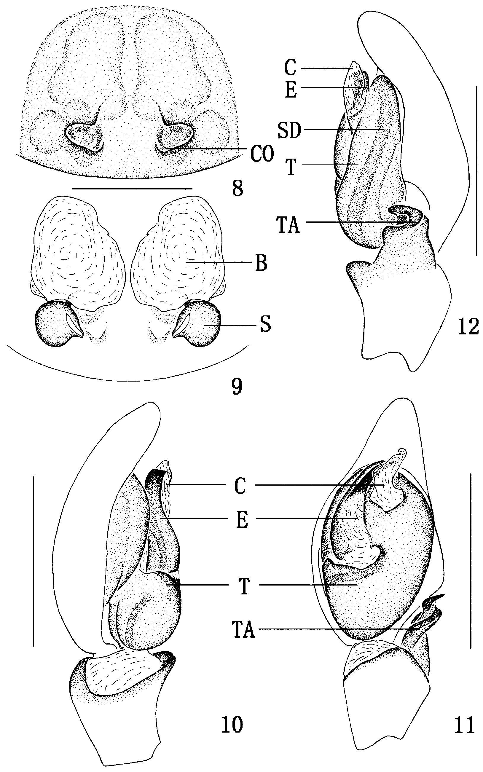
Male palp ( Figs 10–12 View FIGURES 8 – 12 ) relatively simple, tibia with complicated retrolateral apophysis, with broader base and acutely pointed tip from ventral view. Tegulum rounded at base; sperm duct distinctive, encircling long loop, originating from upper part of tegulum and ending at embolus. Spiniform embolus originating from prolateral tegulum centrally, long, the tip reaching beyond tip of tegulum and concealed behind conductor from ventral view. Conductor membranous. Median apophysis absent.
Female (paratype). Carapace colour, eye arrangement, abdominal coloration as for male, but carapace and abdomen with different shape than male ( Figs 1, 6 View FIGURES 1 – 7 ). Total length 10.70; cephalothorax 4.90 long, 3.42 wide; abdomen 5.80 long, 3.15 wide. Eye diameters: AME 0.15, ALE 0.12, PME 0.11, PLE 0.10; eye interdistances: AME–AME 0.18, AME–ALE 0.15, PME–PME 0.22, PME–PLE 0.17, ALE–PLE 0.13; MOA 0.25 long, anterior width 0.36, posterior width 0.35. Clypeal height 0.14, almost equal to the diameter of AME. Chelicerae with three promarginal and two retromarginal teeth. Endites, labium and sternum as for male holotype. Leg spination: femora: I d1-1-1, p1-1-0, r0-0-2; II d1-1-1, p0-2-0, r0-1-2; III d1-1-1, p0-1-1, r1-1-1; IV d1-1-1, p1-1-1, r0-0-1; tibiae: I v2-2 -0; II v0-2-0; III p1-1-0, r1-1-0, v2-2 -2; IV p1-1-0, r1-1-0, v2- 2 -2; metatarsi: II v1 -0-0; III p1-1-1, r1-1-2, v2 -0-2; IV p1-1-2, r1-1-2, v2-2 -2. Leg formula: 4123 ( Table 2).
Epigynal plate weakly sclerotised ( Fig. 8 View FIGURES 8 – 12 ), the epigynal atrium without a distinct sclerotised rim anteriorly; two copulatory openings concave, located postero-medially near the epigastric furrow ( Fig. 9 View FIGURES 8 – 12 ). Bursae situated laterally to the copulatory openings, large sac-shaped; spermathecae globular, distinctly smaller than bursae, situated posteriorly; connecting with the copulatory opening through a thick duct; fertilization ducts short, arising from the postero-lateral ends of the spermathecae.
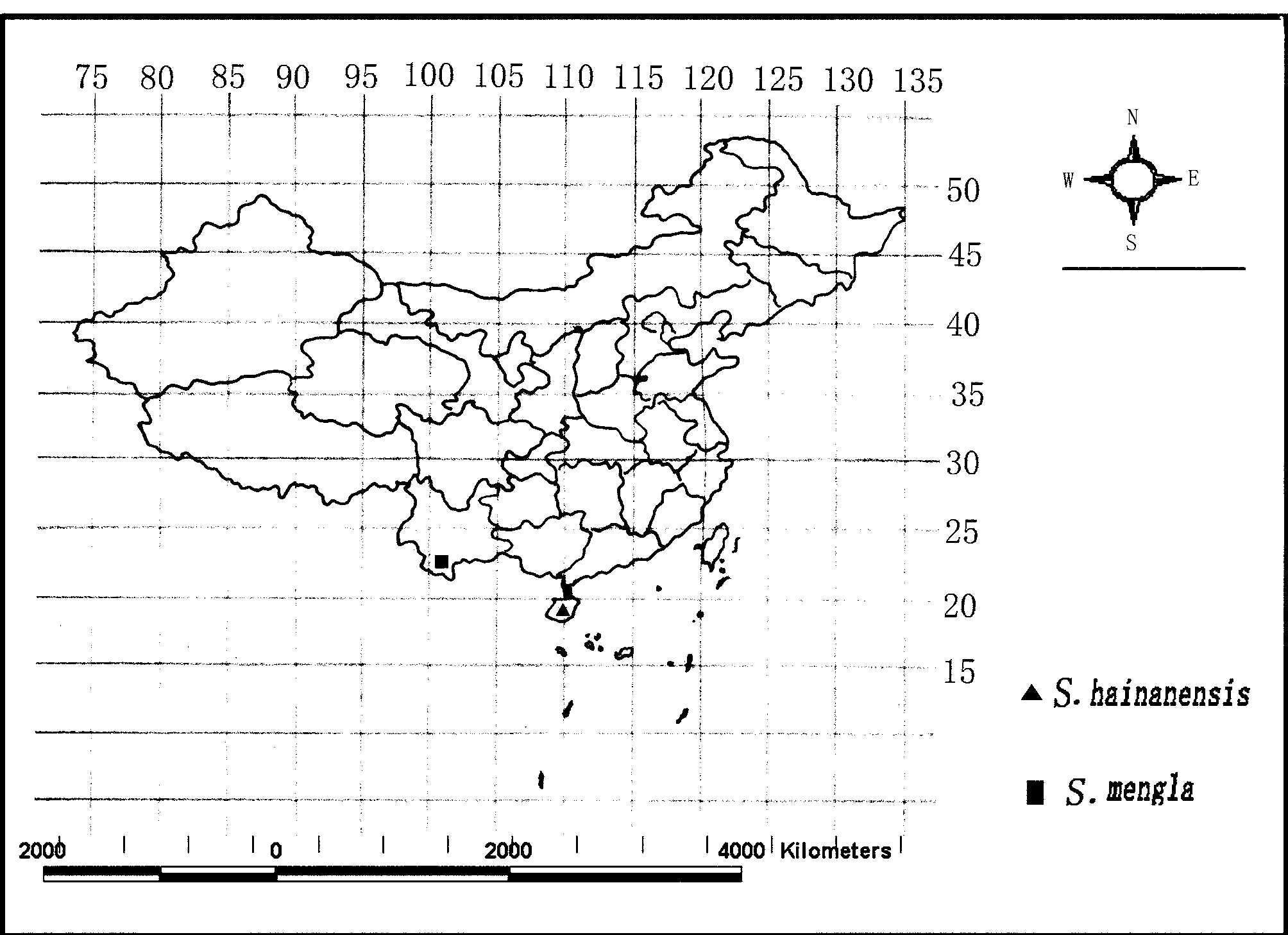
Distribution. Presently known only from the type locality, Hainan Island, China ( Fig. 20 View FIGURE 20 ).
Femur Patella Tibia Metatarsus Tarsus Total I 2.70 1.40 2.61 1.71 0.95 9.37 II 2.65 1.30 2.30 1.67 0.86 8.78 III 2.92 0.90 1.80 1.98 0.81 8.41 IV 3.60 1.35 2.84 3.30 1.62 12.71

No known copyright restrictions apply. See Agosti, D., Egloff, W., 2009. Taxonomic information exchange and copyright: the Plazi approach. BMC Research Notes 2009, 2:53 for further explanation.
|
Kingdom |
|
|
Phylum |
|
|
Class |
|
|
Order |
|
|
Family |
|
|
Genus |