
Styela atlantica ( Van Name, 1912 )
|
publication ID |
https://doi.org/ 10.11646/zootaxa.4114.3.1 |
|
publication LSID |
lsid:zoobank.org:pub:6EA59057-0E05-4AA5-8B84-327CBDB32E5B |
|
DOI |
https://doi.org/10.5281/zenodo.6068919 |
|
persistent identifier |
https://treatment.plazi.org/id/A25D4D00-D653-7606-7BF3-FEDD7D4BF894 |
|
treatment provided by |
Plazi |
|
scientific name |
Styela atlantica ( Van Name, 1912 ) |
| status |
|
Styela atlantica ( Van Name, 1912)
Figures 25 View FIGURE 25 , 26 View FIGURE 26 .
Stations. CP4364; CP 4366; CP4367; CP4368; CP 4374; CP4375; CP 4380; CP4407.
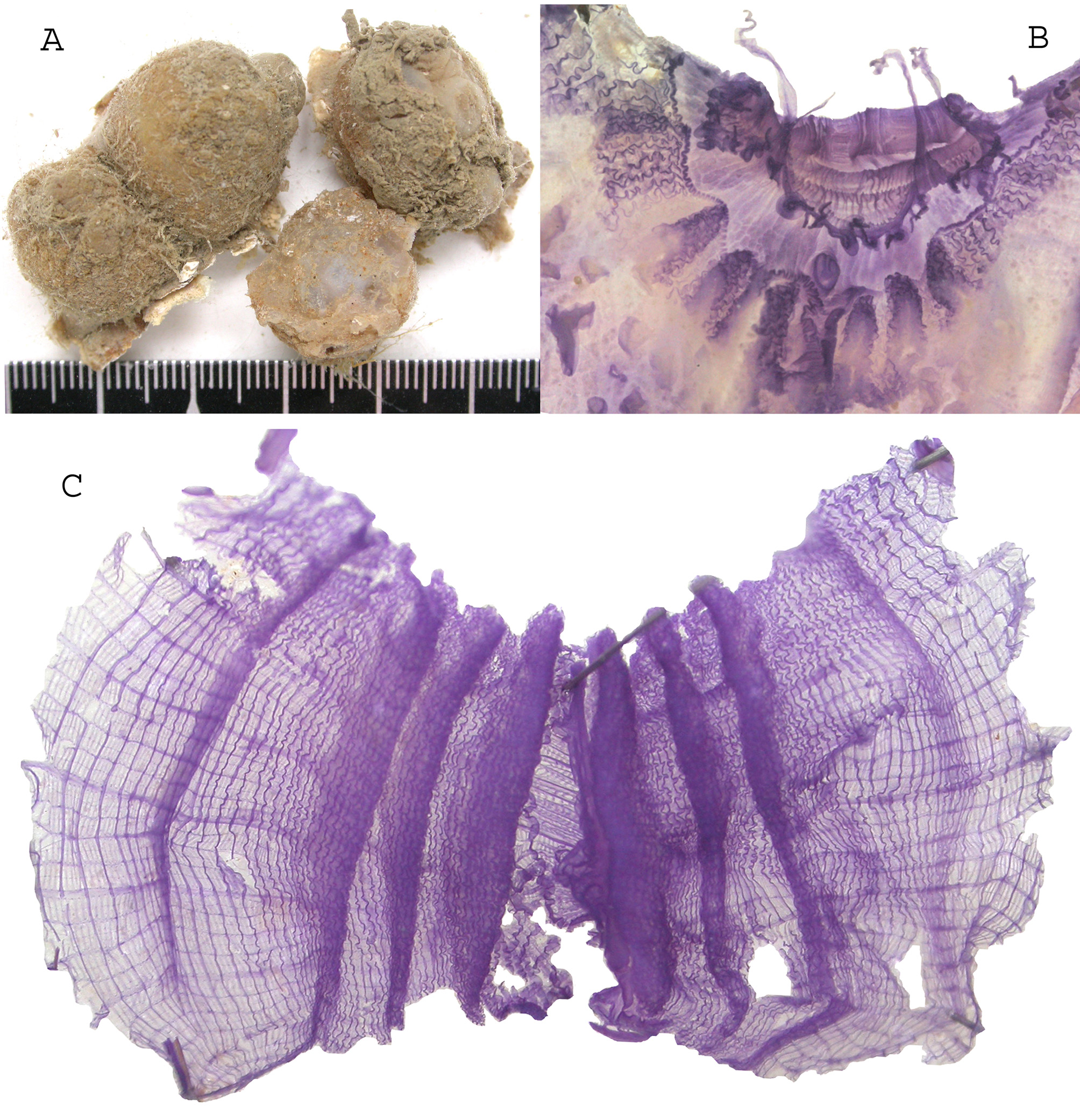
All specimens have the same spherical shape ( Fig. 25 View FIGURE 25 A) fixed by the ventral side sometimes a little expanded on the substrate with a rough tunic retaining some mud. The tunic is thick, resistant but soft. The siphons are close to each other with sharp spinules. The internal layer of the siphons remained yellow in formalin. Extracted from the tunic the body wall is colourless and thin with a weak musculature. Its internal layer is spotted with cells highly stainable ( Fig. 26 View FIGURE 26 A,B). A velum with undulated margin is present inside both siphons. The oral tentacles are in variable number, 4 of them longer ( Fig. 25 View FIGURE 25 B), the others distributed in 3 orders of size. In several specimens the tentacles are coiled. The prepharyngeal band has a single crest not deeply indented dorsally ( Fig. 25 View FIGURE 25 B). The dorsal tubercle is protruding and opens anteriorly in a U. The branchial tissue has a peculiar aspect due to the undulating longitudinal vessels ( Fig. 25 View FIGURE 25 C). There are 4 folds on each side but unequal. The most ventral fold is well apart, thinner than the others and progressively disappears posteriorly ( Fig. 25 View FIGURE 25 C). The space between the dorsal lamina and the first fold on the right enlarges posteriorly ( Fig. 25 View FIGURE 25 C). The branchial formula on the right side in a specimen of 18mm in diameter is:
E-8 (10) 13 (18) 10 (18) 8 (20) 4-DL
and in another specimen: E- 4 (11) 8 (14) 6 (12) 6 (14) 4-DL.
These formulae are somewhat subjective as it is difficult to determine precisely when a vessel belongs or not to a fold. The digestive tube is thick in a single loop ( Fig. 26 View FIGURE 26 A,B). The stomach occupies a large part of the ascending limb and is not well separated from the intestine. Twelve to 14 folds can be seen in transparency on the mesial side. A wide bundle links the stomach to the intestine. The rectum does not draw a secondary loop. The edge of the anus is scalloped. The whole gut is loosely attached to the body wall and to the branchial sac by thin straps. There are 2 gonads on each side, each a long ovary with a basal massive testis in a cap ( Fig. 26 View FIGURE 26 A,B). The short sperm ducts issuing from the testis vesicles converge into a single sperm duct at the distal tip of the ovary which runs over it and ends close to the lobed female papilla. Numerous foliated endocarps are evenly distributed on the body ( Fig. 26 View FIGURE 26 A,B). A ring of dense thread-like tentacles encircles the ventral part of the atrial aperture and extends dorsally in a loop to reach the neural area.
Styela atlantica is a species of cold waters recorded from the east coast of the middle United States ( Van Name 1945), Scandinavia and the Gulf of Gascony ( Monniot C 1969) down to 300m depth. From records from the Pacific Ocean Nishikawa (1991) considered as identical S. atlantica and S. stigma ( Hartmeyer 1906) S. sigma having priority, but he wrote “if verified to be distinct, the Atlantic population may safely be referred to as S. atlantica! Following the descriptions of Nishkawa (1991) and Sanamyan (2000) for Pacific samples it appears that these specimens have more endocarps and 4 equal branchial folds instead of inequal folds in S. atlanica and are presumably different species.
Other Atlantic Styela species have 2 gonads on each side and many endocarps such as S. canopus ( Savigny 1816) or S. partita ( Stimpson 1852) which were recorded in the shallow Caribbean area ( Van Name1945; Sloot 1969; Monniot C. 1983b) and in Brazil ( Rocha et al 2012). These species differ from S. atlantica in having the testis lobes along the both sides of the ovary instead of an apical cluster.
Styela glans Herdman 1881 described from a single specimen from Rio de La Plata was later recorded in several Antarctic areas. It has a cap of testis vesicles but at the top of a short ovary.
Styela eurygaster Millar 1977 was recorded from 3°27’N and 50° 15’W at 60m depth a single specimen having 2 gonads on the right side and only one on the left side with the same shape as in S. atlantica . The branchial folds are said to be rudimentary. The shape of the gut with an enlarged stomach and a caecum differs from S. atlantica and no endocarps are mentioned.
Pyura ocellata n. sp. Figures: 27, 28.
Stations. CP4386 (Type MNHN S2 PYU 496); CP 4358 and 16°38.4’N –61° 31.3W, 69m (coll. Bouchet, 2015).
Etymology. ocellata reminds the tunic design
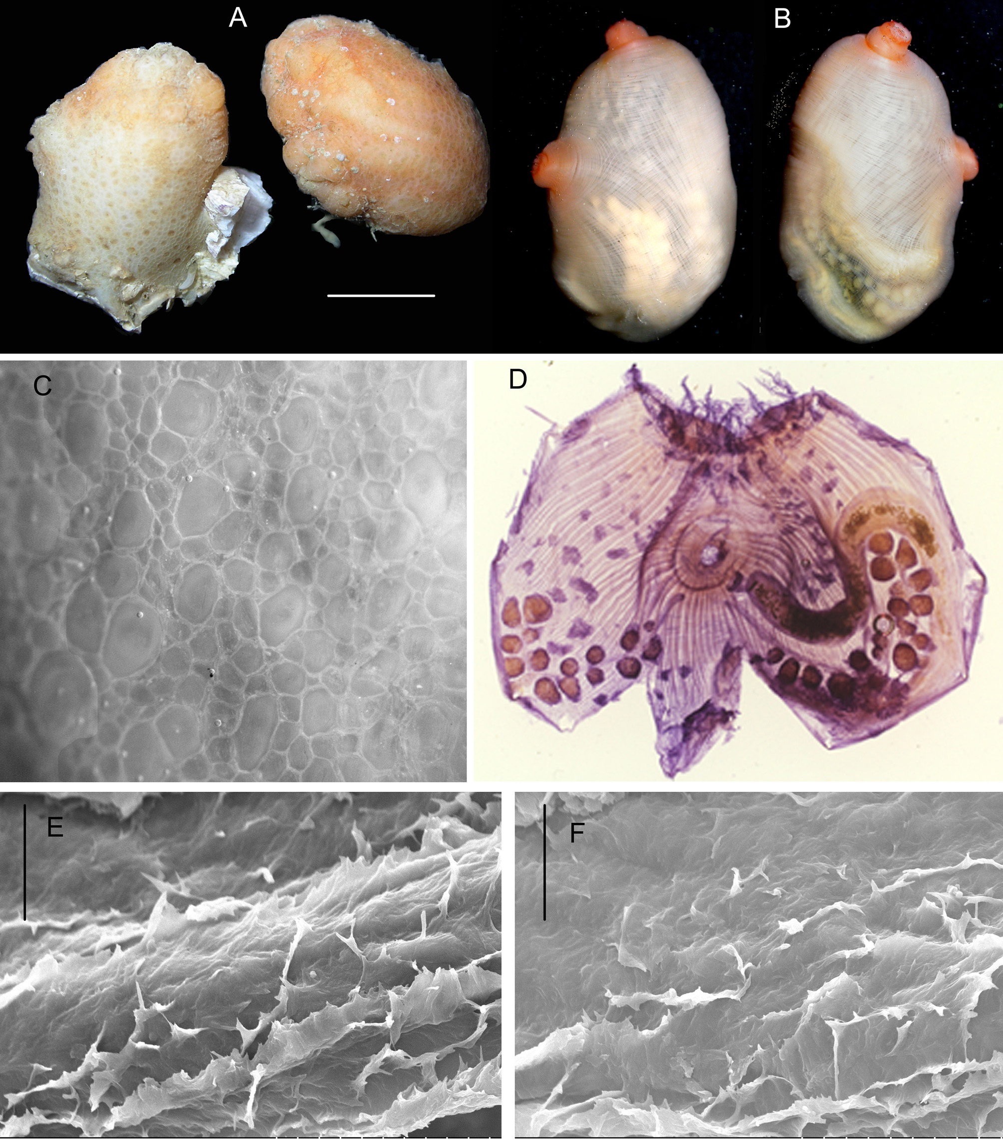
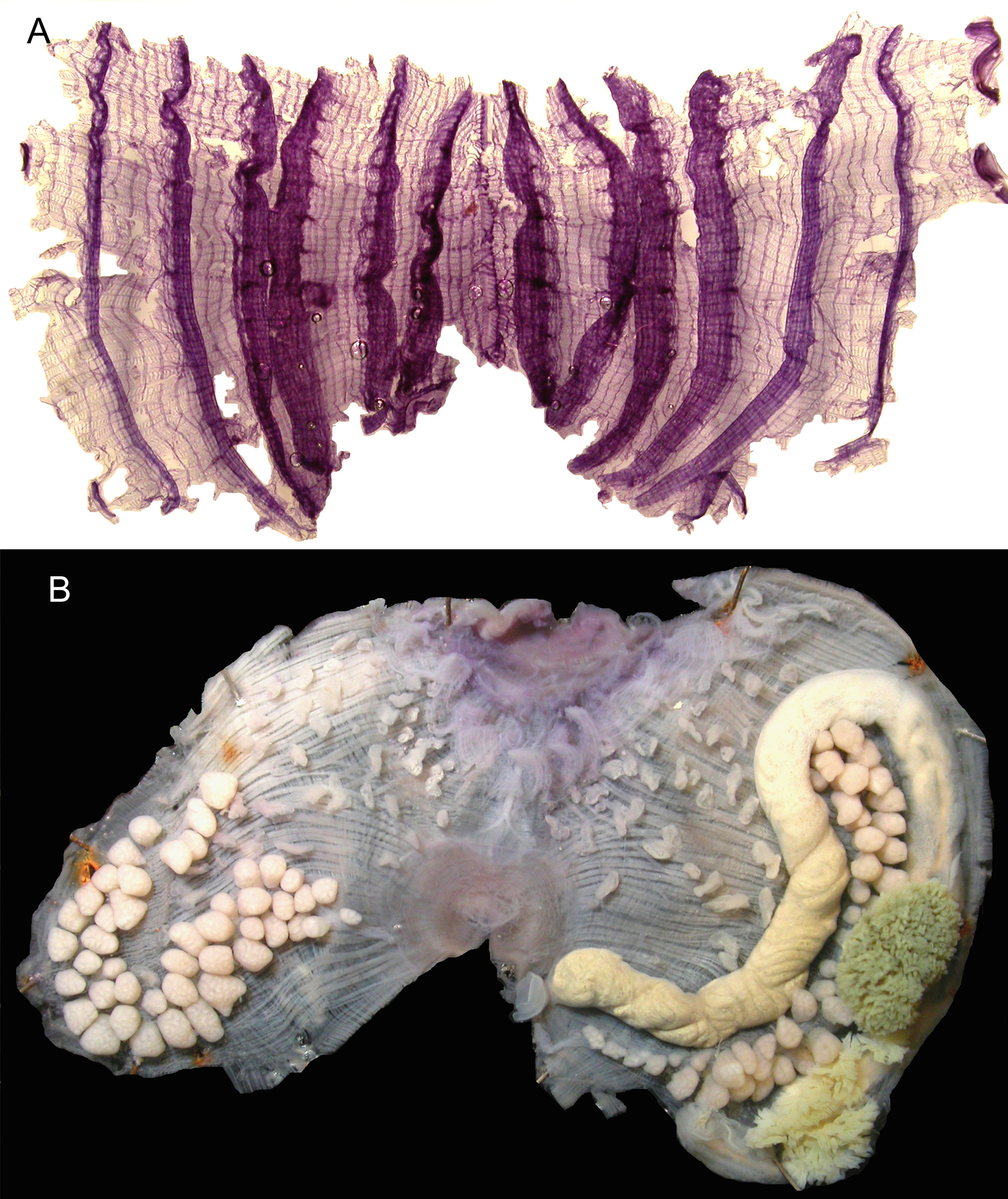
Among the 9 specimens collected from Guiana the largest reaches 2cm in length ( Fig. 27 View FIGURE 27 A). They were attached posteriorly and lacked any epibionts. They are more or less faded in formalin but some red pigment remains at least on the siphons ( Fig. 27 View FIGURE 27 A,B) and more intense on more recent collected Guadeloupe specimens. The most remarkable is the structure of the surface of the tunic carved with round irregular scale-like flat compartments ( Fig. 27 View FIGURE 27 C), some of them with a central brown point. The edge of the siphons bears very small spinules wich are absent from the internal lining, and have a shape which is not the common one found in the genus Pyura ( Fig. 27 View FIGURE 27 E,F). The tunic is thin nacreous internally. The oral siphon is apical, the atrial at 1/3 or ½ of the body length. Devoid of tunic the body is colourless except the red siphons ( Fig. 27 View FIGURE 27 B). The musculature is a continuous sheet of dense but thin fibres issued from the siphons and crossing on the body sides ( Figs 27 View FIGURE 27 D, 28B). There is a short oral velum. About 16 long oral tentacles are intercalated with smaller ones, they are several times ramified. The pre-pharyngeal band is not deeply indented. The dorsal tubercle opens anteriorly in a C. The dorsal lamina is made of thin long languets. Six branchial folds are well spaced on each side ( Fig. 28 View FIGURE 28 A), the most ventral on each side narrower than the rest. There are of 2 to 4 stigmata per mesh on average between the folds. Parastigmatic vessels are present everywhere. Formulae in 2 specimens are:
RE-2(13)4(13)3(17)3(17)3(16)3(18)3-DL-3(18)2(17)3(19)3(17)4(14)4(10)4-LE
RE-2-(10)3(16)3(19)3(19)3(18)2(16)3-DL
The gut forms a long loop reaching the anterior part of the body ( Fig. 28 View FIGURE 28 B). The oesophagus is wide, the stomach the same width but long, followed by an isodiametric intestine curved at the end ( Figs 27 View FIGURE 27 D, 28B). The anus ends in a curious funnel-shaped anus ( Figs 27 View FIGURE 27 D,28B). The hepatic gland consists of 2 main parts; one of a bush of pale papillae on the oesophagus, few more or less isolated papillae on the cardiac part of the stomach and a large cauliflower mass of green papillae on the stomach ( Fig. 28 View FIGURE 28 B). There is one gonad on each side made of a tubular ovary attached to the body wall, fringed on each side with numerous testis lobes(for example: 46 on the right and 44 on the left) ( Fig. 28 View FIGURE 28 B). The left gonad occupies the whole length of the gut loop ( Figs 27 View FIGURE 27 D,28B). The right gonad draws a deep loop along the endostyle ( Fig. 28 View FIGURE 28 B). Male and female ducts are joined in a long papilla. ( Fig. 28 View FIGURE 28 B). Small endocarps are at the top of the testis lobes and the majority of the endocarps are distributed on a large part of the body wall on both sides, dorsally to the gut loop on the left and to the gonad on the right ( Figs 27 View FIGURE 27 D,28B). There are no endocarps on or inside the gut loop. The atrial velum is a large membrane but without thread-like processes.
Pyura ocellata n. sp. differs from all Puyra species of the western Atlantic Ocean in having 6 branchial folds, the presence of numerous endocarps on the body wall but no endocarps along the intestine, minute spinules on the internal siphonal lining, and a peculiar design of the tunic surface.

No known copyright restrictions apply. See Agosti, D., Egloff, W., 2009. Taxonomic information exchange and copyright: the Plazi approach. BMC Research Notes 2009, 2:53 for further explanation.
|
Kingdom |
|
|
Phylum |
|
|
Class |
|
|
Order |
|
|
Family |
|
|
Genus |
Styela atlantica ( Van Name, 1912 )
| Monniot, Françoise 2016 |
Styela eurygaster
| Millar 1977 |
Styela glans
| Herdman 1881 |
S. partita (
| Stimpson 1852 |
S. canopus (
| Savigny 1816 |