
Sphecotypus niger ( Perty, 1833 )
|
publication ID |
https://doi.org/ 10.11646/zootaxa.3814.1.10 |
|
publication LSID |
lsid:zoobank.org:pub:164387ED-CF6F-4AC2-97D5-E682BE1EB2FE |
|
DOI |
https://doi.org/10.5281/zenodo.6123378 |
|
persistent identifier |
https://treatment.plazi.org/id/03DCD762-EC45-2432-CCEC-C0A6FD0EFAE2 |
|
treatment provided by |
Plazi |
|
scientific name |
Sphecotypus niger ( Perty, 1833 ) |
| status |
|
Sphecotypus niger ( Perty, 1833) View in CoL
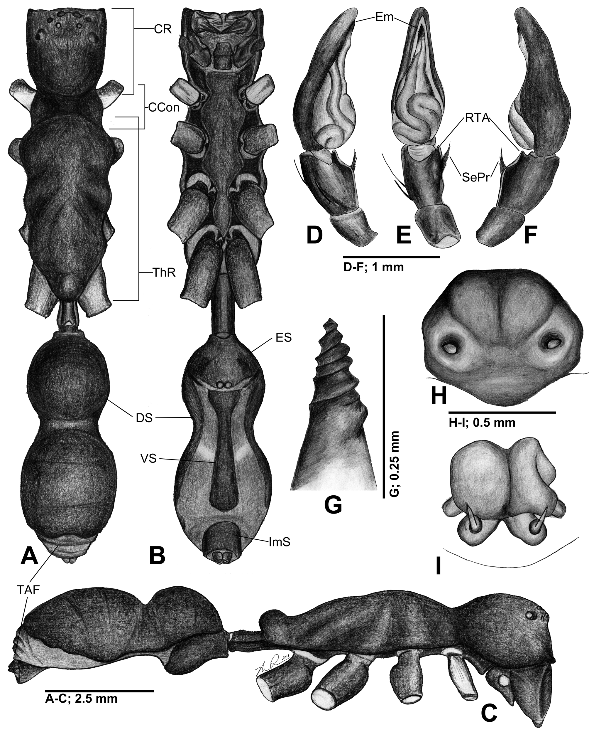
Figure 1 View FIGURE 1 A–I
Myrmecia nigra Perty, 1833: 199 View in CoL , pl. 39, fig. 9; C. L. Koch, 1841: 15, fig. 701; Keyserling, 1891: 80, pl. 2, fig. 40. Sphecotypus formicarius O. Pickard-Cambridge, 1895: 153 , pl. 19, fig. 4.
S. niger, Simon, 1897: 176 View in CoL , figs 168-169, 171; Reiskind, 1969: 282, 232, fig. 290.
Diagnosis. Sphecotypus niger is currently the only member of the genus found in the New World and can be easily distinguished from other members of the genus by: (1) the presence of an abdominal constriction in both sexes, whereas the Asian species lack this constriction, (2) the setal projection on the male pedipalp tibia, and (3) a male embolus that is screw-like in appearance ( Fig. 1 View FIGURE 1 A–I).
Material examined. Nicaragua: Río San Juan: Bartola 10°58.333'N, 84°20.346'W, elev. 36 m 18–23 May 2012, K.B. Miller, M. Leister coll., 1♂ ( MSBA 30573); Región Autónoma del Atlántico Sur: RN Kahka Creek, 12°40.356'N, 83°42.751'W, elev. 40 m, 9 Jun 2011, 2° lowland rainforest, beating vegetation, LLAMA #Go-D- 07-1-01, 1 imm. ( MCZ 125157). Costa Rica: Puntarenas: Carara Reserve (9°47'N, 84°33'W) 7 March 1991, D.M. Olson coll., 1♀ ( MCZ 28119).
Description. Male: body elongate, highly modified ant-like form ( Fig. 1 View FIGURE 1 A–C), legs long, slender; carapace black, elongate, granulose, covered in fine white feathery setae, sparse thin, long, white setae throughout. Eight eyes formed in two recurved rows; PER wider then AER; posterior eyes sub-equal, small, AME largest, roughly twice diameter of PME, ALE small. Carapace with two distinct regions, cephalic region length subequal to width, domed, anterior margin truncate, posteriorly rounded, distinguished from thoracic region by deep constriction; Thoracic region length 2 × width, domed, highest anteriorly, narrowed posteriorly, constricted into three smaller, overlapping, triangular lobes, terminating with small raised dome; thoracic groove absent ( Fig. 1 View FIGURE 1 A–C). Abdomen with dorsum black, elongate, granulose; dorsal sclerite nearly complete, covered in fine white feathery setae, with sparse, long, thin, white setae throughout; abdomen divided into two lobes by strong median constriction, anterior lobe spherical, posterior lobe elliptical, length greater than width; white feathery setae separating dorsal sclerite of posterior lobe into three broad annulations giving segmented appearance; posterior abdomen divided by four transverse folds giving an appearance of segmentation, pseudosegmentation reinforced by patterning of feathery setae ( Fig. 1 View FIGURE 1 A, C); epigastric sclerite length slightly greater than width, protruding from abdomen; ventral sclerite length nearly 2 × width, greatest width anteriorly, posteriorly pointed; inframamillary sclerite length greater than width, enclosing spinnerets ventrally ( Fig. 1 View FIGURE 1 B). Sternum brown-black, elongate, granulose, covered in fine white feathery setae, length approximately 4 × width, anteriorly extending beyond first pair of coxae, tapering posteriorly, extending between coxae, contiguous with precoxal and intercoxal sclerites ( Fig. 1 View FIGURE 1 B). Chelicerae black with straight edges, vertical, with slight concavity on anteromedial surface, covered in long dark setae along anterior surface; with dense setae covering two retromarginal and three promarginal teeth, distal promarginal tooth smallest, medial promarginal tooth largest. Labium black, rectangular, length greater than width, divided with medial transverse suture. Endites black, rectangular, length greater than width ( Fig. 1 View FIGURE 1 B). Coxa I smooth, yellow with brown maculae; coxae II–IV dark brown to black, granulose ( Fig. 1 View FIGURE 1 A–C); trochanters slightly notched; femur I dark brown to black with yellow maculae, tapered distally; patella I dark red-brown; tibia I slender, smooth, distally covered with coarse dark setae; rest of leg I dark red-brown to black; femora II–IV, dark red-brown, granulose, remainder of legs II–IV dark red-brown to black; tibia I ventral spination 3–3. Pedipalp tibia with two spines on lower prolateral surface ( Fig. 1 View FIGURE 1 D, E); RTA small, pointed, continuing as thin lateral sclerotized ridge projecting from middle of tibia, with cluster of stout setae on ridge forming a sharp, pointed setal projection ( Fig. 1 View FIGURE 1 D–F); genital bulb globose, extending as a thick neck ending in a sharp, twisted, sclerotized embolus that is screw-like in appearance ( Fig. 1 View FIGURE 1 G).
Measurements. Based on one male specimen: Body length 13.55. Carapace length 7.20, width 2.25; carapace index 31; cephalic width 2.00; sternum length 4.10, width 1.10; sternum index 26. Pedicel length 1.45, width 0.55. Abdomen length 5.20, width 2.20; abdominal index 42; anterior abdominal lobe length 1.85, width 1.85; posterior abdominal lobe length 3.35, width 2.20. Dorsal sclerite length 4.75, width 2.20. Epigastric sclerite length 1.90, width 1.75. Ventral sclerite length 2.15, width 0.90. Inframamillary sclerite length 0.75, width 1.20. Eyes: AME 0.21; ALE 0.15; PME 0.15; PLE 0.15; AME–AME 0.10; AME–ALE 0.10; ALE–ALE 1.00; ALE–PLE 0.50; PME–PME 0.30; PME–PLE 0.55; PLE–PLE 1.70. Leg formula: IV, I, II, III; Leg I, 12.75 (1.20, 0.40, 2.95, 0.80, 3.10, 2.50, 1.80); Leg II, 10.95 (1.10, 0.40, 2.75, 1.00, 2.30, 2.20, 1.20); Leg III, 10.80 (1.35, 0.35, 2.70, 1.00, 2.10, 2.20, 1.10); Leg IV, 15.15 (1.50, 0.40, 4.20, 1.00, 3.20, 3.55, 1.30); Pedipalp, 3.95 (0.30, 1.15, 0.55, 0.70, 1.25).
Female: body, color, shape, form as in male (Total body length 14.80); dorsal sclerite restricted to anterior abdominal lobe; ventral sclerite absent. Pedipalp dark red-brown to black, smooth, setose, terminating in single smooth tarsal claw. Tibia I ventral spination 3 – 3. Epigynum ventrally with two circular openings facing posteriorly; dorsally with spermathecae asymmetrical, medially contiguous, round, sac-like with short thick posterior necks pointing slightly lateral ( Fig. 1 View FIGURE 1 H, I).
Natural history. A juvenile specimen from Nicaragua was examined that has an overall yellow-orange coloration with dorsal and ventral abdominal patterning of transverse dark bands. Legs are patterned with dark, lateral, longitudinal stripes, and black tarsi. The specimen is smaller (total body length = 4.80) but otherwise nearly identical in shape and form to adult specimens. Based on morphological similarities to adults of S. niger , and a known adult specimen from Nicaragua, this specimen most likely represents an immature S. niger .
Adult S. niger View in CoL are believed to be batesian mimics using the large ponerine ant, Pachycondyla villosa (Fabricius, 1804) View in CoL as the model ( Oliveira 1986, 1988; Cushing 1997). The juvenile has morphological characters that also clearly mimic ants. It shares with the adults the same elongate and constricted carapace and abdomen. The observed patterning of abdominal stripes resembles the abdominal tergites on ants. Evidently, juveniles also mimic ants, but the smaller size and difference in color and pattern suggest a separate, distinct model from the adult, and, therefore, transformational mimicry ( Mathew 1934).
The mimicry of S. niger View in CoL is very convincing. When the first author collected the male from the wall of a building in Nicaragua it was initially mistaken for a large ponerine ant. The pedipalps were held anteriorly against the chelicerae, giving the appearance of ant mandibles, and, while at rest, leg I was held forward and bent at the patellar joint above the carapace, giving the distinct geniculate appearance of ant antennae. The constriction between the cephalic and thoracic region gave the impression of a distinct and separate ant head and thorax. The observed specimen was behaviorally similar to ants, also, as it walked in an erratic fashion with legs I raised and with constant alternating movements similar to antennal movements in ants. Similar observations in the field have been made for this species by others ( Oliveira 1988). When frightened, however, the spider immediately abandoned the behavioral mimicry and utilized all legs for running. Similar behavior has also been observed in another species of Castianeirinae , Myrmecotypus iguazu Rubio & Arbinio, 2009 View in CoL , from Argentina ( Rubio et al. 2013). In Brazil, S. niger View in CoL was seen in close proximity to P. villosa View in CoL , yet avoided direct contact with them ( Oliveira 1988).
| MCZ |
Museum of Comparative Zoology |
No known copyright restrictions apply. See Agosti, D., Egloff, W., 2009. Taxonomic information exchange and copyright: the Plazi approach. BMC Research Notes 2009, 2:53 for further explanation.
|
Kingdom |
|
|
Phylum |
|
|
Class |
|
|
Order |
|
|
Family |
|
|
Genus |
Sphecotypus niger ( Perty, 1833 )
| Leister, Matthew & Miller, Kelly 2014 |
S. niger
| Reiskind 1969: 282 |
| Simon 1897: 176 |
Myrmecia nigra
| Pickard-Cambridge 1895: 153 |
| Keyserling 1891: 80 |
| Koch 1841: 15 |
| Perty 1833: 199 |