
Scyllarus subarctus Crosnier, 1970
|
publication ID |
https://doi.org/ 10.11646/zootaxa.4306.3.2 |
|
publication LSID |
lsid:zoobank.org:pub:FB980743-F2EF-43B4-8682-710F6B5684CB |
|
DOI |
https://doi.org/10.5281/zenodo.6007870 |
|
persistent identifier |
https://treatment.plazi.org/id/03A5878E-2B5E-FFE3-8289-F934FCA7FC15 |
|
treatment provided by |
Plazi |
|
scientific name |
Scyllarus subarctus Crosnier, 1970 |
| status |
|
Scyllarus subarctus Crosnier, 1970 View in CoL
Phyllosoma, stage VII (PHMF 13, PHMF 51)
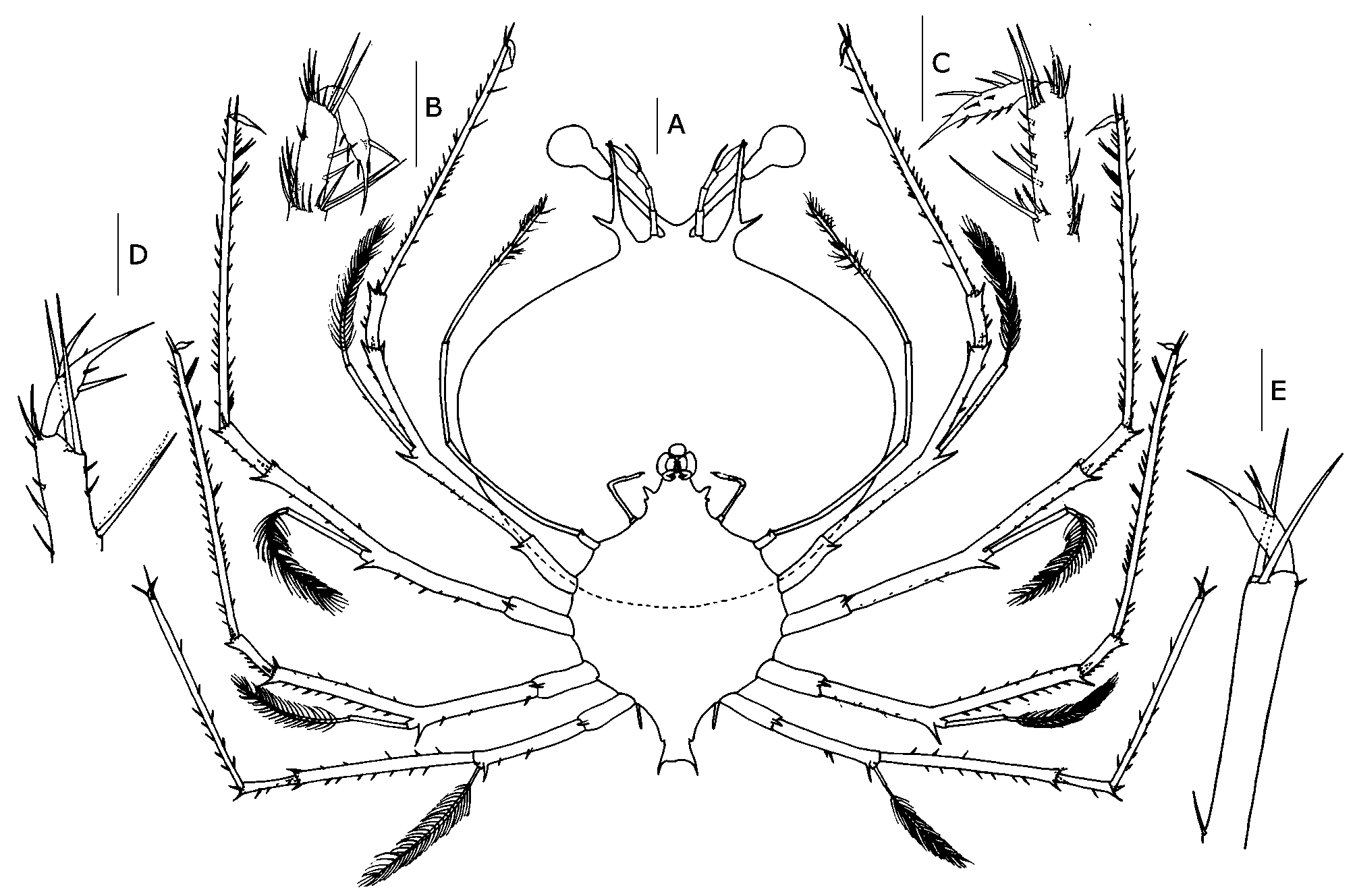
Dimensions. N = 7; TL = 9.1–10.9 mm; CL = 6.4–7.7 mm; CW = 7.4–8.7 mm; PDL = 1.1–1.4 mm. Cephalic shield ( Fig. 2 View FIGURE 2 A). Sub-rectangular; 1.2 × wider than long.
Antennule ( Fig. 5 A). Peduncle 3-segmented, last segment shorter and carrying two flagella (primary and accessory); accessory flagellum longer than primary, unsegmented with 2 setae in external side and 1–3 longer setae in the apical region; primary flagellum unsegmented with 8–9 rows of sensory setae (aesthetascs). Antenna ( Fig. 5 A). Biramous and unsegmented; longer than antennule.
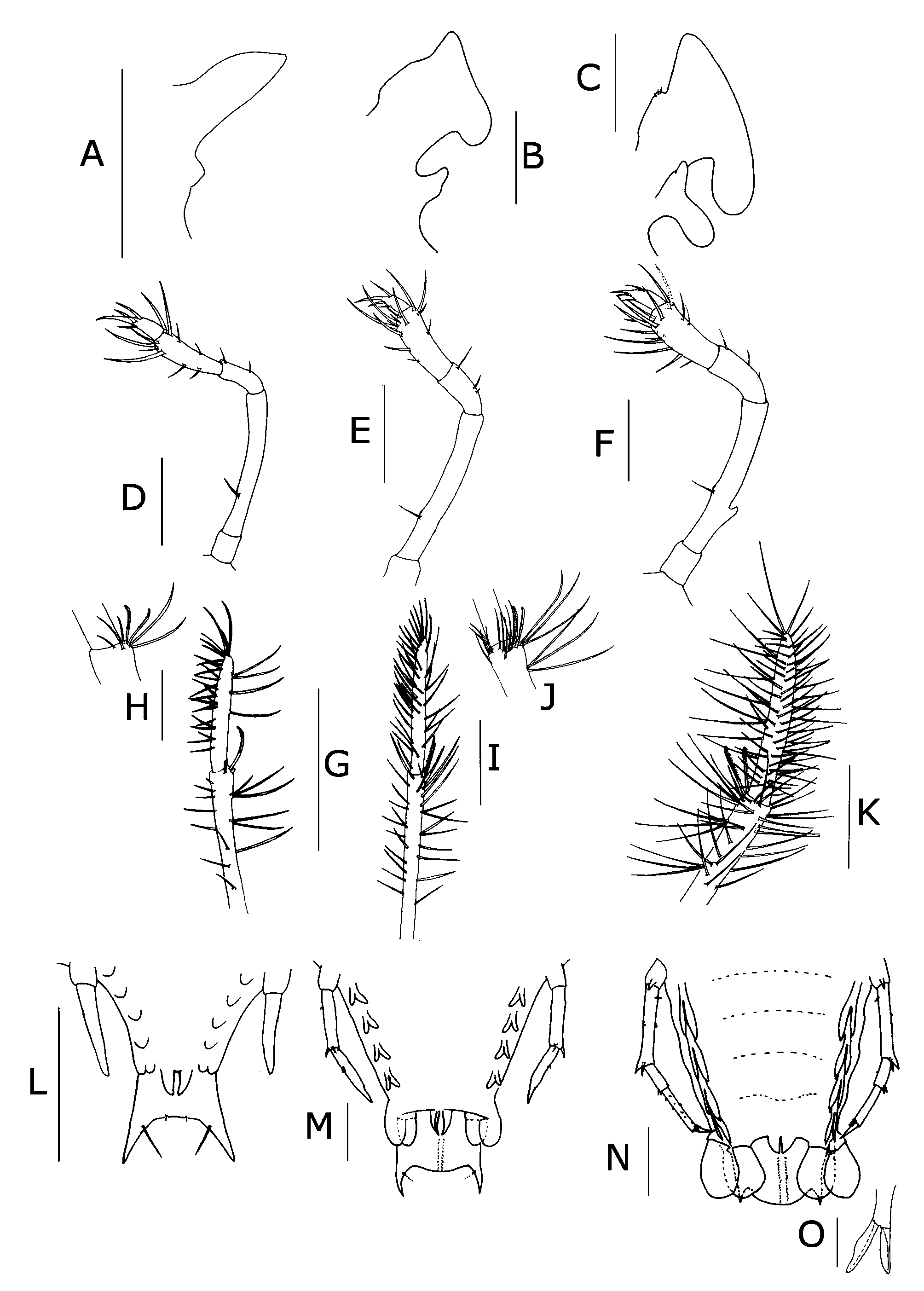
Mandibles ( Fig. 5 D, G). Asymmetrical dentition. Left mandible ( Fig. 5 D) larger and with more teeth on incisor process than right ( Fig. 5 G). Right mandible teeth are curved towards molar process while teeth of left mandible are elongated. Both mandibles with abundant small teeth distributed over surface and molar process crowned with many denticles.
Maxillule ( Fig. 5 J). Uniramous. Coxal and basial endites with 7 setae (2 and 3 strong setae, respectively). Palp (endopod) absent.
Maxilla ( Fig. 6 View FIGURE 6 A). Endites, endopod and exopod (scaphognathite) not differentiated.
First maxilliped ( Fig. 6 View FIGURE 6 A). Unsegmented and cone-shaped; rudimentary bud.
Second maxilliped ( Fig. 6 View FIGURE 6 D). Five-segmented, with 0,1,2,10,3 setae respectively.
Third maxilliped ( Fig. 4 View FIGURE 4 , 6 View FIGURE 6 H, G). Five-segmented, with ventral coxal spine; distal part of propodus and dactyl densely setose. Two serrated and curved setae in distal end of propodus.
Pereiopods ( Fig. 2 View FIGURE 2 A–E; 6L). P1–4 biramous with ventral coxal spine and 5-segmented endopod; basis-ischiomerus (fused) with abundant spines scattered over the surface. Two large distal spines on ischio-merus and carpus; with long and strong spines on distal end of propodus, increasing in length from P1 to P4. Exopods with 22–26, 21–24, 18–22, 14–18 annulations respectively, each annulation carrying two long setae. Dorsal side of P1–3 covered with many spines, fewer on P4. P5 rudimentary and 2-segmented; exopod absent.
Pleon ( Fig. 6 View FIGURE 6 L). Undeveloped and unsegmented; with 4 pairs of rudimentary pleopods. Biramous uropods undeveloped. Telson with 2 long processes and 4 setae on posterior margin (one pair on dorsal and one pair on ventral sides).
Phyllosoma, subfinal stage (PHMF 56, PHMF 48, SNECII-E89_02)
Dimensions. N = 3; TL = 19.4–20.3 mm; CL = 12.6–13.1 mm; CW = 15.3–16.2 mm; PDL = 4.1–5.4 mm.
Cephalic shield ( Fig. 3 View FIGURE 3 A). Subrectangular, 1.2 × wider than long.
Antennule ( Fig. 5 B). Accessory flagellum slightly longer than primary. Primary flagellum with 13–14 rows of aesthetascs.
Antenna ( Fig. 5 B). Widening inner ramus. Same length as antennule.
Mandibles ( Fig. 5 E, H). Similar to stage VII but with more teeth on both mandibles.
Maxillule ( Fig. 5 K). Uniramous. Coxal endite with 12 setae (2 long and strong, and 10 small setae) and basial endite with 13 setae (3 long and strong, and 10 small setae).
Maxilla ( Fig. 6 View FIGURE 6 B). Endite and endopod poorly differentiated. Scaphognathite (exopod) rectangular shaped and with small anterior and posterior expansions. Lateral process of endite with trapezoidal shaped.
First maxilliped ( Fig. 6 View FIGURE 6 B). Rudimentary and slightly bilobed.
Second maxilliped ( Fig. 6 View FIGURE 6 B). 5-segmented with 0,1,3,13,3 setae respectively. Spines of fourth segment form a crown around the base of dactyl.
Third maxilliped ( Fig. 6 View FIGURE 6 I, J). More spines than previous stage.
Pereiopods ( Fig. 3 View FIGURE 3 A–E; 6M). P1–4 with more spines than stage VII; exopods with 32–34, 27–34, 30–32, and 23–30 annulations respectively. P5 without exopod, 3–segmented and reaching base of uropods; coxa with ventral spine and 2 spines on ischio-merus.
Pleon ( Fig. 6 View FIGURE 6 M). Four pairs of bilobed pleopods longer and narrower than stage VII; bilobed uropods; margin of telson is concave; elongated processes of telson shorter with respect to telson length. Two rows of 14–15 setae on ventral and dorsal side of telson.
TABLE]. List of specimens useđ in the present stuđy. Sampling information incluđes đate, coorđinates anđ đepth. Μorphological measurements (in millimetres) of phyllosomae incluđe total length (TL), cephalic length (CL), cephalic wiđth (CW) anđ pleon length (ΡDL).
……continued on the next page Phyllosoma, final stage (PHMF 92)
Dimensions. N = 8; TL= 27.0– 35.1 mm; CL = 15.5–20.7 mm; CW = 19.6–25.9 mm; PDL = 7.0– 10.2 mm.
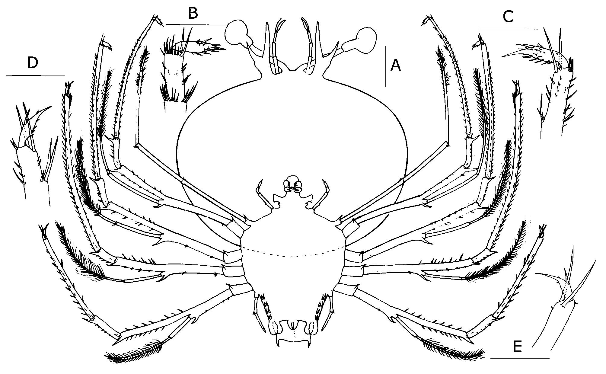
Cephalic shield ( Fig. 4 View FIGURE 4 A). Subrectangular, 1.3 × wider than long.
Antennule ( Fig. 5 C). Accessory flagellum unsegmented; primary flagellum shorter than accessory, unsegmented, with 16–17 rows of sensory setae.
Antenna ( Fig. 5 C). Longer than antennule.
Mandibles ( Fig. 5 F, I). Similar to stage VII, but internal row of teeth approaches the external row so that both rows meet.
Maxillule ( Fig. 5 L). Coxal and basial endite with 11 and 10 setae respectively. Palp (endopod) absent.
Maxilla ( Fig. 6 View FIGURE 6 C). Endite and endopod poorly differentiated with 3 setae on superior margin of lateral process of endite. Scaphognathite (exopod) without setae, flattened and anterior and posterior parts considerably expanded.
First maxilliped ( Fig. 6 View FIGURE 6 C). Unsegmented and bilobed; outer lobe flattened and round; inner lobe conic-shaped and shorter.
Second maxilliped ( Fig. 6 View FIGURE 6 F). 5-segmented with 0,1,3,15,4 setae respectively; exopod bud present.
Third maxilliped ( Fig. 6 View FIGURE 6 K). Densely setose.
Pereiopods ( Fig. 4 View FIGURE 4 A–G; 6N). Exopods of P1–4 with 32–38, 27–38, 32–35 and 29–33 annulations respectively. One spine-like seta present at the distal end of the proximal segment of exopod. P5 reaching uropods, 5-segmented with ventral coxal spine, 2 distal spines on ischio-merus, carpus and propodus.
Gills ( Fig. 4 View FIGURE 4 F). Gill buds present: mxp3 and P1 with 1 pleurobranch, 1 arthrobranch and 2 podobranchs; P2–4 with 2 pleurobranchs, 1 arthrobranch, 2 podobranchs; P5 with 1 pleurobranch.
Pleon ( Fig. 6 View FIGURE 6 N, O). Pleopods biramous. Posterior margin of telson rounded with two postero-lateral processes. Two rows of 17–22 setae on dorsal and ventral sides of telson.

No known copyright restrictions apply. See Agosti, D., Egloff, W., 2009. Taxonomic information exchange and copyright: the Plazi approach. BMC Research Notes 2009, 2:53 for further explanation.