
Rochinia granulosa, Ng, Peter K. L. & Forges, Bertrand Richer De, 2013
|
publication ID |
https://doi.org/ 10.11646/zootaxa.3718.4.5 |
|
publication LSID |
lsid:zoobank.org:pub:50BBC8C0-C684-4738-8E0D-795805CC0BEF |
|
DOI |
https://doi.org/10.5281/zenodo.6148800 |
|
persistent identifier |
https://treatment.plazi.org/id/038A87DF-2B61-322C-FF05-FB6EFB1CF1DD |
|
treatment provided by |
Plazi |
|
scientific name |
Rochinia granulosa |
| status |
sp. nov. |
Rochinia granulosa View in CoL n. sp.
( Figs. 3B View FIGURE 3. A, E ?D, F, 4E, F)
Material examined. Holotype: female (9.7 × 6.7 mm) (MNHN-IU- 2011-2944 a), stn DW 3754, Papua New Guinea, near Bougainville Island, 615– 532 m, coll. BIOPAPUA, RV Alis, 13 October 2010. Paratypes: 1 male (6.4 × 4.7 mm) (MNHN-IU- 2011-2944 b), 1 female (9.9 × 7.0 mm) (ZRC), same data as holotype.
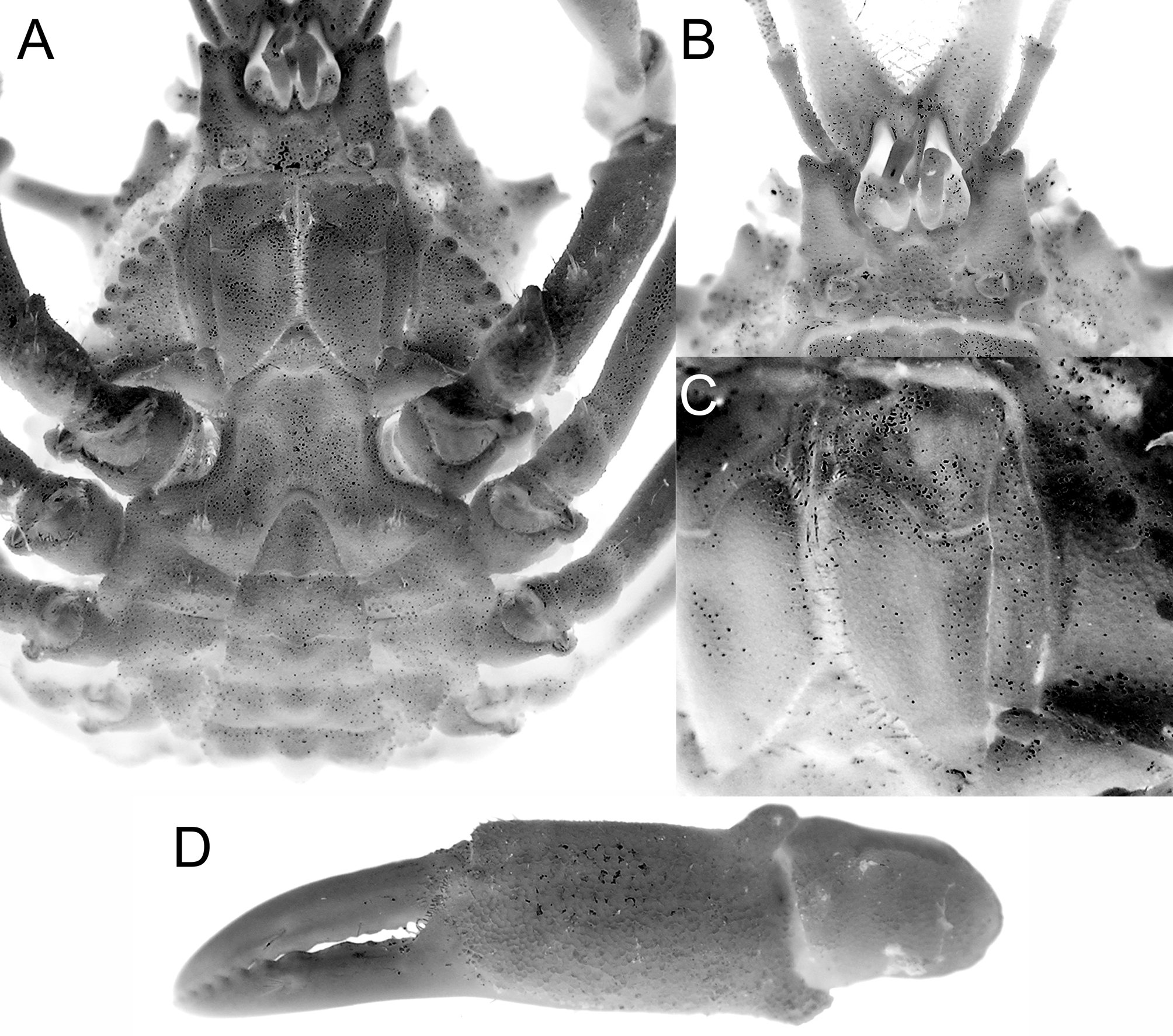
Diagnosis. Dorsal surface of carapace with relatively large, rounded granules, forming groups on gastric, cardiac areas ( Fig. 3B, C View FIGURE 3. A, E ). Pseudorostral spines long, straight, diverging ( Fig. 3B, C View FIGURE 3. A, E ). Ocular peduncules short with rounded cornea ( Fig. 3B, C View FIGURE 3. A, E ). Supraocular eave narrow forming a triangular tooth, directed anteriorly; postocular tooth broadly triangular ( Fig. 3B, C View FIGURE 3. A, E ). Hepatic spine triangular, short, with rounded or relatively blunt tip ( Fig. 3B, C View FIGURE 3. A, E ). Lateral margin with relatively large granules, with prominent lateral spine directed sublaterally ( Fig. 3B, C View FIGURE 3. A, E ). Basal antennal article broad, with distal blunt tooth ( Fig. 3D View FIGURE 3. A, E ). Ambulatory legs, especially meri, relatively short ( Fig. 3B, C View FIGURE 3. A, E ). Anterior male thoracic sternum relatively broad transversely ( Fig. 3D View FIGURE 3. A, E ). Male abdomen triangular ( Fig. 3D View FIGURE 3. A, E ). Female abdomen rounded; surface smooth.
Description. Carapace pyriform; dorsal surface with regions weakly defined, covered by relatively large rounded granules, forming groups on gastric, cardiac areas, without large tubercles or spines; cardiac region convex ( Figs. 3B, C View FIGURE 3. A, E ). Pseudorostral spines long, slender, straight, subcylindrical in cross-section, prominently diverging outwards ( Figs. 3B, C View FIGURE 3. A, E ). Supraocular eave relatively narrow, forming distinct anteriorly directed acutely triangular tooth; postocular tooth relatively short, broadly triangular, outer margin convex or almost straight ( Fig. 3B, C View FIGURE 3. A, E ). Hepatic spine triangular, short, with rounded or blunt tip; directed obliquely outwards ( Fig. 3B, C View FIGURE 3. A, E ). Lateral carapace margin with relatively large granules, lateral branchial spine well developed, directed sublaterally ( Fig. 3B, C View FIGURE 3. A, E ). Intestinal region with 2 rounded tubercles (may be low) just before posterior carapace margin ( Fig. 3B, C View FIGURE 3. A, E ).
Ocular peduncle short; cornea round, pigmented ( Fig. 3B, C View FIGURE 3. A, E ). Suborbital margin confluent with lateral margin of basal antennal article. Basal antennal article broad, with distal blunt tooth; flagellum relatively short, slender, positioned outside orbit ( Fig. 3D View FIGURE 3. A, E ). Antennules folding almost vertically ( Fig. 3D View FIGURE 3. A, E ). Epistome longitudinally narrow; posterior margin crenulated ( Fig. 3D View FIGURE 3. A, E ). Pterygostomial area almost smooth, separated from sub-branchial region by row of 3 or 4 low tubercles ( Fig. 3D View FIGURE 3. A, E ). Third maxillipeds completely covering buccal cavity when closed; surfaces smooth, covered by short pubescence; ischium with distinct median sulcus; merus quadrate, anterolateral margin auriculiform; exopod relatively stout, reaching to middle of merus, with long flagellum ( Fig. 3D View FIGURE 3. A, E ).
Chelipeds subequal, chelae not inflated; merus trigonal in cross-section, with low distal tooth on dorsal margin; carpus short; surface of palm almost smooth; fingers slightly shorter than palm, cutting margins gently serrulated, more prominent along distal half ( Fig. 3B View FIGURE 3. A, E ?D). Ambulatory legs relatively short; first leg longest, fourth leg shortest; surfaces with scattered short setae; meri short, unarmed; carpus short with lateral sulcus; dactylus tapering, surface with short setae except for corneous tip ( Fig. 3B, C View FIGURE 3. A, E ).
Anterior male thoracic sternum relatively broad transversely ( Fig. 3D View FIGURE 3. A, E ); surface almost glabrous. Sternites 1, 2 completely fused, forming small triangular plate, separated from sternite 3 by distinct suture; sternite 3, 4 completely fused except for lateral clefts; sternite 3 medially almost flat, lateral margins concave ( Figs. 3D View FIGURE 3. A, E ).
Abdomen triangular, with 6 free somites, telson; somites 3, 4 trapezoidal; somites 5, 6 subrectangular, somite 6 slightly longer; telson broadly triangular, longer than somite 6, with gently concave lateral margins ( Figs. 3D View FIGURE 3. A, E ).
Adult G1 not known, juvenile G1 relatively straight, flattened laterally along distal quarter; distal part subtruncate ( Fig. 4 View FIGURE 4 E, F). G2 very short.
Female abdomen rounded, dome-shaped, covering entire thoracic sternum; all somites and telson free; outer surfaces smooth. Sterno-abdominal cavity deep; vulva large, not raised, flushed with rest of sternal surface, opening large, subcircular ( Fig. 3 View FIGURE 3. A, E F).
Etymology. The species is named after the large granules covering the carapace.
Remarks. Superficially, Rochinia granulosa n. sp. is closest to Samadinia longispina n. sp. in the general shape, and in the presence of many granules on the dorsal surface of the carapace, which has weakly defined regions. It differs, however, by the following characters: the adult size is relatively small, with females 6.7 mm in carapace width already mature (the single adult female of S. longispina n. sp. is 12.9 × 8.9 mm, MNHN-IU-2013- 1678); the male thoracic sternum is proportionally broader ( Fig. 3D View FIGURE 3. A, E ) (cf. S. longispina n. sp., Fig. 2 View FIGURE 2 A); the carapace and margin are covered by a relatively smaller number of proportionally larger granules ( Fig. 3B, C View FIGURE 3. A, E ) (large number of small rounded granules in S. longispina n. sp., Figs. 1 View FIGURE 1 A, 3A); the pseudorostral spines are relatively shorter, straight and less divergent ( Fig. 3B, C View FIGURE 3. A, E ) than in S. longispina n. sp. ( Figs. 1 View FIGURE 1 A, 3A); the spines on the hepatic and lateral branchial regions are relatively short and blunt ( Fig. 3B, C View FIGURE 3. A, E ) (long and sharp in S. longispina n. sp.: Figs. 1 View FIGURE 1 A, 3A); the ambulatory legs (notably the meri) are relatively shorter ( Fig. 3B, C View FIGURE 3. A, E ) (cf. longer in S. longispina n. sp., Figs. 1 View FIGURE 1 A, 3A); the male abdomen is markedly less broad ( Fig. 3D View FIGURE 3. A, E ) (cf. broader in S. longispina n. sp., Fig. 2 View FIGURE 2 A); and the abdomen of the ovigerous female is rounded and smooth (with a raised median ridge of tubercles in S. longispina n. sp.).
The only male is a pre-adult specimen with the G1 ( Fig. 4 View FIGURE 4 E, F) not yet fully chitinised.


No known copyright restrictions apply. See Agosti, D., Egloff, W., 2009. Taxonomic information exchange and copyright: the Plazi approach. BMC Research Notes 2009, 2:53 for further explanation.