
Ripersiella kelloggi Ehrhorn and Cockerell
|
publication ID |
https://doi.org/ 10.5281/zenodo.5179281 |
|
publication LSID |
lsid:zoobank.org:pub:B7DA7357-19D4-4877-BF9E-04E1B0D2FD7F |
|
persistent identifier |
https://treatment.plazi.org/id/0388645A-A259-9051-558C-AAA6FB3F82E7 |
|
treatment provided by |
Felipe |
|
scientific name |
Ripersiella kelloggi Ehrhorn and Cockerell |
| status |
|
Ripersiella kelloggi Ehrhorn and Cockerell View in CoL
Diagnostic features and descriptions of the adult female of R. kelloggi were given by Hambleton (1946), Ferris (1953), McKenzie (1967), Williams and Granara de Willink (1992), Kosztarab (1996) and Kozár and Konczné Benedicty (2007).
Description. McKenzie (1967) described the insects in life as follows: “According to Ehrhorn (1906), the adult female of this species produces a small quantity of white, cottony secretion which generally encases the body. Its body its creamy-white, and its shape is broadly oval, sometimes pyriform. The mealybug was described as from the roots of ‛bunch grass.’ Ferris (1953) states that specimens he collected were attached to very small rootlets, at some distance from the base of the plant itself.”
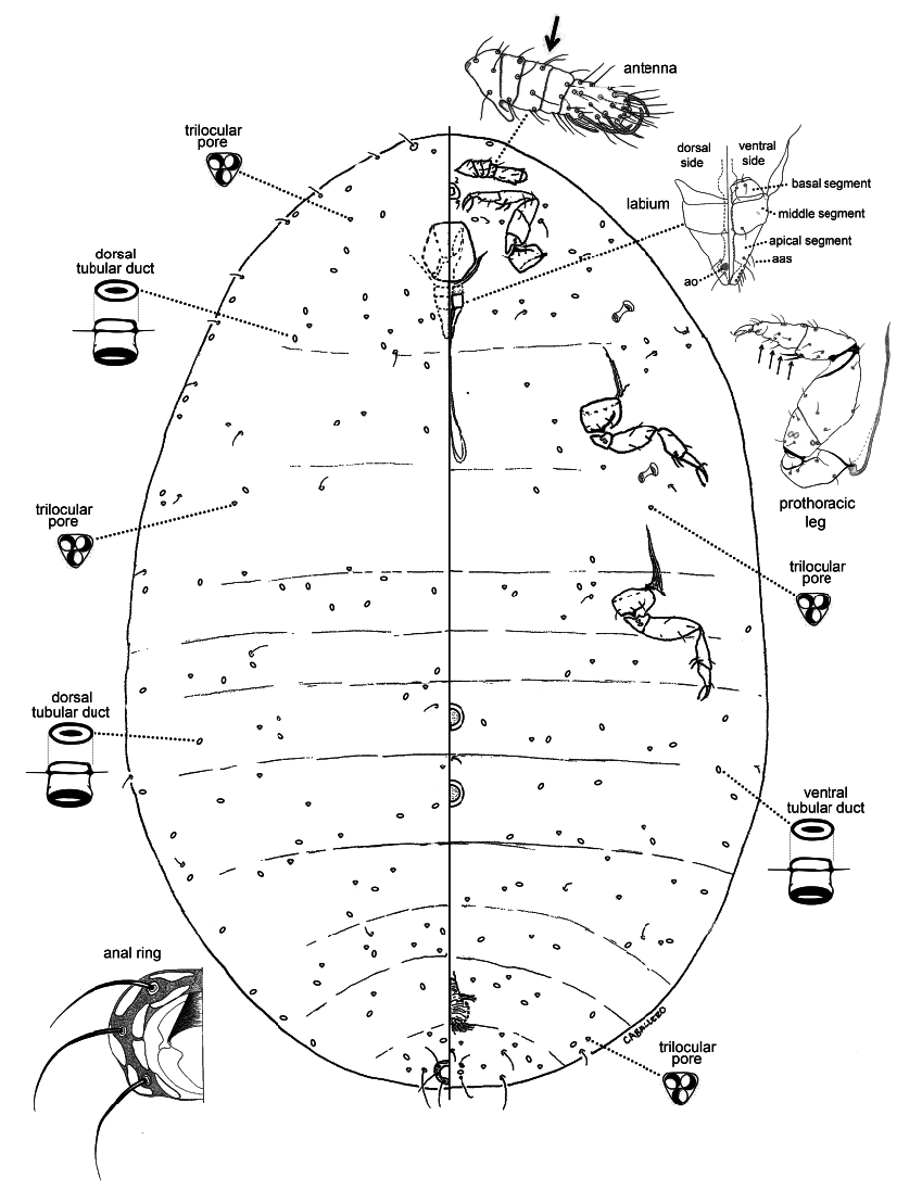
Body of slide-mounted adult female oval to elongate oval, 0.79–1.19 mm long, 0.68–0.86 mm wide.
Dorsum. Dorsal cerarii absent. Trilocular pores present, rather few; each trilocular pore composed of three circular orifices, grouped in the shape of a triangle and almost always of same size. Small dorsal tubular ducts present, each with an elliptical pore opening, rim strongly sclerotized, scarcely longer than wide, with rim slightly protruding from surface of cuticle; ducts abundant, with more than two hundred on both surfaces, evenly distributed. Body setae sparse, with apparently 2 pairs marking position of anal lobes, other setae scattered on dorsum. Anal ring present on dorsal surface, either close to apex or well removed from apex of abdomen; anal ring complete, with 16–20 anal ring pores (each half with 8–10 anal ring pores distributed in 2 rows); with 6 short setae, length of each seta scarcely exceeding diameter of ring.
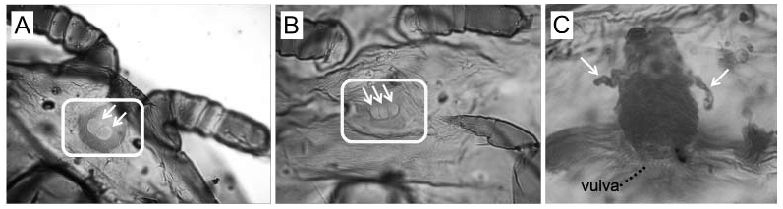
Venter. Trilocular pores similar to those on dorsum, very sparsely distributed on abdomen and submarginally on thorax and head. Tubular ducts similar in size and shape to those on dorsum, sparsely distributed on venter. A small round circulus present medially on each of fourth and fifth abdominal segments. Legs well developed, appearing small in older specimens due to enlargement of body. Prothoracic leg: tibia slightly expanded subapically towards inner margin, with 2 spur-like subapical setae; tarsus slightly shorter than tibia, ratio of length of tibia to tarsus 1.2; with 2 spur-like setae at inner margin, each much shorter than those on tibia (see arrows Fig. 1 View Figure 1 ); claws elongate, curved, sharply pointed, length nearly 4 times width, ratio of length to width 3.7; with 2 knobbed claw digitules, each digitule longer than claw. Mesothoracic leg: tibia bulky, shorter than femur, about 1.5 times longer than wide, with 2 spur-like subapical setae at inner margin; tarsus 1.5 times longer than wide, almost same length as tibia, ratio of length of tibia to tarsus 1.1, with 2 spur-like setae at inner margin, much shorter than those on tibia; claws elongate, curved and sharply pointed, with a pair of knobbed claw digitules, each longer than claw. Metathoracic leg: tibia bulky, length twice its width; with 2 spur-like subapical setae at inner margin; tarsus stout, elongate, almost as long as tibia; with 2 spur-like setae at inner margin, much shorter than those on tibia; claws similar to those of mid leg. See Table 1 for measurements of the leg segments. Cephalic plate subtriangular or subquadrate in shape, 22.4–30.0 μm long, 20.3–30.9 μm wide, present on area between antennal bases and mouthparts ( Fig. 2 View Figure 2 ), with 2 ( Fig. 2A View Figure 2 ) or 3 circular vacuoles ( Fig. 2C View Figure 2 ) present distally, each vacuole 5.0–7.3 μm in diameter. Eight out of 10 specimens had 2 vacuoles, and 2 out 10 had 3 vacuoles. Labium 3-segmented; base of labium appearing membranous, both middle and apical segments slightly sclerotized; length of apical segment almost twice length of middle one; length of middle + apical segment 39.0–40.0 μm long, 35.6–42.7 μm wide at base; apical segment possessing a paired of apical anterior setae (aas) on each side next to apical organ (ao); apical organ oval, wider than long. Antennae each 5- segmented, geniculate, third antennal segment with a single row of 5 flagellate setae (see arrow on Fig. 1 View Figure 1 ).
Size of antennal segments as follows: segment I: 22.3–37.2 μm long, 12.2–23.1 μm wide; segment II: 18.2–25.0 μm long, 11.2–14.2 μm wide; segment III: 20.2–24.5 μm long, 10.4–13.0 μm wide; segment IV: 20.1–24.2 μm long, 7.2–9.6 μm wide; segment V: 18.7–21.8 μm long, 33.3–37.4 μm wide. Spiracles normal. Eyes absent. Female genitalia sclerotized, longer than wide, with 2 well-developed elongate vaginal glands; slightly constricted at insertion of vaginal glands and posteriorly ( Fig. 2C View Figure 2 , see arrows). This is the first description of the female genitalia in R. kelloggi .
Note. The present description of R. kelloggi differs from that of McKenzie (1967) as follows (character states by McKenzie (1967) in parenthesis): (1) dorsal trilocular pores present ( McKenzie (1967) described R. kelloggi as having no trilocular pores on the dorsum, however, his drawing clearly shows trilocular pores on the dorsal surface); (2) dorsal tubular ducts abundant (few in number); (3) dorsal tubular ducts without a median partition (ducts with suggestion of a median partition); (4) anal ring located at dorsal apex or on dorsum well removed from apex (located on dorsum, well removed from apex); (5) anal ring with 18–20 pores (few relatively large anal ring pores); and (6) spiracles normal in size (extremely small). The above differences may be due to differences in interpretation, and whether the observations were made on younger or older individuals. McKenzie (1967) described the circuli as being present on each of segments IV and V, however, this is because he likely followed the interpretation of body segmentation proposed by Ferris (1950), which considered the abdomen as having 10 segments, but the loculi are located on segments III and IV when the current interpretation of mealybug segmentation is followed, e.g., Beardsley (1965), and Williams and Granara de Willink (1992). The syntype studied, although in rather poor condition, matches well our concept of the species. We believe that McKenzie (1967) based his description on old enlarged adult female specimens. Furthermore, there is also the possibility that among the material studied by McKenzie (1967) there were specimens belonging to other closely related species, but this needs to be further studied. The material studied by McKenzie (1967) was from six localities in California and is detailed in his book. The drawing by McKenzie (1967) was based on specimens collected on Stipa sp. from Santa Clara County, California, the same county as the syntype, however, there is no evidence that McKenzie (1967) studied the type material. The material studied by McKenzie (1967) is deposited at the Bohart Museum of Entomology, held at the University of California, Davis. Reviewing the material studied by McKenzie (1967) was out of the scope of this study.
The first author studied a single specimen labeled as type and could not locate other type specimens at the USNM. The original description by Cockerell (1901) is very brief and does not mention details about how many specimens were studied. Hambleton (1946), in his redescription of R. kelloggi (as Radicoccus kelloggi ) referred to the studied type material as “type specimens” in plural, indicating that there should be other slide-mounts. As expected, 2 other syntypes of R. kelloggi were found recently misplaced in the collection, thus there is a total of 3 slides each with an adult female at the USNM (G. Evans, personal communication).
Diagnosis. Ripersiella kelloggi is a rather distinctive species that can be differentiated from other species of Ripersiella known from Colombia by the following combination of features: (1) antenna geniculate, five segmented, third segment rectangular, wider than long, with a single row of five flagellate setae ( Fig. 1 View Figure 1 ), (2) bi- or tri-tubular ducts absent, (3) tubular ducts with an elliptical pore opening, (4) outer and inner rims of tubular ducts strongly sclerotized, and (5) multilocular pores absent. Ripersiella kelloggi has been previously recorded from the Nearctic region, from the United States ( Ben-Dov 1994; Ben-Dov et al. 2014; Cockerell 1901; Ferris 1953; Kosztarab 1996; McKenzie 1960) and Mexico ( Ben-Dov 1994; Williams and Granara de Willink 1992). The current record is the third for the New World and the first one for the Neotropics. We found no records of its economic importance.
No known copyright restrictions apply. See Agosti, D., Egloff, W., 2009. Taxonomic information exchange and copyright: the Plazi approach. BMC Research Notes 2009, 2:53 for further explanation.
|
Kingdom |
|
|
Phylum |
|
|
Class |
|
|
Order |
|
|
Family |
|
|
Genus |