
Rhynchozoon splendens Hayward, 1988
|
publication ID |
https://doi.org/ 10.1080/00222930601062771 |
|
persistent identifier |
https://treatment.plazi.org/id/3C0487C6-FF81-940A-BAC2-C224FB183984 |
|
treatment provided by |
Felipe |
|
scientific name |
Rhynchozoon splendens Hayward, 1988 |
| status |
|
Rhynchozoon splendens Hayward, 1988 View in CoL
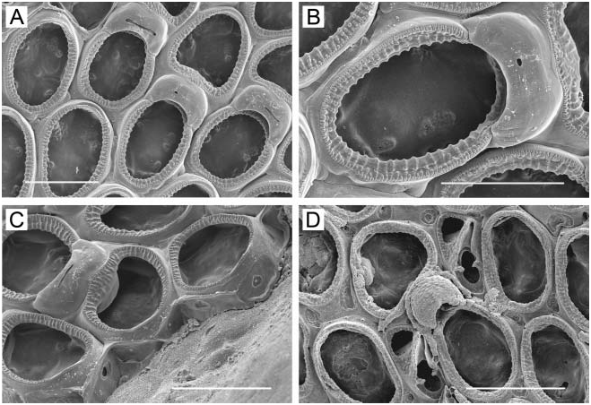
( Figure 15A–C View Figure 15 )
Rhynchozoon splendens Hayward 1988, p 335 View in CoL , Plate 13f, g.
Rhynchozoon splendens: Ryland and Hayward 1992, p 294 View in CoL , Figures 31f, 32a, b; Tilbrook et al. 2001, p 100, Figure 22A, B.
? Rhynchozoon rostratum: Winston and Heimberg 1986, p 38 View in CoL , Figures 95–98.
Measurements
ZL, 0.48–0.70 (0.569¡0.061). ZW, 0.28–0.48 (0.386¡0.065). OrL, 0.09–0.10 (0.098¡0.004). OrW, 0.10–0.11 (0.108¡0.006). OvL, 0.175. OvW, 0.225 (ovicell n 51).
Description
One colony found; encrusting, multilaminar, light tan in colour, covered by shiny ectocyst; growing on a serpulid tube; 9 mm × 15 mm in extent. Zooids distinct only at margin; oval, hexagonal, or irregular in outline; oral spines lacking. Frontal wall ( Figure 15A, C View Figure 15 ) moderately convex in young zooids, rugose, with interareolar buttresses between large areolae. Zooidal boundaries indistinct with age; frontal wall rugose; areolar openings enlarged. Primary orifice ( Figure 15B View Figure 15 ) slightly broader than long, with low, broad, Ushaped sinus demarcated by condylar shelves; condyles rounded, conspicuous; orificial rim with around 19 denticles; becoming deeply immersed in peristome. Secondary orifice ( Figure 15C View Figure 15 ) with up to six processes, usually conical or nodular but sometimes long and acute. Oral avicularium ( Figure 15A View Figure 15 ) large, on a raised chamber, situated immediately proximolateral to orifice; rostrum often angled 60 ° or more from frontal plane of zooid, hooked at tip; mandible long-triangular, directed laterally or distolaterally; with age, oral avicularium immersed in peristome. Many zooids have a frontal avicularium ( Figure 15C View Figure 15 ) pointing in any direction, the mandible equilateral or nearly so. Many of the frontal avicularia have a diamond-shaped rostrum, meaning that the proximal end of the rostrum is acute, as is the mandibular end. Ovicell ( Figure 15A, C View Figure 15 ) completely immersed, opening low in peristome; endooecium showing as granulated, circular tabula in peristome.
Distribution
Broadly distributed throughout the Indo-West Pacific ( Tilbrook et al. 2001). This is the first record for the Hawaiian Islands.
Class STENOLAEMATA Borg, 1926
Order CYCLOSTOMATIDA Busk, 1852
Suborder CERIOPORINA Hagenow, 1851
Family DENSIPORIDAE Borg, 1944
Genus Disporella Gray, 1848
Disporella pristis ( MacGillivray, 1884) View in CoL
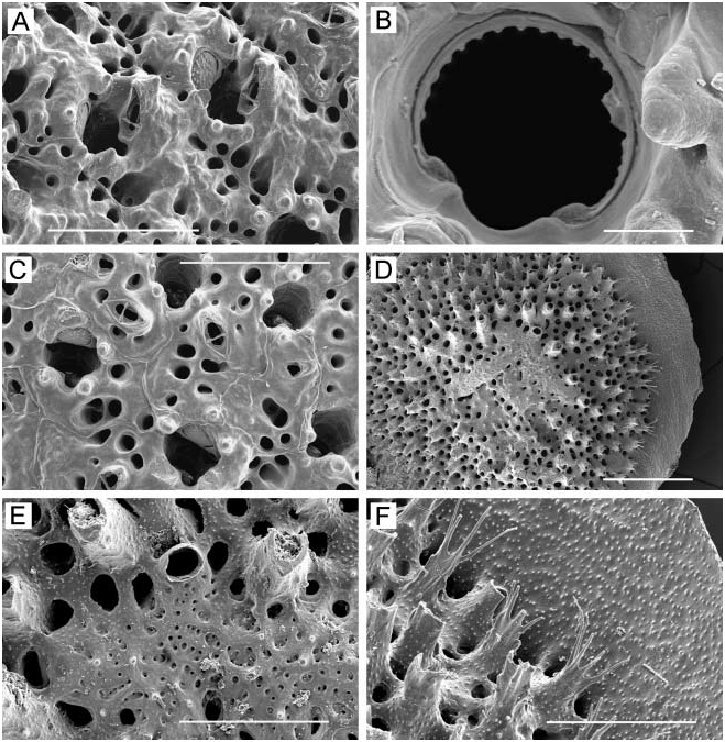
( Figure 15D–F View Figure 15 )
Discoporella pristis MacGillivray 1884, p 126 , Figure 3, 3a, b View Figure 3 .
Disporella pristis: Gordon and Taylor 2001, p 260 View in CoL , Figures 22–30.
Measurements
Diameter of peristome at colony margin 0.05–0.07 (0.060¡0.006); diameter of kenozooidal opening near colony margin 0.04–0.07 (0.056¡0.009).
Description
Based on a single circular colony 3.5 mm in diameter. Colony ( Figure 15D View Figure 15 ) mound-shaped, adnate, the lamellar margin slightly raised; whitish in colour. Peristomes distinct, not connate, but organized in approximately radial columns with the zooids offset from one column to the next, so that they are quincuncially arranged. Entire surface of colony finely granulated ( Figure 15E, F View Figure 15 ), including the marginal lamella, which shows no radial striae. Peristomes with elevated extension on macular side that is semicircular in transverse section and in marginal zooids is often prolonged at end into as many as six long, sharp processes ( Figure 15F View Figure 15 ). Interior walls of kenozooids with sparse, small, sharp denticles. Centre of colony occupied by a brood chamber ( Figure 15D View Figure 15 ) that is about one-third of colony diameter in extent, consisting of two elongate, irregular lobes; surface of chamber irregularly porous, in the process of becoming thickened with reticulate calcification. One oeciostome present ( Figure 15E View Figure 15 ), located at abmacular edge of brood chamber, the opening oval, raised on a low peristome; internal surface of oeciostome covered with minute spines.
Remarks
Our specimen agrees well with the description by Gordon and Taylor (2001) of small, neanic colonies of this species. According to these authors, colonies can become quite large, up to 6 cm in extent, and with more than a hundred maculae.
Distribution
Broadly distributed in the Indo-West Pacific from Japan through Indonesia, New Zealand, the Great Barrier Reef, southern Australia, eastern and southern Africa (referenced in synonymies by Gordon and Taylor 2001). This is the first record for the Hawaiian Islands.
No known copyright restrictions apply. See Agosti, D., Egloff, W., 2009. Taxonomic information exchange and copyright: the Plazi approach. BMC Research Notes 2009, 2:53 for further explanation.
|
Kingdom |
|
|
Phylum |
|
|
Class |
|
|
Order |
|
|
Family |
|
|
Genus |
Rhynchozoon splendens Hayward, 1988
| Dick, Matthew H., Tilbrook, Kevin J. & Mawatari, Shunsuke F. 2006 |
Disporella pristis: Gordon and Taylor 2001 , p 260
| Gordon DP & Taylor PD 2001: 260 |
Rhynchozoon splendens:
| Tilbrook KJ & Hayward PJ & Gordon DP 2001: 100 |
| Ryland JS & Hayward PJ 1992: 294 |
Rhynchozoon splendens
| Hayward PJ 1988: 335 |
Rhynchozoon rostratum:
| Winston JE & Heimberg BF 1986: 38 |
Discoporella pristis
| MacGillivray PH 1884: 126 |