
Rhyacophila fasciata delici Kučinić & Valladolid, 2020
|
publication ID |
https://doi.org/ 10.11646/zootaxa.4885.1.3 |
|
publication LSID |
lsid:zoobank.org:pub:6C730E09-02B0-44CA-915D-A1311461FE02 |
|
DOI |
https://doi.org/10.5281/zenodo.4324293 |
|
persistent identifier |
https://treatment.plazi.org/id/03CA8D07-F91F-7762-FF1B-AEC0ED8CF87E |
|
treatment provided by |
Plazi |
|
scientific name |
Rhyacophila fasciata delici Kučinić & Valladolid |
| status |
|
Rhyacophila fasciata delici Kučinić & Valladolid (ssp. nov.)
This subspecies has been found widely distributed in Croatia and it is also present in Bosnia and Herzegovina.
Etymology. The specific name is the genitive singular of Delić, given in honour of Dr. Antun Delić, naturalist and biologist, retired professor of the Teachers Faculty, University of Zagreb.
Type material. Holotype ♂: CROATIA, Roški slap (Roški waterfall), National Park “Krka” (43.90625º N 15.975º E, 80 m a.s.l.), 10/v/19 (M. Kučinić & A. Delić) [ R1 , no. 4444 ( TCK)]. GoogleMaps
Paratypes: 1 ♀, same locality as holotype, 19/v/19, (M. Kučinić & A. Delić) [ R2 , no. 4445 ( TCK)]. GoogleMaps 2 ♂, spring of the river Čabranka, 21/vi/15 (D. Cerjanec) [C1–C2, nos. 4446–4447 ( MKC)] GoogleMaps 2 ♂, same locality, 05/v/15 (D. Cerjanec) [C3, no. 4448 ( MKC), C4 , no. 1110 ( UMBH)]. GoogleMaps 1 ♂, spring of river Bijela rijeka, National Park “Plitvice Lakes”, 16/iii/18 (I. Sivec, M. Kučinić & S. Žalac) [BI1, no. 4449 ( MKC)]. GoogleMaps 1 ♂, Brkljača channel, Udovičić, 20/vi/15 (S. Žalac & M. Kučinić) [BR 1, no. 4450 ( MKC)] GoogleMaps . 1 ♀, tributary of river Cetina, Civljani , 07/vi/15 (M. Kučinić & A. Delić) [CT1, no. 4451 ( MKC)]. 1 ♂ and 1 ♀, river Butišnica, Golubić , 06/viii/15 (A. Delić & A. Ćukušić) [BT1–BT2, nos. 4452–4453 ( MKC)] . 1 ♀, Brušane, Gospić, Ličko-Senjska county, Road 25, 10-15/vii/16 (G. Galli) [BU1, ID Data number of Access Catalog 33789 ( MCSN)]. 1 ♂ and 1 ♀, spring of the river Dretulja , 09/ix/14 (D. Cerjanec & M. Kučinić) [D1–D2, MNCN _ Ent 269370, MNCN _ Ent 269371 ( MNCN)]. 1 ♂ and 1 ♀, Vukovića spring, river Cetina, 09/v/17 (A. Delić & M. Kučinić) [ V1 – V2 , MNCN _ Ent 269372, MNCN _ Ent 269373, ( MNCN)]. 2 ♂, middle part of river Gacka , Otočac, 11/v/15 (D. Cerjanec & M. Kučinić) [G1–G2, MNCN _ Ent 269374, MNCN _ Ent 269375, ( MNCN)] .
Description of the imago. Holotype (R1): Length from front of head to distal edge of segment IX 8.82 mm, each forewing 11.42 mm, each hind wing 9.65 mm.
Males: Length 7.21–9.92 mm (x = 8.61, n = 12), each forewing 10.07–13.09 (x = 11.15, n = 12), each hind wing 8.29–11.17 mm (x = 9.66, n = 12).
Females: Length from front of head to distal edge of segment VIII 8.96–11.61 mm (x = 10.69, n = 6), each forewing 10.48–14.15 mm (x = 12.28, n = 6), each hind wing 9.77–12.7 mm (x = 10.93, n = 6).
In ethanol-preserved specimens colour generally pale brown and yellowish, with golden brown setae. Head, thorax, and abdomen dorsally pale brown, with small black and irregular darker brown spots in dorsal area, abdomen ventrally yellowish; females generally darker than males, anterior 2/3 of abdominal segments pale brown or brown in darker specimens. Legs light, with spurs reddish brown. Forewings brown, spotted lighter than background; hind wings pale.
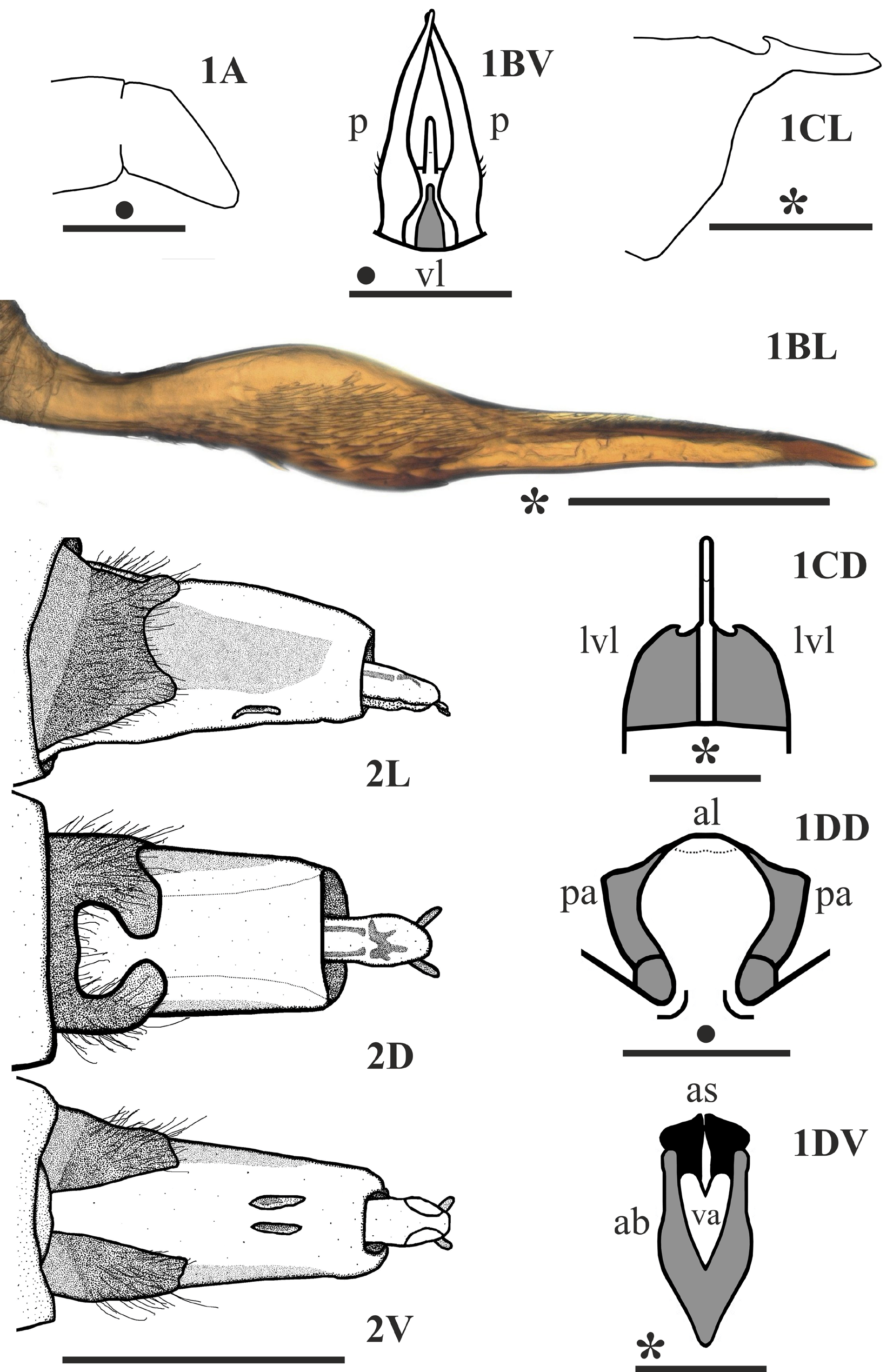
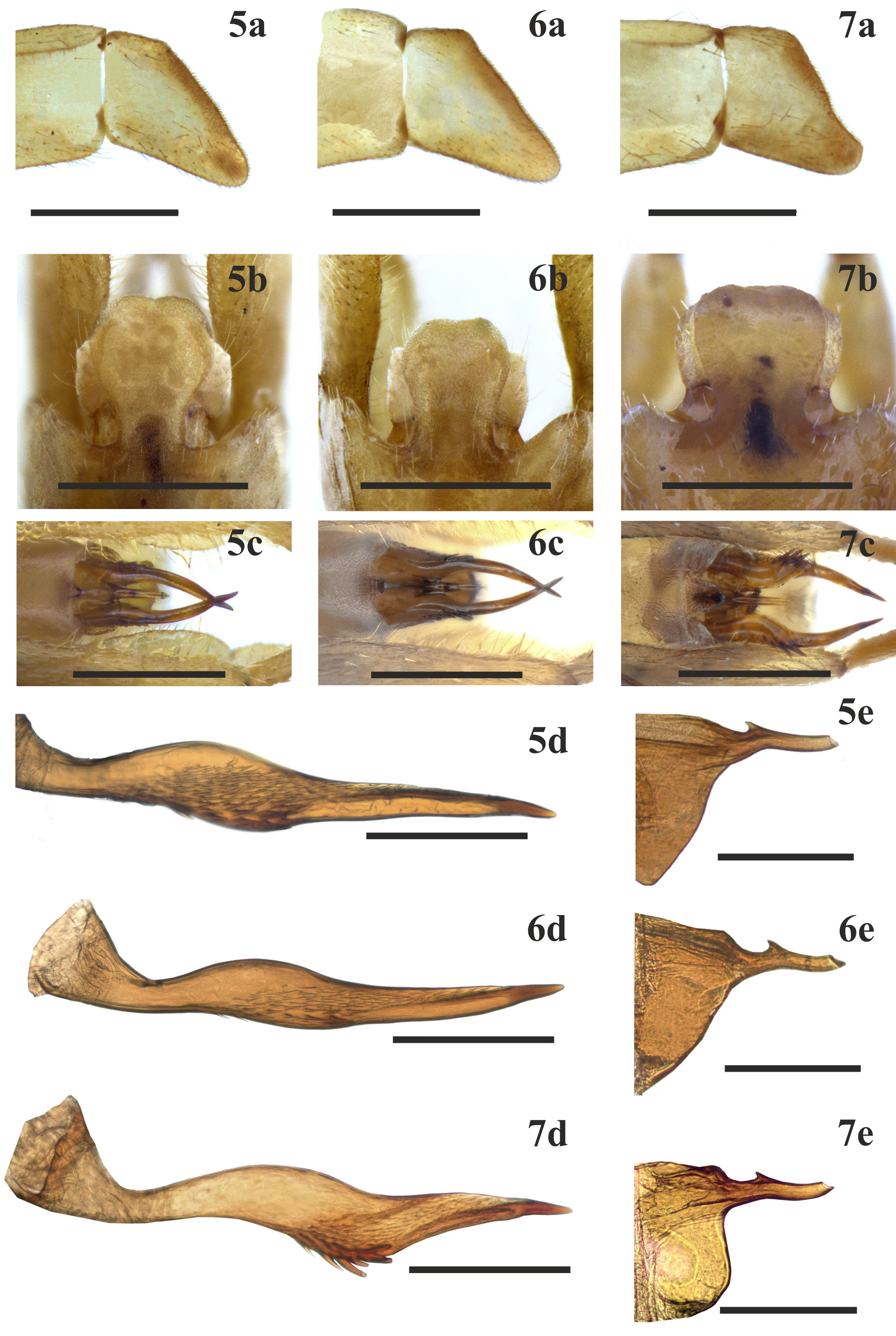
Male genitalia ( Figs 1 View FIGURES 1, 2 , 5 View FIGURES 5–7 a–5e): Apical segment of each inferior appendage ( Figs 1A View FIGURES 1, 2 , 5a View FIGURES 5–7 ) with basal and distal edges diverging, posterior edge oblique straight or slightly convex, ventral edge slightly concave, at least two times longer that dorsal edge. Apicodorsal vertex slightly angular, apicoventral angle projecting as thick lobe narrowing progressively to round apex ( Fig 5a View FIGURES 5–7 ).
Parameres ( Figs 1 View FIGURES 1, 2 BV–1BL, 5c, 5d) in ventral view curved posteromesad in apical half ( Figs 1 View FIGURES 1, 2 BV p, 5c). In lateral view ( Figs 1 View FIGURES 1, 2 BL, 5d) each slender at base, dilated in middle, with almost parallel dorsal and ventral margins in central area, pointed at apex; few thick spines on midventral margin and posterior midventral area, parallel to surface ( Figs 5c, 5d View FIGURES 5–7 ); midlateral surface almost completely covered by thin spicules or setae, reaching from anteroventral edge to posterodorsal edge of paramere, decreasing in size from ventral to dorsal margin, absent on middorsal edge.
Aedeagus (phallicata) in lateral view ( Figs 1 View FIGURES 1, 2 CL, 5e) with anterodorsal margin straight, then slightly concave with posterior corner of concavity hooked anterad, upper posterior edge slightly concave, then straight caudad, ventral apex round, ventral edge curved upward anteriorly to slight angle below hook. Lateroventral lobes of phallus ( Fig 1 View FIGURES 1, 2 CD lvl) convex, narrowing progressively towards apex, apicolateral margins slightly pointed to inside, posterior edge of each lobe concave. Ventral lobe of aedeagus subtriangular, pointed progressively backwards ( Fig 1 View FIGURES 1, 2 BV vl).
Apicodorsal lobe of segment IX ( Figs 1 View FIGURES 1, 2 DD, 5b) dilated subapicolaterally, nearly circular, ratio of posterior maximum width to anterior width 2:1; preanal appendages ( Fig 1 View FIGURES 1, 2 DD pa) shorter than apicodorsal lobe ( Fig 1 View FIGURES 1, 2 DD al) of segment IX laterally, with concave posterior edges. In ventral view ( Fig 1 View FIGURES 1, 2 DV), apical band V-shaped, inner and outer edges almost parallel in posterior third, rounded apically, longer than wide ( Fig 1 View FIGURES 1, 2 DV ab); posterior edge of non-sclerotized ventral area round, with apicomesal incision ( Fig 1 View FIGURES 1, 2 DV va), anal sclerites triangular ( Fig 1 View FIGURES 1, 2 DV as).
No known copyright restrictions apply. See Agosti, D., Egloff, W., 2009. Taxonomic information exchange and copyright: the Plazi approach. BMC Research Notes 2009, 2:53 for further explanation.
|
Kingdom |
|
|
Phylum |
|
|
Class |
|
|
Order |
|
|
Family |
|
|
Genus |