
Rhopalaea circula, Monniot & Monniot, 2001
|
publication ID |
https://doi.org/ 10.5281/zenodo.5391440 |
|
persistent identifier |
https://treatment.plazi.org/id/F57D87A3-FF81-3158-E815-FCD6FB5E1380 |
|
treatment provided by |
Marcus |
|
scientific name |
Rhopalaea circula |
| status |
sp. nov. |
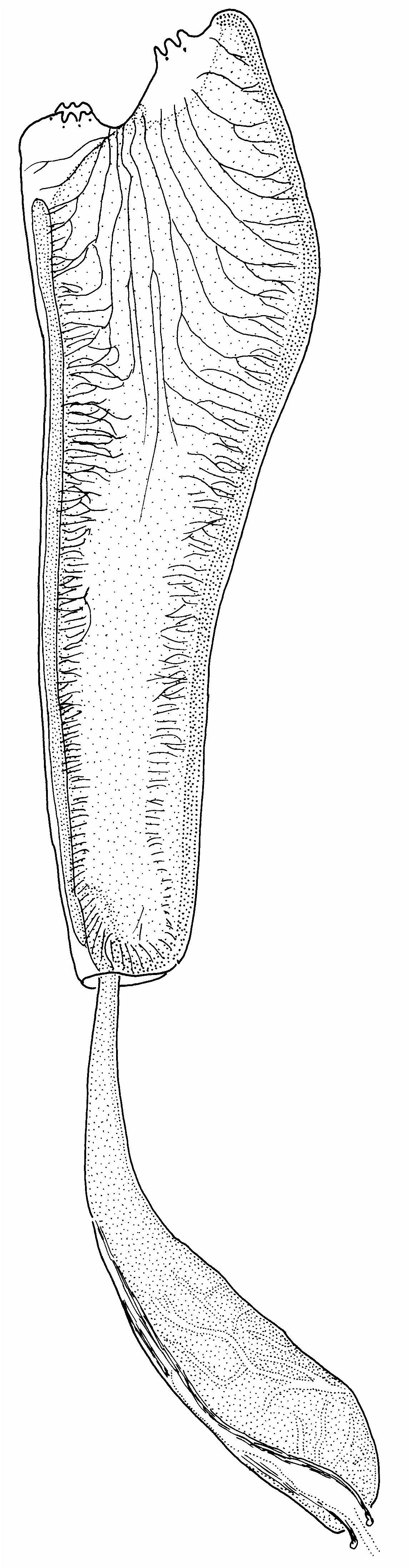
Rhopalaea circula View in CoL n. sp. ( Figs 75 View FIG ; 124C View FIG )
TYPE MATERIAL. — Federated States of Micronesia.
Pohnpei, NE lagoon, Tehpehk Island, bath sponge farm, growing on nylon lines, 6°57.27’N, 158°19.57’E, 8 m, 15.X.1996 ( MNHN P1 RHO.A 23).
ETYMOLOGY. — From the Latin circulus: circle.
OTHER MATERIAL EXAMINED. — Palau. Koror, Ngerikuul Pass, wall, 7°19.22’N, 134°29.24’E, 7 m, 17.VI.1994 (MNHN P1 RHO.A 16). — Koror, Malakal Harbour, rock island near Ngerchaol Island, 7°20.04’N, 134°27.27’E, 16.VII.1993 (Sample: CRRF).
Papua New Guinea. Milne Bay Province, N side of E Cape, Hiliwau, 10°15.65’S, 150°42.75’E, 18 m, 28. V.1998 (Sample: CRRF). — Milne Bay Province, Reef SE of Drawari Island, 10°18.03’S, 151°03.94’E, 16 m, 6. VI.1998 (Sample: CRRF). — Milne Bay Province, Samarai Island, 10°36.98’S, 150°39.77’E,
9 m, 10. VI.1998 (Sample: CRRF).
Mariana Islands. Guam, Apra Harbour, Jade Shoals, 6-18 m, 5. VI.1997, coll. Paulay( MNHN P1 RHO.A 30).
DESCRIPTION
The zooids are in “bushes” united at their base by a mass of tunic containing numerous vascular ampullae ( Fig. 124C View FIG ). A bush is made of large and small zooids. It is possible to separate some of the zooids, but for others the tunic is fused and a connection exists with the vascular ampullae. In life this species is transparent bluish, and with some rings of a deep blue ( Fig. 124C View FIG ) (hence the species name). It is possible to see the musculature through this transparency. All pigments disappear after fixation in formalin. The oral siphon is terminal. The cloacal siphon is smaller and lateral. The six lobes of the siphons are not visible in life. There is a pigment spot between each lobe of both siphons. On the thorax the tunic is thin, soft and naked. Around the abdomen it is hard, often constituted of successive rings, and covered with epibionts. Filamentous algae are partially included into the tunic. The abdomen lies within a fibrous tissue that is difficult to tear; the abdomen is narrow at its upper end, and the base of the long thorax covers it in a circular fold. The oesophagus entrance is located clearly above this fold and not at the posterior extremity of the branchial sac.
The thoracic musculature ( Fig. 75 View FIG ) is made of a few longitudinal bundles, about seven issuing from the oral siphon and six from the intersiphonal space. They are prominent on the anterior part of the thorax and become inconspicuous posteriorly, ramifying and ending perpendicular to the dorsal lamina and medio-ventral line. Some muscles extend onto the abdomen in two bundles on each side of the heart. They terminate abruptly below the posterior abdominal extremity. There are about 40 oral tentacles in three or four orders at increasing distance from a high crest, the largest of them making basal ribs. The prepharyngeal band has a single blade. It makes a slight dorsal curve. The dorsal tubercle protrudes and has an antero-posterior slit. The dorsal lamina consists of sharp languets inserted on one per two to five transverse vessels. In the posterior part of the thorax the endostyle turns up to reach the oesophagus entrance.
The branchial sac is conical. We counted up to 180 transverse vessels and about 60 longitudinal vessels per side in the anterior part and only 30 longitudinal vessels per side at the oesophagus entrance. There are about 30 transverse vessels posteriorly of the oesophagus entrance. The sinuses are held on flat, high papillae. The elongated meshes contain one to three stigmata.
The internal anatomy of the abdomen is not visible, as it is embedded in an opaque tissue. The stomach is at the bottom of the gut loop. It bears a network of blood sinuses. The rectum is long, attached to the dorsal lamina; it ends at the level of the 50 th stigmata row which is at the base of the cloacal siphon, with an anus that has a thick, plain rim. The oviduct opens in a small flat papilla; the sperm duct extends beyond the anus with an elongated papilla. The gonads could not be seen.
REMARKS
This species is close to R. tenuis ( Sluiter, 1904) sensu Kott (1990) as indicated by the presence of muscles on the abdomen. But other characters indicate a different species: a small size, yellow lines on the siphons, the thoracic muscles ( R. tenuis “has about 20 fine longitudinal muscles, about half from the branchial siphon and half from the atrial siphon” Kott writes [1990: 30]), and only 60 rows of stigmata and 12 longitudinal sinuses per side.
The Japanese species R. macrothorax Tokioka, 1953 has a soft and transparent thin tunic on the thorax, 150 rows of stigmata, similar muscles ( Nishikawa 1991, revision), ocelli between the oral lobes, and a thickened rim of the anus. It looks much like R. circula .
Rhopalaea crassa ( Herdman, 1880) ( Figs 76 View FIG ; 124D View FIG )
Ecteinascidia crassa Herdman, 1880: 723 ; 1882: 240, pl. 36, figs 12-14. Type locality: Kii Islands.
Rhopalaea crassa View in CoL (part) – Kott 1990: 26, specimens with hard tunic.
Rhopalaea crassa View in CoL – Monniot C. 1997b: 558, fig. 1A-C, Mozambique.
MATERIAL EXAMINED. — Philippines. Mindanao, Davao, off NE Samal Island, SW Ligit (Big Cruz) Island, 7°09.63’N, 125°47.05’E, 10 m, 30.III.1996 ( MNHN P1 RHO.A 25). — Cebu, E Mactan Island, cave, 10°15.62’N, 123°59.11’E, 25 m, 15.II.1994 ( MNHN P1 RHO.A 17).
Papua New Guinea. Milne Bay Province, Fringing reef west of China Straits, Kuiaro Bay, 10°35.17’S, 150°39.08’E, 16 m, 10. VI.1998 (Sample: CRRF).
Mariana Islands. Guam, Apra Harbour, Jade Shoals, 6-18 m, 5. VI.1997 ( MNHN P1 RHO.A 29).
DESCRIPTION
This species, always solitary ( Fig. 124D View FIG ), lives most often with the abdomen dug into crevices or between coral branches so that only its cylindrical thorax emerges from the substrate. The tunic is hard, opaque, 2 to 5 mm thick. The siphons are simple round holes or with six low lobes. The colour of the tunic varies from yellow-green to orange-yellow. It generally has no epibionts. The colour fades in alcohol or formalin, but traces of yellow always remain.
If the specimens were expanded when fixed, the body wall is thin and transparent and the musculature appears oblique ( Fig. 76A View FIG ). About 20 muscle bundles coming from the oral siphon are ramified at their contact with the endostyle. The muscles arising between the siphons end at the bottom of the branchial sac. Those coming from the cloacal siphon remain parallel and make parallel bundles on the abdomen. (In the specimens of this collection, the base of the abdomen was not collected, so it is not possible to specify to what level these bundles extend). The oral siphon has six lobes which only appear when the siphon is contracted. The largest oral tentacles (about 12) are not much longer than the distance between them. Between each of them, there are one or two much shorter ones at the top of a high crest. The largest tentacles are planted behind the crest but linked to it by their base ( Fig. 76B View FIG ). The prepharyngeal band has a single high blade distinctly curved in a dorsal V ( Fig. 76B View FIG ). The dorsal tubercle is protruding, and opens in a vertical slit ( Fig. 76B View FIG ). The circular neural ganglion lies against the dorsal tubercle. The dorsal lamina begins only at the level of the fifth transverse vessel. It is made of languets as long as eight to 10 rows of stigmata. They arise from one of each two transverse vessels.
The branchial sac is thin and flat. We counted 105 rows of stigmata and 70 longitudinal vessels on each side. The laterally flattened papillae are as high as a row of stigmata. They do not exceed the level of the longitudinal vessels. There is an average of four stigmata in a mesh.
No detail of the abdomen is visible externally, as it is wrapped in an opaque tissue. The bottom of the gut loop is missing in these specimens. The rectum is long, follows the dorsal lamina to which it is attached, and ends in an anus with many triangular lobes ( Fig. 76C View FIG ). The oviduct and sperm duct open by small papillae at a short distance behind the anus.
REMARKS
We use the name R. crassa even though Herdman’s description (1882) is very insufficient. It mentions a tunic that is “very strong, cartilaginous and very thick” and says the colour in alcohol is “warm grey, slightly yellowish in places” (p. 240). Ecteinascidia (? Rhopalopsis ) solida Herdman, 1906 from Sri Lanka may belong to this species.
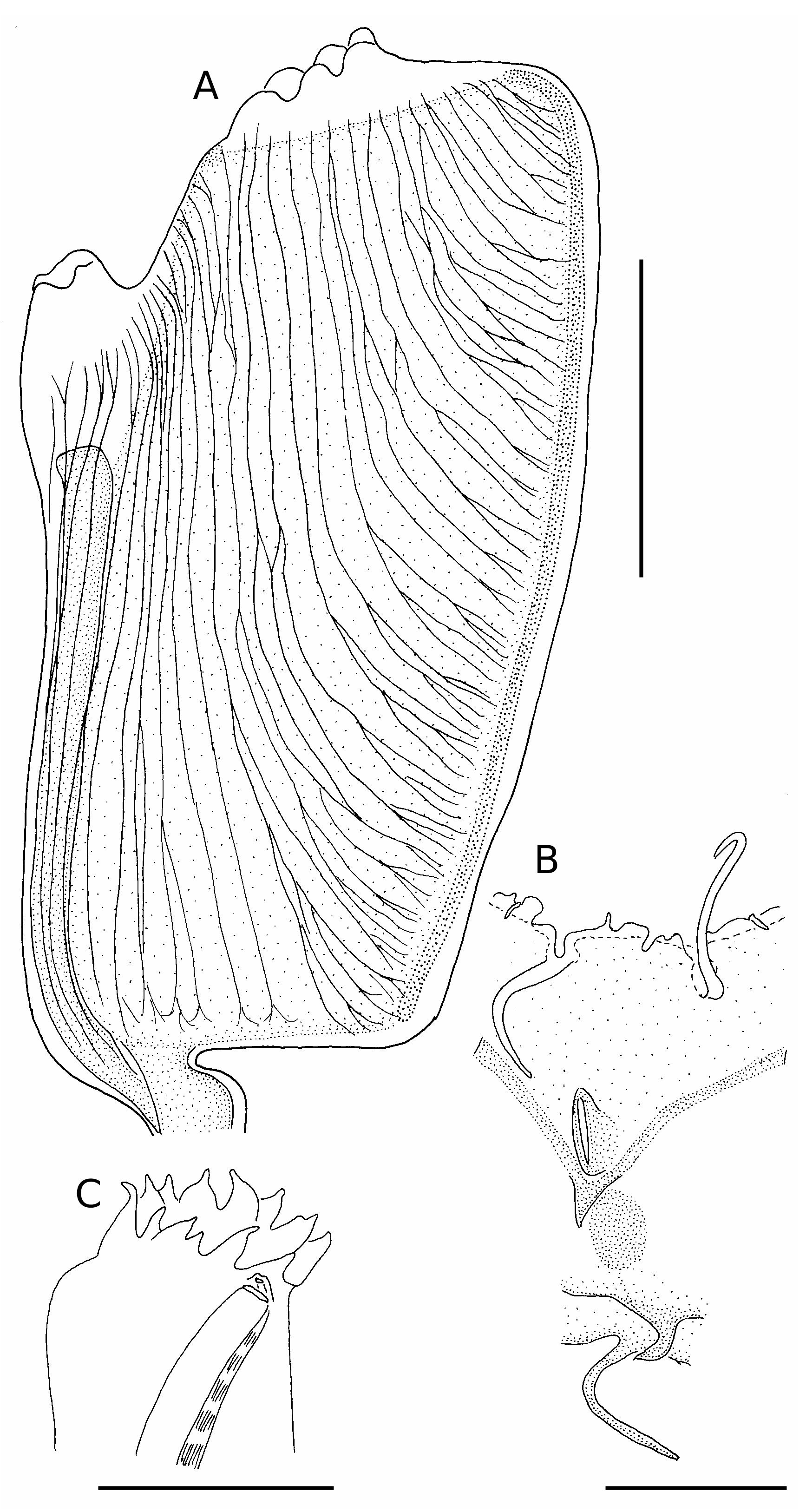
Rhopalaea fusca ( Herdman, 1880) ( Figs 77 View FIG ; 124E View FIG )
Ecteinascidia fusca Herdman, 1880: 723 ; 1882: 241, pl. 36, figs 7-11. Type locality: Banda Sea.
Rhopalopsis fusca View in CoL – Sluiter 1904: 13, pl. 2, fig. 6, Indonesia, numerous localities.
? Rhopalopsis crassa View in CoL – Van Name 1918: 126, pl. 23, figs 14; 82-84, Philippines.
? Rhopalaea crassa View in CoL – Millar 1975: 242, fig. 44, Philippines and Japan.
MATERIAL EXAMINED. — Philippines. Sulu Sea, SE of Puerto Princessa, Jessie Beazley Reef, 9°02.73’N, 119°48.77’E, 8 m, 19.IV.1995 ( MNHN P1 RHO.A 27). — Bohol Sea, Camiguin Island, 9°13.73’N, 124°38.56’E, 7 m, 19.IV.1997 ( MNHN RHO.A 26). Indonesia. N Sulawesi, West of Manado, 1°27.07’N, 124°44.59’E, 10 m, 7. V.1993 ( MNHN RHO.A 14).
DESCRIPTION
In this species the zooids are either isolated or grouped in small bushy clumps. The colour is characteristic and concentrated in the tunic. The thoraces are of an intense blue ( Fig. 124E View FIG ), while the siphons are rimmed with a thin orange line and an irregular blackish band. The specimens of this collection were living in a complex mass of sponges, alcyonids, and didemnid ascidians. The abdominal part of the body is encircled by vascular processes containing blood ampullae that remain blue in fixatives while the body becomes colourless and transparent. In specimens making a bush, the vascular processes of the different individuals form a common mass with numerous blood ampullae. It is impossible to separate the zooids without tearing the tunic. The thorax length reaches 4 cm, the abdomen length 1.5 cm. The thoracic tunic is firm and thick. Around the abdomen it is thick and covered with epibionts. The anterior part of the oesophageal peduncle is encircled by a ring of harder tunic around which the base of the thorax makes a fold to produce a collar ( Fig. 77H View FIG ).
The musculature ( Fig. 77A, H View FIG ) is characteristic. In the anterior half of the thorax, it comprises eight to 10 bundles issuing from the oral siphon and three to five bundles coming from the space between the siphons. Well-separated anteriorly, they part and ramify to form fibres perpendicular to the body axis. These fibres do not reach the dorsal lamina or the endostyle. In the posterior half of the thorax, the longitudinal fibres become thin and disappear as the transverse fibres become preponderant. The muscles do not extend onto the abdomen.
In living or relaxed specimens, the siphons are not lobed ( Fig. 77A View FIG ) but six lobes appear with contraction ( Fig. 77H View FIG ). The oral tentacles, 24 in three orders, stand well apart from each other. Those of the first two orders are long and may even extend out of the siphon. They are somewhat withdrawn behind those of third order.
Smaller crests than in other species link the bases of the large oral tentacles. The prepharyngeal band has a single thick crest. It makes a dorsal V. The dorsal tubercle is a simple slit. The globular neural ganglion is under the dorsal tubercle. The dorsal lamina is made of large flat languets corresponding to one of two transverse vessels ( Fig. 77F View FIG ). On the left side, the transverse vessels that do not correspond to a large rapheal papilla generally end by a small digitation.
The branchial sac is cylindrical, thin and regular. We counted more than 110 rows of stigmata with up to 60 longitudinal vessels on each side. Most of the longitudinal vessels are complete. The branchial papillae are large and transversally flattened. There are three to five long stigmata per mesh. There are no parastigmatic vessels and the tissue is flat.
The gut is below the branchial sac ( Fig. 77H View FIG ). The short oesophagus opens into an elongate stomach whose internal wall has numerous folds ( Fig. 77D View FIG ). The stomach stripes that can be observed externally have no correspondence with the internal folds of the stomach but rather are large blood sinuses ( Fig. 77H View FIG ). There is a well- defined, short post-stomach. The intestine is totally embedded in a mass of spongy tissue, which contains the testes ( Fig. 77E View FIG ). The ovary, more external, covers a part of the right side of the intestine. The rectum is long with a transparent wall. The lobed anus opens at the base of the cloacal siphon at the level of the 35 th stigmata row. The genital ducts accompany the rectum. The oviduct opens below the anus, but the sperm duct is prolonged by a papilla ( Fig. 77G, I View FIG ) with a length of at least four rows of stigmata. This papilla is attached to the dorsal lamina.
REMARKS
An erect form with a cylindrical thorax and a firm tunic is typical, according to Herdman’s figure (1882) and also clearer with the blue colour in Sluiter’s figure (1904). This spectacular species is one of the most photographed ascidians in guides to the Indo-Pacific fauna.
Rhopalaea respiciens Monniot C., 1991 ( Fig. 124F View FIG )
Rhopalaea respiciens Monniot C., 1991a: 494 View in CoL , fig. 2. Type locality: New Caledonia.
MATERIAL EXAMINED. — Papua New Guinea. Milne Bay, Alotau, 10°19.30’S, 150°27.68’E, 12 m, 9. VI.1998 (Sample: CRRF). — Milne Bay Province, Samarai Island, 10°36.98’S, 150°39.77’E, 9 m, 10. VI.1998 (Sample: CRRF).
DESCRIPTION
The species has a thorax that is distinctly larger than the abdomen. The gut loop is short with the stomach located close to the bottom of the loop. The ovary is massive, partly under the digestive tract. The zooids are isolated from their base. The body is bluish with a brown collar around the oral siphon ( Fig. 124F View FIG ) – a characteristic pigmentation.


| MNHN |
Museum National d'Histoire Naturelle |
| V |
Royal British Columbia Museum - Herbarium |
| VI |
Mykotektet, National Veterinary Institute |
No known copyright restrictions apply. See Agosti, D., Egloff, W., 2009. Taxonomic information exchange and copyright: the Plazi approach. BMC Research Notes 2009, 2:53 for further explanation.
|
Kingdom |
|
|
Phylum |
|
|
Class |
|
|
Order |
|
|
Family |
|
|
Genus |
Rhopalaea circula
| Monniot, Françoise & Monniot, Claude 2001 |
Rhopalaea crassa
| KOTT P. 1990: 26 |
Rhopalaea crassa
| MILLAR R. H. 1975: 242 |
Rhopalopsis crassa
| VAN NAME W. G. 1918: 126 |
Rhopalopsis fusca
| SLUITER C. P. 1904: 13 |
Ecteinascidia crassa
| HERDMAN W. A. 1882: 240 |
| HERDMAN W. A. 1880: 723 |
Ecteinascidia fusca
| HERDMAN W. A. 1882: 241 |
| HERDMAN W. A. 1880: 723 |