
Rhabdias japalurae Kuzmin, 2003
|
publication ID |
https://doi.org/ 10.11646/zootaxa.3639.1.1 |
|
publication LSID |
lsid:zoobank.org:pub:32584FBD-212B-4042-BCEF-04C698D71117 |
|
DOI |
https://doi.org/10.5281/zenodo.5262587 |
|
persistent identifier |
https://treatment.plazi.org/id/039087A9-FFEE-FFD1-09F0-F895AFB4C887 |
|
treatment provided by |
Felipe |
|
scientific name |
Rhabdias japalurae Kuzmin, 2003 |
| status |
|
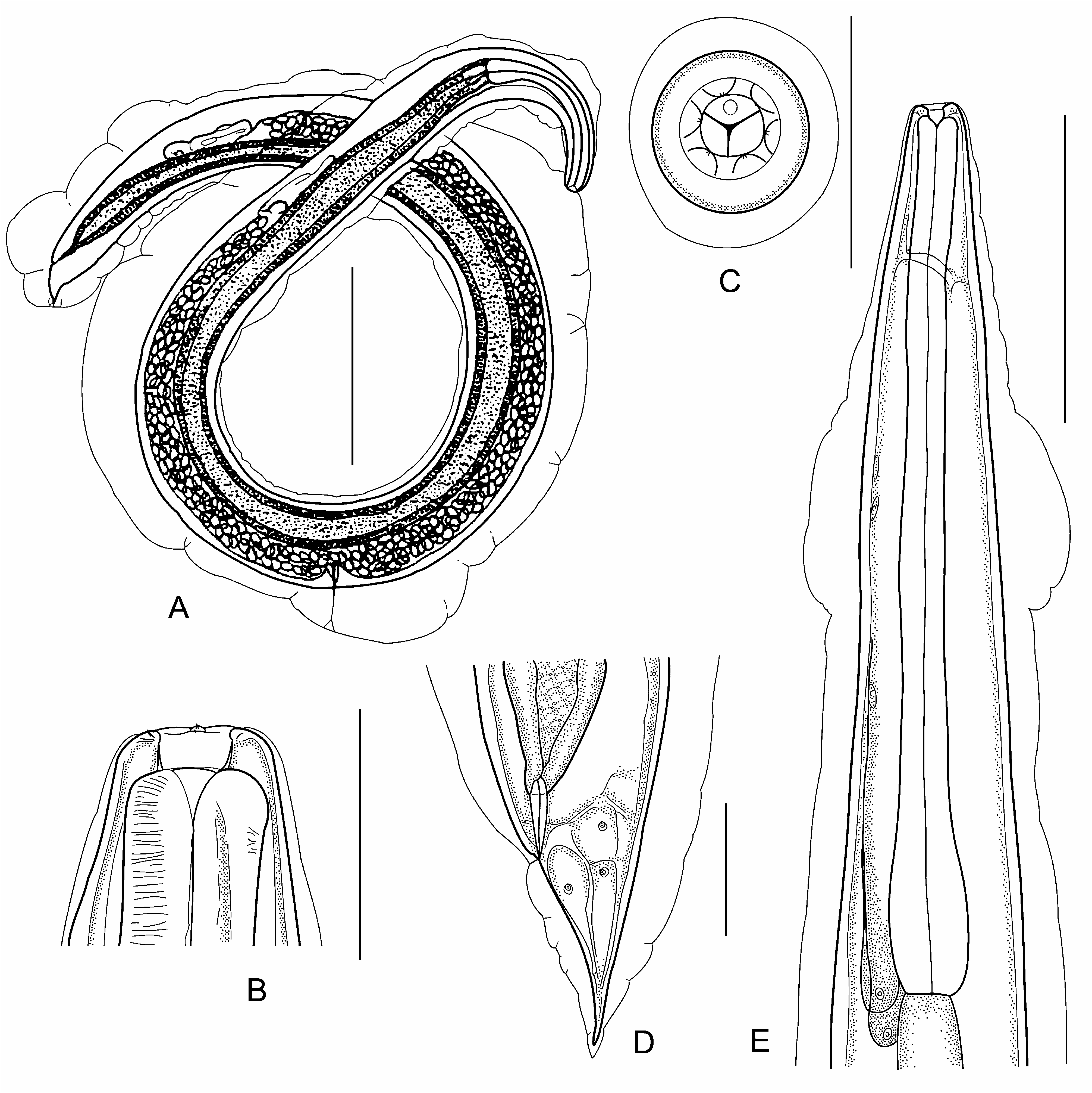
Rhabdias japalurae Kuzmin, 2003 View in CoL
( Fig. 36 View FIGURE 36 )
Hosts: Japalura polygonata , J. swinhonis ( Reptilia: Sauria : Agamidae ).
Site: lungs.
Distribution: Okinawa Island, Japan; Taiwan.
Description (after Kuzmin 2003, modified). Comparatively large species. Body length 13.790 (9.775–16.0) mm, width 479 (423–600). Head end truncated, tail end tapered. Body cuticle swollen and covered with irregular transverse folds. Cuticle on anterior-most body part relatively thin and clearly separated from the following wide cuticular swelling in region of oesophagus midlength. Oral opening round. Six circumoral lips small, equal in shape and size. Dense circumoral cuticular ring present. Buccal capsule cup-like, 28 (26–30) wide, 18 (14–20) deep. Oesophagus elongated, club-shaped, 1.263 (0.921 –1.530) mm long (9.4 [8.0–12.1] % of body length). Muscular part of oesophagus relatively short. Width of oesophagus anterior end 57 (52–65), width of posterior bulb 114 (100–125). Nerve ring situated 226 (191–282) from oesophagus anterior end (18.2 [15.8–20.7] % of oesophagus length). Vulva transverse, slit-like, vulva lips reduced. Distance from anterior end to vulva 7.237 (4.850 –8.713) mm (52.4 [49.6–54.5] % of body length). Uteri thin-walled, filled with numerous (more than 100) eggs. Egg size 112–115 × 55–65 (after Hasegawa and Iwatsuki 1984). Most eggs containing fully developed larvae. Tail conical, 281 (207–326) long (2.1 [1.8–2.4] % of body length). Tail end tapered. Swollen cuticle covering entire surface of tail.
Material studied: 13 specimens ( SIZK, USNPC) including the type series .
References: Hasegawa and Iwatsuki (1984), Kuzmin (2003).
Genus Neoentomelas Hasegawa, 1989
Type species: N. asatoi Hasegawa, 1989 .
Diagnosis (after Hasegawa 1989). Cephalic end hemispherical. Massive dorsoventral lips (pseudolabia) with well-developed muscular and hypodermal tissues present. Each lip with 2 double papillae and 2 minute single papillae; amphids near corners of mouth. Posterior extremity of each labium projected outwards forming 1 median and 2 lateral round lobes. Buccal capsule globular, well-developed, thick-walled, and slightly indented laterally. Onchia absent. Oesophagus club-shaped. Eggs thin-shelled and uncleaved at deposition. Parasitic in the lungs of reptiles.
No known copyright restrictions apply. See Agosti, D., Egloff, W., 2009. Taxonomic information exchange and copyright: the Plazi approach. BMC Research Notes 2009, 2:53 for further explanation.
|
Kingdom |
|
|
Phylum |
|
|
Class |
|
|
Order |
|
|
Family |
|
|
Genus |