
Reliquantha variipes, Roháček, 2013
|
publication ID |
https://doi.org/ 10.5281/zenodo.5740784 |
|
publication LSID |
lsid:zoobank.org:pub:17F1D510-AA62-4279-B592-2768D9B5D24E |
|
persistent identifier |
https://treatment.plazi.org/id/AB613866-4E14-FFB9-47EC-FEE8E818FCDC |
|
treatment provided by |
Marcus |
|
scientific name |
Reliquantha variipes |
| status |
sp. nov. |
Reliquantha variipes View in CoL sp. nov.
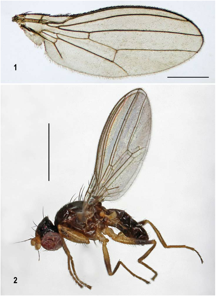
( Figs 1–22 View Figs 1–2 View Figs 3–6 View Figs 7–12 View Figs 13–16 View Figs 17–22 )
Type material. HOLOTYPE: J, labelled: ‘GLAMORGAN, Oxwich Wood, SS5085/5086, 5.vii.2009, P. J. Chandler’ and ‘ HOLOTYPUS J, Reliquantha variipes sp.n., J. Roháček det. 2013’ (red label); left antenna of the holotype broken off (lost during dissection), both mid legs and left wing detached and all (except for one mid leg being glued on plastic bricket below specimen) preserved together with cleared abdomen and dissected genitalia in glycerine in a sealed plastic tube pinned below specimen.
PARATYPE: ♀, labelled: ‘Bracket fungus, Elm 15 VII 75, Oxford GCV’ (pencil handwriting), ‘Oxford University Museum of Natural History ( OUMNH) ’ and ‘ PARATYPUS ♀, Reliquantha variipes sp.n., J. Roháček det. 2013’ (yellow label); abdomen detached and genitalia dissected and all parts preserved in glycerine in plastic tube pinned below specimen).
Both specimens deposited in Oxford University Museum of Natural History, Hope Entomological Collections, Oxford, England, U.K (= OXUM).
Description. Male. Total body length 2.14 mm; general colour dark brown, with extremities and extreme anterior part of head mostly yellow; thorax and abdomen distinctly shining despite sparse greyish brown microtomentum ( Fig. 2 View Figs 1–2 ).
Head distinctly higher than long (almost 1.3 times as high as long), dorsally very slightly wider than thorax; dorsal part of occiput distinctly concave. Occiput blackish brown, subshining, with dark grey microtomentum. Frons moderately broad, slightly tapering anteriorly, orange-yellow in anterior fourth, brown to blackish brown posteriorly, microtomentose up to ocellar triangle. Orbit brown (paler anteriorly), densely microtomentose and dull anteriorly, with sparse microtomentum and distinctly shining posteriorly (behind posterior ors). Frontal triangle relatively long, reaching to anterior fourth of frons, dark brown and largely (including entire ocellar triangle) dark grey microtomentose but not dull, with only a horseshoe-shaped area surrounding (anteriorly and laterally) ocellar triangle bare and lustrous. Ocellar triangle distinctly elevated and ocelli large. Frontal lunule small but distinct, yellow. Face (praefrons) narrow, medially concave, dirty whitish yellow and microtomentose. Parafacialia and gena whitish yellow, with silvery white microtomentum and ochreous- to brown-bordered; this border darker and wider dorsally on parafacialia but very narrow and lighter ventrally on gena. Postgena pale brown ventrally, darker and less densely microtomentose dorsally. Cephalic chaetotaxy: pvt relatively short, convergent but not crossed; vti distinctly shorter than vte (longest cephalic seta) and oc, but slightly longer than posterior ors; 3 relatively short ors (the right foremost lost in the holotype), the hindmost ors longest (but distinctly shorter than oc), the middle somewhat shorter, the foremost small (only half of middle ors); there is 1 orbital microsetula in front of the foremost ors and 4 pairs of microsetulae medially, between anterior point of frontal triangle and anterior margin of frons; postocular setulae (9–10) in a single row, none of them enlarged; postgena with 2 (1 longer) ventral setae and about 3 short setulae; vi relatively long (almost as long as posterior ors) and also subvibrissa well developed (threefourths of vi length) being twice longer than 5 short peristomals. Eye large, covering most of head in profile, with longest diameter (about 1.3 times as long as shortest) subvertical. Gena relatively short (low); its height 0.10 times as long as shortest eye diameter. Palpus short and slightly clavate, distally with 3–4 dark setulae, the subapical markedly longer. Mouthparts pale yellow, palpus whitish. Antenna geniculate, dark yellow with 1st flagellomere light yellow, the latter strongly laterally compressed and very shortly whitish ciliate on anterior margin. Arista pale ochreous, 1.9 times as long as antenna, with small and slender basal segment and very short cilia (yet shorter than those on 1st flagellomere).
Thorax very slightly narrower than head, dark brown, with small paler brown areas (humeral callus, anterior part of notopleural line, around suture) and pale ventral corner of sternopleuron being distally grading to ochreous-yellow. Mesonotum relatively shining despite sparse grey to brownish grey microtomentum; no bare areas on scutum or scutellum; pleural part of thorax more densely microtomentose and, particularly ventrally, duller. Thoracic chaetotaxy: 1 relatively short hu (shorter than posterior npl), 2 npl (anterior distinctly longer), 1 sa (slightly shorter than pa), 1 pa (relatively long), 1 distinct prs (only as long as sa); 2 dc (both postsutural), anterior longer than half of posterior, the latter long and strong, 6–7 dc microsetae in front of anterior dc; ac microsetae short but not very dense, in 4 rows, posteriorly only reaching slightly beyond level of anterior dc; 2 sc, apical strong and slightly longer than posterior dc (hence longest of thoracic setae), laterobasal much shorter and weaker but about as long as scutellum length; 1 minute upcurved ppl; 2 relatively long stpl (anterior only slightly shorter) and 1 microseta in front of them; only 2 (1 long) curved setae on ochreous-yellow ventral corner of sternopleuron. Scutellum rounded triangular and strongly convex dorsally; postscutellum well developed.
Legs yellow and brown variegated ( Fig. 2 View Figs 1–2 ), with all coxae, trochanters, basal parts of femora and all tarsi (except for at least partly brownish apical segments) yellow to dark yellow. All femora with distal third (f 1) or half (f 2, f 3) brown to dark brown, otherwise (basally and on knees) yellow ( Figs 20–22 View Figs 17–22 ). Tibiae also somewhat variegated but this variegation less contrasting than that on femora; t 1 and t 2 with small proximal and distal parts yellow and large pale-brown darkened middle section; t 3 similarly coloured but with lighter annulus in the middle of darkened section in addition ( Fig. 22 View Figs 17–22 ). f 1 with ctenidial spine entirely lacking ( Fig. 20 View Figs 17–22 ), with only usual rows of long thin but relatively sparse posterodorsal and posteroventral setae; f 2 simply setulose; f 3 with posteroventral row of 8 erect setae, 4–5 of which in apical half shortened and more or less thickened ( Fig. 22 View Figs 17–22 ). t 1 and t 3 uniformly short-setulose; t 2 with short ventroapical seta and 3 short thickened setulae adjacent to it ( Fig. 21 View Figs 17–22 ). Tarsi without peculiarities; mid basitarsus long and slender, fore and hind basitarsus with a few slightly longer setulae proximoventrally.
Wing ( Fig. 1 View Figs 1–2 ) moderately wide, hyaline, membrane and veins pale ochreous brown. C with slightly thicker (and thus rather indistinct), short and sparse setulae among usual fine hairs on Cs 2. Sc fused with R 1 apically to form a distinct preapical kink. R 2+3 bent, parallel to C and only apically straighter, ending slightly farther from wing apex than does M. R 4+5 very slightly bent, distally slightly convergent to M. Discal (dm) cell moderately long and narrow; its distal part (beyond r-m) slightly widened distally; anterior cross-vein (r-m) situated in the middle of discal cell. CuA 1 almost reaching wing margin, A 1 ending far from it. Terminal section of CuA 1 about 1.7 times as long as posterior cross-vein (dm-cu). Alula small but relatively broad. Wing measurements: length 2.48 mm, width 0.83 mm, Cs 3: Cs 4 = 1.15, r-m\dm-cu: dm-cu = 2.64. Haltere yellow, knob yellowish white.
Abdomen dark brown and more shining than thorax despite sparse greyish brown microtomentum. All preabdominal terga rather sparsely and shortly setose. T1 dorsally distinctly delimited, only laterally fused with T2 , with only a few short setulae. T2 – T5 large and broad ( T3 widest), extended ventrolaterally ; T2 shorter than T3 , others subequal in length but becoming slightly narrower caudally. Preabdominal sterna brown and moderately broad ; pleural membrane between terga and sterna narrower than in female. S1 damaged in holotype and therefore not described. S2 as long as wide, slightly shorter and narrower than S3; S3–S5 almost subequal in length but becoming distinctly wider posteriorly. S3 as long as wide, S4 slightly wider than long, S5 largest, markedly wider than long and hence transverse, trapezoidal (posteriorly wider). S2–S5 simply shortly setulose.
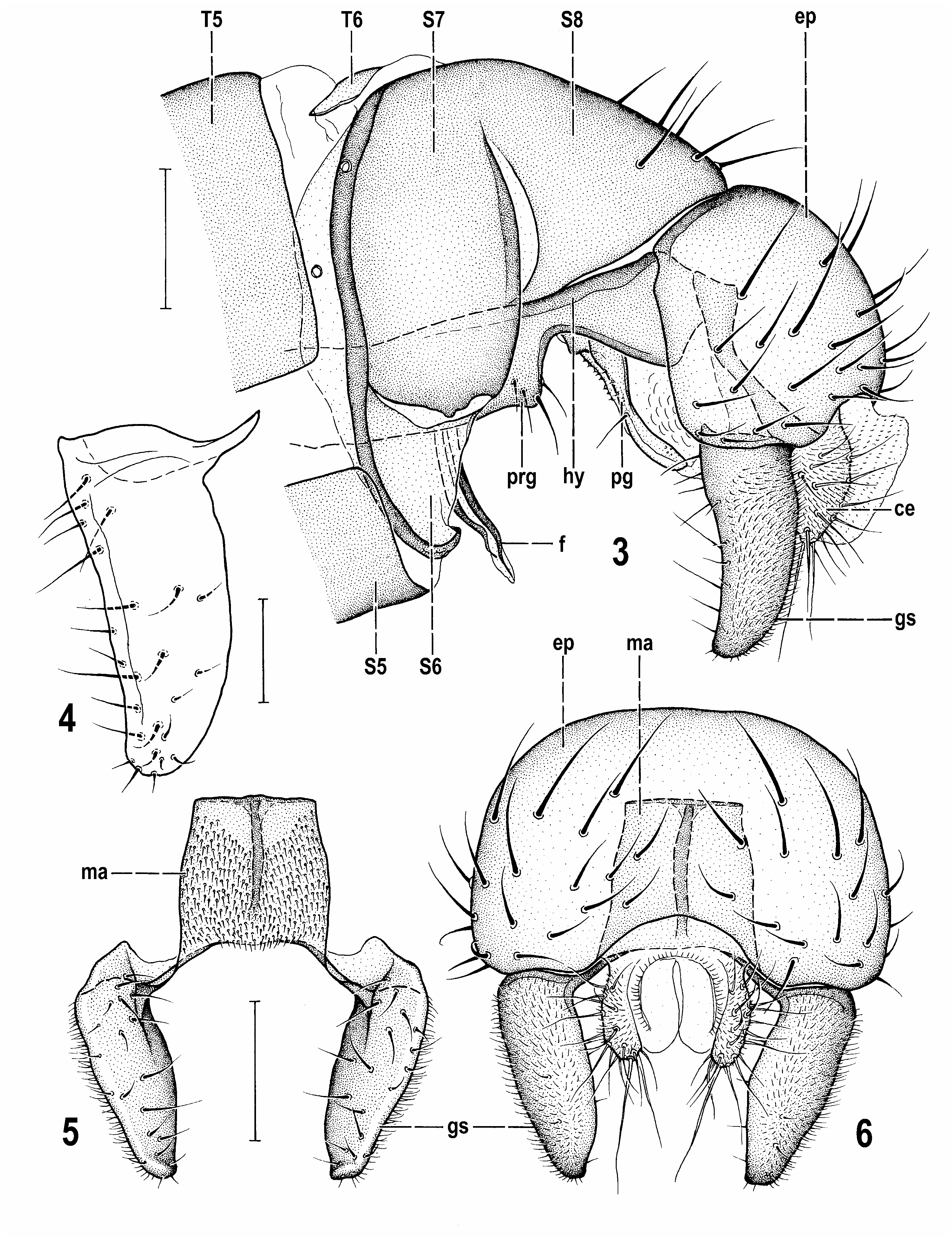
Postabdomen ( Fig. 3 View Figs 3–6 ) strongly sclerotized, more shining because of reduced microtomentum. T6 distinctly sclerotized but lighter brown than other sclerites, medially undivided, forming a simple, strongly transverse dorsal sclerite. S6, S7 and S8 partly coalesced but their borders distinct. S6 the shortest, of distinctive form, strongly asymmetrical, band-like tapered on left and right side, and its largest (middle) part situated rather ventrally and separated horizontally from S7 ( Fig. 3 View Figs 3–6 ), pale-pigmented to membranous except for dark, sclerotized marginal ledge on the border with S7; S7 longer, slightly asymmetrical, situated on left side of postabdomen, dark brown with yet darker anterior bordering ledge (fused with that of S6). Both S6 and S7 without setae. S8 longest, dark and heavily sclerotized, slightly asymmetrical (longer on left side) and situated dorsally, with sparse moderate setae in posterodorsal half. 6th spiracle situated laterally in anterior membranous part of S6, 7 th spiracle laterodorsally in dark bordering ledge between S6 and S7 ( Fig. 3 View Figs 3–6 ).
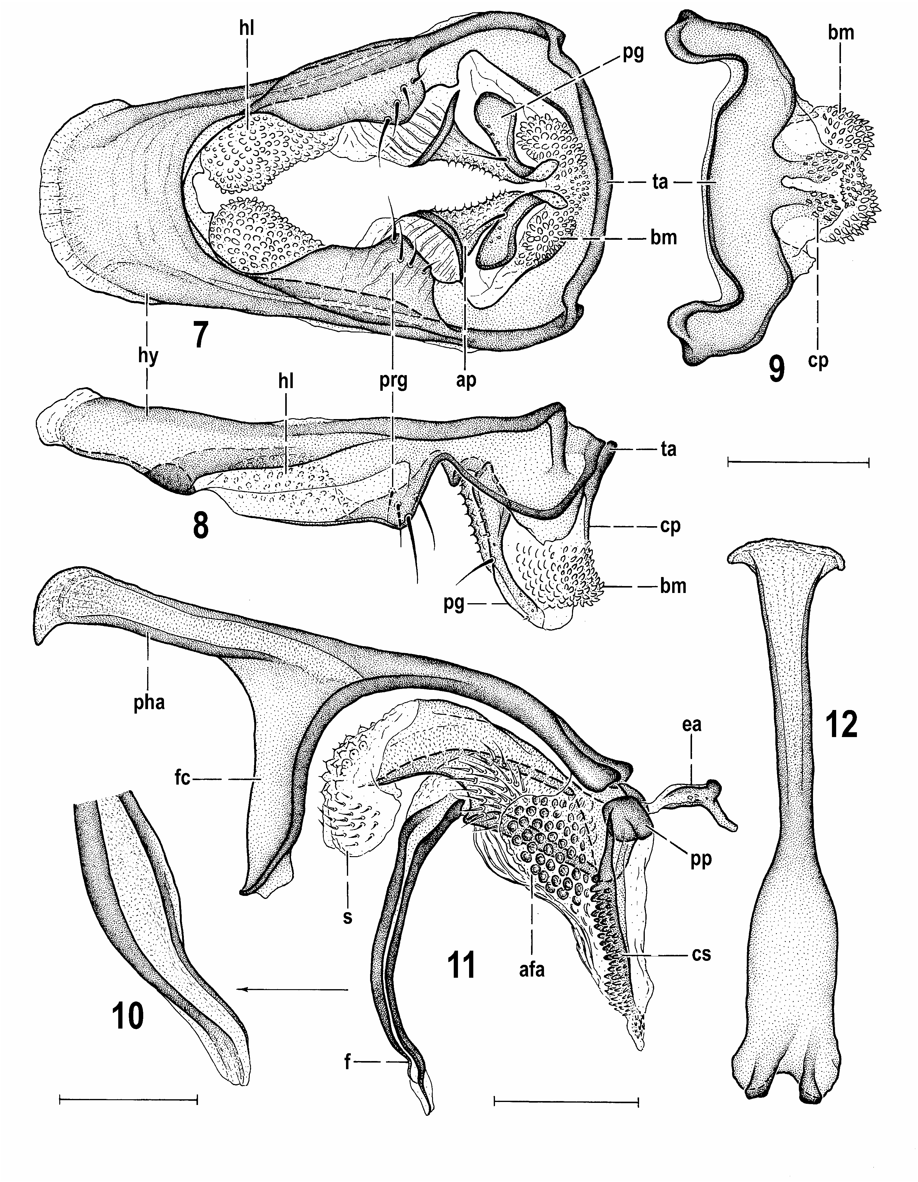
Genitalia. Epandrium ( Figs 3, 6 View Figs 3–6 ) not very large, globose, somewhat wider than high ( Fig. 6 View Figs 3–6 ), shining blackish brown, without particularly enlarged setae, although some (3–4 pairs) are slightly longer and more robust than others. Anal fissure reduced ( Fig. 6 View Figs 3–6 ), unusually low (yet smaller than in Fungomyza spp. , most resembling that of fossil Lacrimyza lacrimosa ); cercus about half length of gonostylus, laterally somewhat flattened, pale-pigmented including setae, apical of which longest ( Figs 3, 6 View Figs 3–6 ). Medandrium relatively high (long), of almost rectangular outline, only very slightly narrower dorsally, with dark medial ridge and very dense short setosity on anterior (internal) surface (see Fig. 5 View Figs 3–6 ); its ventral arms short, fused with posterodorsal, internally projecting corners of gonostyli. Gonostylus ( Figs 3–6 View Figs 3–6 ) darker brown than epandrium, relatively simple, almost as long as epandrial height, elongate, slightly bent in profile, broadest proximally and slightly gradually tapered apically, with apex broadly rounded but not inclinate. Outer convex side of gonostylus ( Fig. 3 View Figs 3–6 ) largely covered by dense long micropubescence (leaving only anterior margin bare) and bearing only a few small setulae. Most setae (some relatively long) are inserted on inner concave and otherwise bare side at anterior margin of gonostylus ( Figs 4, 5 View Figs 3–6 ). Hypandrium ( Figs 7, 8 View Figs 7–12 ) forming together with transandrium usual frame-shaped structure, moderately robust, with distinct and weakly sclerotized, flat but peculiarly tuberculate anterior internal lobes ( Fig. 7 View Figs 7–12 ) being posteriorly also appended to pregonites. Posterior part of hypandrium separated from pregonite by deep ventral incision and its ventral side somewhat projecting as small pale flat lobe (visible on Figs 8, 9 View Figs 7–12 ). Transandrium robust ( Fig. 9 View Figs 7–12 ), with dark dorsal marginal ledge, ventromedially projecting in flat, forked and distally spinose caudal process ( Fig. 9 View Figs 7–12 , cp); basal membrane below caudal process medially provided with a group of small tuberculiform spines; its lateroventral bulging lobes armed by dense short spines ( Figs 8, 9 View Figs 7–12 ). Pregonite relatively large, anteriorly flat and fused to hypandrium, incurved, but without setae; posteriorly ventrally angular, dark and heavily sclerotized, separated from posterior part of hypandrium by deep notch and with 4, mostly internal, setae ( Figs 7, 8 View Figs 7–12 ). Postgonite relatively long and slender ( Figs 7, 8 View Figs 7–12 ), very slightly bent, proximally broader and darker, apically blunt, with 1 seta in the middle of anterior margin, 3 microsetulae in proximal half and 1 subapically and several fine grain-like sensillae on outer surface. In front of postgonite there is another unusual structure – an anteriorly striated membranous lobe continued posteroventrally as a sclerotized, pigmented, distally tapered projection provided with spinulose tubercles (see Fig. 7 View Figs 7–12 , ap). This structure is obviously not homologous with the “basal sclerite” of Amygdalops species (see ROHÁČEK 2004, 2008) because it is not attached to basal part of postgonite, and could be a secondarily sclerotized outer part of the folding apparatus whose inner aedeagal part (see Fig. 11 View Figs 7–12 , afa) is attached to the base of the aedeagus and phallapodeme and covered by dense but pale, rounded tubercles and (anteriorly) dark striae. Connecting sclerite strongly sclerotized and dark pigmented, largely overgrown by dense blunt spines being distally smaller and more numerous ( Fig. 11 View Figs 7–12 , cs). Phallapodeme ( Figs 11, 12 View Figs 7–12 ) relatively slender, distinctive due to laterally dilated and flattened and only shortly forked basal part; its ventral fulcrum slender and apex slightly bicuspidate. Aedeagus ( Fig. 11 View Figs 7–12 ) with small phallophore only posterodorsally dark and strongly sclerotized, anteroventrally paler and connected with ventrobasal sclerite of distiphallus; distiphallus bifid as usual, composed of relatively small, distally membranous saccus and slender, long sclerotized filum. Saccus internally reinforced with slightly bent elongate sclerite and also its proximal part more or less sclerotized and with left side ( Fig. 11 View Figs 7–12 ) covered by distinctive spine-like setae (similar to those in Carexomyza spp. ); smaller apical membranous part of saccus provided with pale rounded tubercles each having a microspine on apex, and (more distally) with pale short setulae. Filum of primitive form, slender, relatively long and composed of two dark, band-like sclerites; apex of filum ( Fig. 10 View Figs 7–12 ) simple, narrowed, membranous, with attenuated band-like sclerites terminating in narrowed membranous apex. Ejacapodeme on very short duct, but of usual shape, with slightly sinuous digitiform process and dark clubbed distal end ( Fig. 11 View Figs 7–12 , ea).
Female. Similar to male unless mentioned otherwise. Total body length 1.98 mm. Frons darker, brown also anteriorly, with yellowish colour restricted to narrow anterior margin of frons and lower orbit in front of anterior ors. Antenna distinctly darker than in male, with 1st flagellomere dorsally brownish, yellow only on ventral half of outer side to ventral third of inner side and arista brown. Face, parafacialia and gena as in male but palpus yellow. Pedal chaetotaxy as in male but f 3 without posteroventral thickened setae. Tibiae of all legs less darkened but this may be caused by greater age of the female paratype because the dark femoral pattern is also somewhat faded in this specimen. Anterior stpl shorter than in male holotype. Wing with r-m situated more distally, slightly beyond middle of dm cell. Wing measurements: length 2.28 mm, width 0.79 mm, Cs 3: Cs 4 =1.28, r-m\dm-cu: dm-cu = 2.29, distal section of CuA 1 1.76 times as long as dm-cu cross-vein. Abdomen with T2 and, particularly, T3 – T5 broader and shorter (hence more transverse) than in male but of similar colour and microtomentose pattern. T1 distinctly separate from T2 , fused only at lateral margins. T2 – T4 subequal in length, T4 broadest ; T5 distinctly longer than T4 but narrower, very slightly tapered caudally. All preabdominal terga bent lateroventrally far onto sides of abdomen. S1 and S2 torn off in female paratype and, therefore, not described ; S3–S5 much narrower than terga, dark brown and well sclerotized, with short sparse setosity; membranous pleural areas between sclerites relatively large. S3–S5(S6) becoming distinctly wider posteriorly; S3 slightly longer than wide, S4 and S5 wider than long and somewhat transverse, of rounded trapezoidal shape.
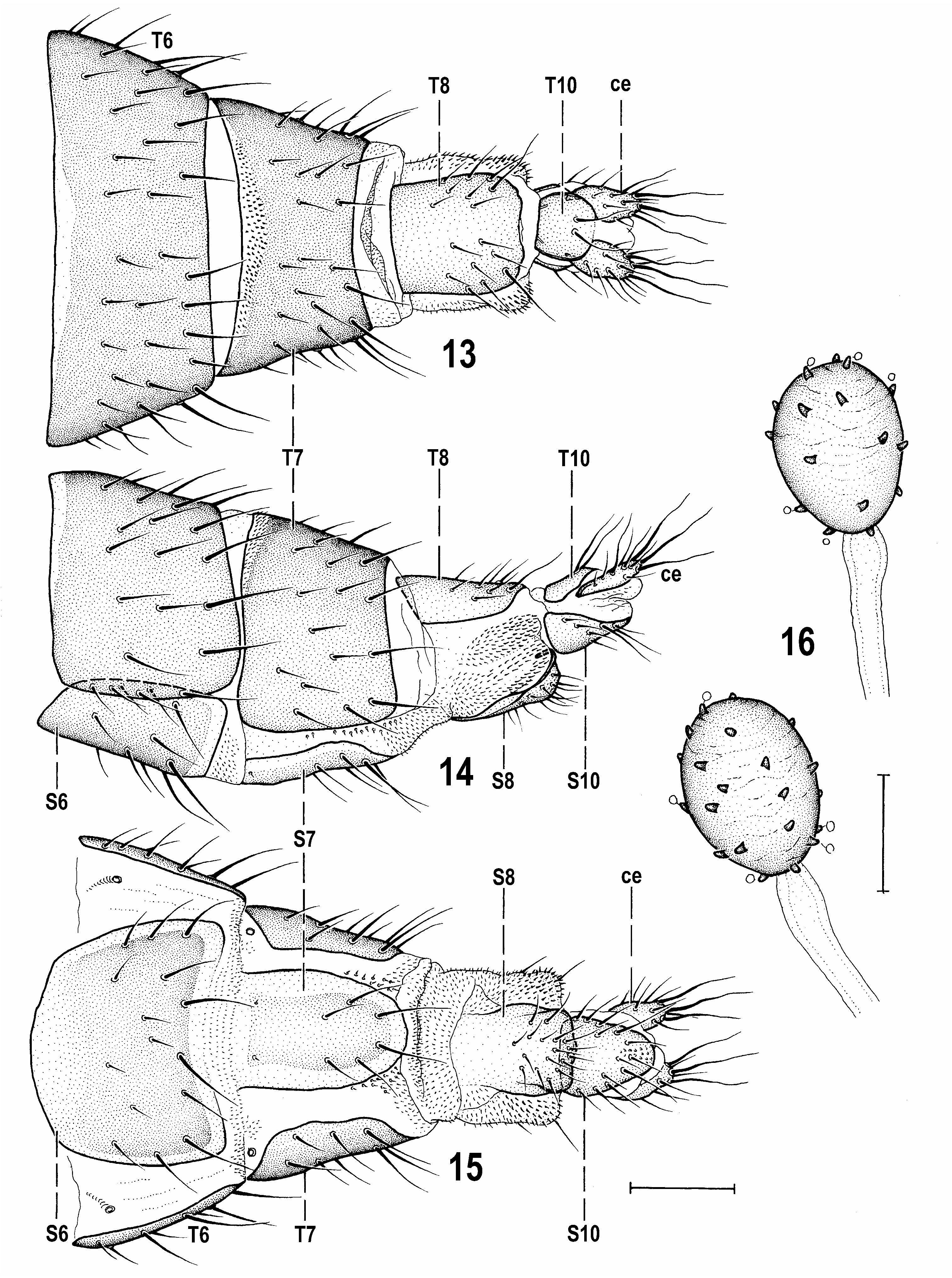
Postabdomen ( Figs 13–15 View Figs 13–16 ) telescopically retractable, relatively long, gradually tapered caudally. T6 broad, transverse, reaching far onto ventral side of abdomen, dark brown pigmented with pale anterior marginal stripe (medially longest) and moderately setose. S6 ( Fig. 15 View Figs 13–16 ) wider than long, larger than S5, posteriorly slightly emarginate, brown but with pale (almost unpigmented) margins, posteriorly and laterally distinctly contrasting with dark remainder of sclerite surface; setae sparse, arising in posterior half, mainly submarginally. T7 and S7 not fused, separate, although T7 reaching far onto ventral side of 7th segment. T7 ( Figs 13, 14 View Figs 13–16 ) distinctly narrower than T6, dark and heavily sclerotized, caudally tapered, having anterior pale marginal stripe covered by strong micropubescence and remaining dark surface with relatively short setae. S7 ( Fig. 15 View Figs 13–16 ) of distinctive ligulate shape, much longer than broad, pale-pigmented only centrally, marginally broadly unpigmented, without micropubescence and with only 6 setae in posterior half apart from a pair of usual setulae (sensillae) near anterior margin. 7th spiracle situated anterolaterally ( Fig. 15 View Figs 13–16 ), in front of anterior corner of T7. Membrane between T7 and S7 largely without micropubescence but posteriorly, along margin of S7, with distinctive short microsetulae. Intersegmental membrane between 7th and 8th segment longer ventrally where distinctly pubescent. T8 ( Fig. 13 View Figs 13–16 ) relatively large though much narrower than T7, longer than broad, convex, pale brownish pigmented, entirely lacking micropubescence and with setae in posterior two-thirds. S8 ( Figs 15 View Figs 13–16 , 17 View Figs 17–22 ) also peculiar, elongate, undivided, and, like S7, anteriorly poorly delimited from membrane. Posterior marginal part of S8 curved dorsally and somewhat invaginated into 8th segment (see Fig. 17 View Figs 17–22 ); its surface completely without micropubescence and with short setae restricted to posterior half. Internal structures of the female genital chamber very weakly developed; there is only a relatively small, pale-pigmented, slightly transversely irregular annular sclerite in centre of ventral side of genital chamber ( Figs 17, 19 View Figs 17–22 ). Accessory glands ( Fig. 17 View Figs 17–22 , ag) small with minute globuli on surface, and with ducts somewhat dilated but with plain middle part. Ventral receptacle membranous ( Fig. 17 View Figs 17–22 , vr), unpigmented, very short, with plain surface, tapered distally to form a beak-like but apically blunt projection ( Fig. 18 View Figs 17–22 ), set on broad short duct. Spermatheace (1+1) small, of primitive simply ovoid form ( Fig. 16 View Figs 13–16 ), brown, with relatively plain surface irregularly overgrown by darker brown, short blunt spines some of which have minute stalked globuli on tips; spermathecal ducts very short ( Fig. 17 View Figs 17–22 ), entirely membranous, without distinct collar. T10 ( Fig. 13 View Figs 13–16 ) small (smaller than S10), pale, about as long as broad, rounded laterally, bare (lacking micropubescence), with only a pair of medial setae. S10 ( Fig. 15 View Figs 13–16 ) of distinctive, elongately triangular but posteriorly broadly rounded shape, with relatively long marginal setae, otherwise bare. Cercus ( Figs 13–15 View Figs 13–16 ) also without micropubescence, relatively short and robust, dorsoventrally somewhat flattened, with rich setae, 3 of which (1 apical, 2 subapicals) are markedly longer than others.
Discussion. This new species is distinguished by yellow and brown variegated femora and (less distinctly) tibiae ( Figs 2 View Figs 1–2 , 20–22 View Figs 17–22 ). Hitherto, the partly (distally) brown-coloured femora (all or only some of them) have only been known in some Afrotropical (see ROHÁČEK 2004) and Oriental ( ROHÁČEK 2008) species of Amygdalops and in species of Fungomyza . While the Amygdalops species differ markedly from R. variipes sp. nov. in having a long-pectinate arista, only 2 long and widely spaced ors, strongly convex elongately suboval eyes, the pleural part of thorax pale yellow with only the dorsal longitudinal band dark, different wing venation and ornamentation and many other characters, members of Fungomyza resemble the new species in general external appearance, colouration of the body and wings. However, only the Nearctic F. buccata Roháček & Barber, 2004 has dark markings on all femora as in R. variipes sp. nov. but differs from the latter (as in both remaining Fungomyza species ) by lacking a distinct subvibrissa and having a ctenidial spine on f 1, longer prs, more setae on the ventral corner of the sternopleuron, the r-m situated distal to the middle of the dm cell and a number of characters in the male genitalia and female terminalia (see discussion above under genus Reliquantha ).
Biology. Poorly known. Both type specimens originate from woodland habitats and were caught in the first half of July. The holotype male was collected in an ash woodland on a limestone hill overlooking the coast of the Gower Peninsula in South Wales (P. J. Chandler, pers. comm., 2013). The paratype female was collected by G. C. Varley in Oxford on a bracket fungus on elm ( Ulmus sp. ), probably during his research on the insects associated with elms suffering from Dutch elm disease (J. W. Ismay and J. Hogan, pers. comm., 2006). Therefore, ROHÁČEK (2009) considered this (then undescribed) species a potential feeder of tree fungi. It is to be stressed that in Anthomyzidae only species of Fungomyza have, to date, been known to be associated with fungi (DELY- DRASKOVITS 1972; CHANDLER 1978, 2010; ROHÁČEK 1999, 2009; ROHÁČEK & BARBER 2004; ROHÁČEK & ŠEVČÍK 2013) and the development of larvae in macrofungal sporocarps has only been demonstrated for F. albimana (Meigen, 1830) .
Etymology. The species is named variipes to reflect its yellow and brown variegated legs.
Distribution. The species is hitherto known only from Great Britain ( Wales, England).

No known copyright restrictions apply. See Agosti, D., Egloff, W., 2009. Taxonomic information exchange and copyright: the Plazi approach. BMC Research Notes 2009, 2:53 for further explanation.
|
Kingdom |
|
|
Phylum |
|
|
Class |
|
|
Order |
|
|
Family |
|
|
Genus |