
Pygmarrhopalites plethorasari, Zeppelini, Douglas, Taylor, Steven J. & Slay, Michael E., 2009
|
publication ID |
https://doi.org/ 10.5281/zenodo.189689 |
|
DOI |
https://doi.org/10.5281/zenodo.5613113 |
|
persistent identifier |
https://treatment.plazi.org/id/03A12C18-4323-C154-CC83-18BAB307FA22 |
|
treatment provided by |
Plazi |
|
scientific name |
Pygmarrhopalites plethorasari |
| status |
sp. nov. |
Pygmarrhopalites plethorasari sp. nov.
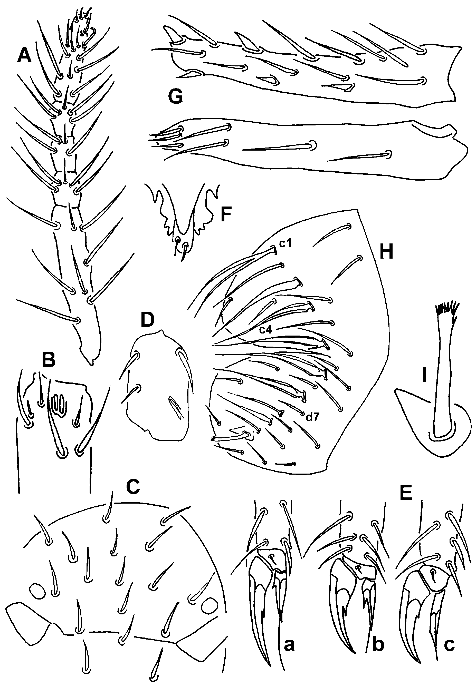
pygmaeus -group s. str. ( Fig. 2 View FIGURE 2 )
Description. No traces of pigment, dorsal body setae short on anterior, longer on posterior part of great abdomen, posterior setae about same length as third unguis ( Table 2 View TABLE 2 ). Ant. IV of holotype 1.32 times cephalic diagonal, with five subsegments ( Fig. 2 View FIGURE 2 A), apex with capitate sense rod. Ant. III not swollen basally; sense organ ( Fig. 2 View FIGURE 2 B) with 2 parallel sense rods in single socket; seta Aai club-shaped, acuminate; Api and Ape short, slender and acuminate; Ae, Ap and Ai normal, elongate setae. 1+1 eyes. Dorsal cephalic setae not spine-like, M5 present, L1–2 not seen ( Fig. 2 View FIGURE 2 C). Metatrochanteral organ elongate ( Fig. 2 View FIGURE 2 D). Seta FSa present on all tibiotarsi. All ungues with inner tooth, no tunica. First and second unguiculi with conspicuous corner tooth, third unguiculus slender, lanceolate, with tiny corner tooth in distal third, all unguiculi with apical filament exceeding unguis tip in first and second claw pairs ( Fig. 2 View FIGURE 2 E). Corpus tenaculum with two setae ( Fig. 2 View FIGURE 2 F). Dens with 7 dorsal E setae, E1 and E3 strongly spine-like, other E setae normal; L1–3 strongly spine-like, 4 ventral setae rows (3,2,1,1) present ( Fig. 2 View FIGURE 2 G), dental chaetotaxy in Table 3. Mucro narrow, gutter-like, with spoon shaped or globular tip, both edges serrate. Anal valve without cuticular spines ( Fig. 2 View FIGURE 2 H); setae C1 forked, C2 swollen, C3–4 slightly lamellate, C4 branched at base, C5–6 lamellate, D5 present, chaetotaxy in Table 4 View TABLE 4 . Female subanal appendage fringed at tip ( Fig. 2 View FIGURE 2 I).
Dental chaetotaxy
Ant. iv Ant. iii Eyes per Ceph. Id ve ve Species subd. basal side spines E1 E2 E3 E6 E7 2 -3 L1 L2 L3 L4 1 5 P. ashcraftensis 6 - 1 - S + S + + + S S s + + - P. leonardwoodensis 5 - 1 - S + S + + + S S S - + - P. buffaloensis 6 - 1 - S + + + + + S + + - + - P. youngsteadtii 7 - 1 - S + S + + + S s - - + - P. plethorasari 5 - 1 - S + S + + + S S s - + - P. shoshoneiensis 7 - 1 v - S + + + + + S s s - + -
+= present, -= absent, S= strongly spine-like, s= spine-like, v= vestigial.
Type material. Holotype (female) (number 1 in the slide): USA, MISSOURI, Pulaski Co., Big Freeman Cave, 9-i-04, S. Taylor, M. Slay leg. ( INHS). Paratypes: female in the same slide with holotype and 3 females: USA, MISSOURI, Pulaski Co., Ramsey Cave, 9-i-04, S. Taylor, M. Slay leg. ( INHS). 8 paratypes in alcohol, same locality as the latter ones ( INHS).
Etymology. An anagram of the name of the type genus of the family, Arrhopalites .
Remarks. Pygmarrhopalites plethorasari was found in caves of the Fort Leonard Wood military installation, close to each other in the physiographic province Salem Plateau of the Ozark Plateaus, the climate is Cfa. This species resembles P. hubbardi from Colorado in the apical filament of feet complexes, dental chaetotaxy, and the forked C1 seta on the anal valve. The fourth antennal subsegmentation and the basal swelling of the third antennal segment of P. hubbardi, the female subanal appendages, and the lamellate C setae on anal valve of P. plethorasari differentiate these species from each other.
| INHS |
Illinois Natural History Survey |
No known copyright restrictions apply. See Agosti, D., Egloff, W., 2009. Taxonomic information exchange and copyright: the Plazi approach. BMC Research Notes 2009, 2:53 for further explanation.