
Pseudosinella triocellata, Nunes & Bellini, 2018
|
publication ID |
https://doi.org/ 10.11646/zootaxa.4420.1.4 |
|
publication LSID |
lsid:zoobank.org:pub:88F9AB23-046F-465E-A2CA-A7A45B522C11 |
|
DOI |
https://doi.org/10.5281/zenodo.5987491 |
|
persistent identifier |
https://treatment.plazi.org/id/F13087AF-FFD0-2C57-FF63-0E36FAF7F841 |
|
treatment provided by |
Plazi |
|
scientific name |
Pseudosinella triocellata |
| status |
sp. nov. |
Pseudosinella triocellata View in CoL sp. nov. Nunes & Bellini
Figs 21‒43
Type material. Holotype female on slide, Brazil, Piauí State, Piracuruca municipality, Parque Nacional de Sete
Cidades (04°05’56.94”S; 41°42’33.42”W), Caatinga (transitional zone between Caatinga and Cerrado biomes), 12‒14.v.2015, R.C. Nunes coll. Paratypes on slides, three males and seven females, same data as holotype. Type material deposited at CC/ UFRN (holotype and paratypes: two males and six females) and INPA (paratypes: one male and one female).
Description. Total length (head + trunk) of type series ranging between 0.57‒0.91 mm (holotype 0.63 mm). Habitus typical of the genus ( Fig. 21). Specimen with blue pigment covering most of the body, over a yellowish background; eyepatches dark ( Fig. 21). Weakly ciliate apically rounded scales covering ventral and dorsal head, trunk, ventral manubrium and dens. Antennae, legs, collophore and dorsal face of furcula without scales.
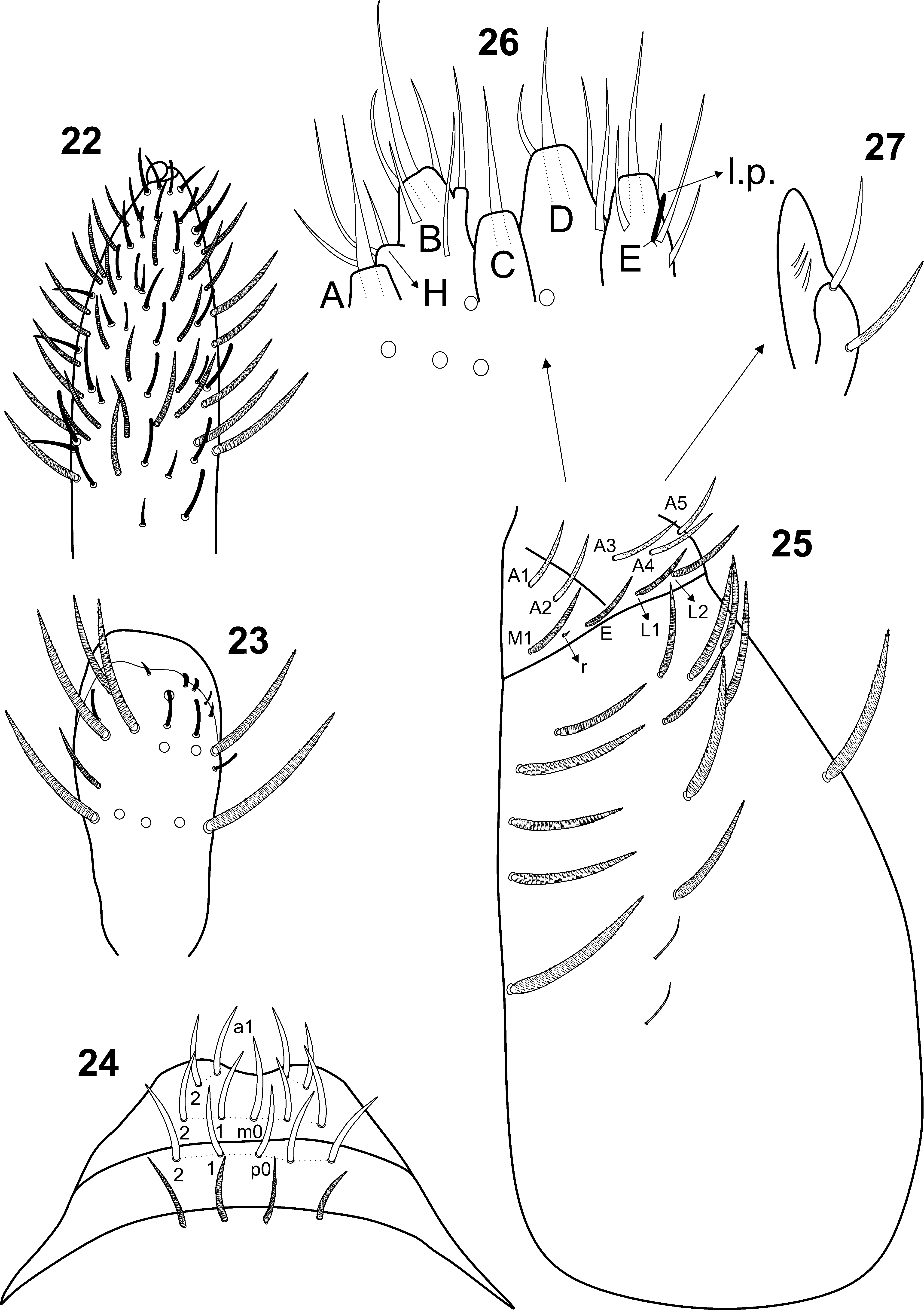
Head ( Figs 22‒28 View FIGURES 22–27 View FIGURE 28 ). Antennae shorter than body length, antennal ratio as I: II: III: IV = 1: 1.2‒2.4: 1.33‒2.1: 2.33‒4.09 (holotype 1: 2.4: 2: 3.5). Apical bulb of Ant. IV simple and membranous, subapical sense organ absent, with at least three types of chaetae: blunt sensilla, pin sensilla and ciliated chaetae ( Fig. 22 View FIGURES 22–27 ). Ant. III sense organ with 2 rods, 3 surrounding guard sensilla (2 smaller pin-like), plus some surrounding blunt sensilla and ciliated chaetae ( Fig. 23 View FIGURES 22–27 ). Four prelabral ciliated chaetae. Labral formula 4 (a1–2), 5 (m0–2), 5 (p0–2), all smooth chaetae ( Fig. 24 View FIGURES 22–27 ). Ventral post-labial chaetotaxy with about 13 ciliated chaetae of different lengths plus two smaller smooth chaetae; cephalic groove with 4+4 surrounding ciliated chaetae with subequal sizes plus 4+4 scales ( Fig. 25 View FIGURES 22–27 , scales not represented). Labial basolateral and basomedian fields with chaeta r reduced , M1, E, L1–2 ciliated, A1–5 weakly ciliated ( Fig. 25 View FIGURES 22–27 ). Labial palp with five smooth proximal chaetae. Labial palp papillae (and guard chaetae) formula as: H(2), A(0), B(5), C(0), D(3), E(4) + l.p.; lateral process finger-shaped not reaching the papilla base ( Fig. 26 View FIGURES 22–27 ). Outer maxillary lobe with basal and distal chaetae weakly ciliated and smooth respectively, and subequal; sublobal plate with three appendages, subequal in length, all smooth ( Fig. 27 View FIGURES 22–27 ). Eyes 3+3, subequal in size. Dorsal chaetotaxy with 6 antennal (An), 5 anterior (A0–4), 5 medio-ocellar (M0–4), 3 sutural (S2, S4, S6), 3 post-sutural (Ps2–3, Ps5), 5 postoccipital anterior (Pa1–3, Pa5–6), 2 postoccipital medial (Pm1, Pm3) and 5 postoccipital posterior (Pp1–5) chaetae; S2 as mac ( Fig. 28 View FIGURE 28 ).
Thorax chaetotaxy ( Figs 29‒30 View FIGURES 29–33 ). Central mac formula from Th. II to Abd. IV as 1,0/0,2,2,2. Th. II with 1 ms, 1 anterolateral sens (al), 1 anterior (a5), 3 medial (m4, m5? plus one unnamed near the pseudopore) and 7 posterior (p1–6e) chaetae; p3 as mac ( Fig. 29 View FIGURES 29–33 ). Th. III with 1 anterolateral sens (al), 6 anterior (a1–4, a6–7), 5 medial (m2, m4–7) and 6 posterior (p1–6) mic ( Fig. 30 View FIGURES 29–33 ).
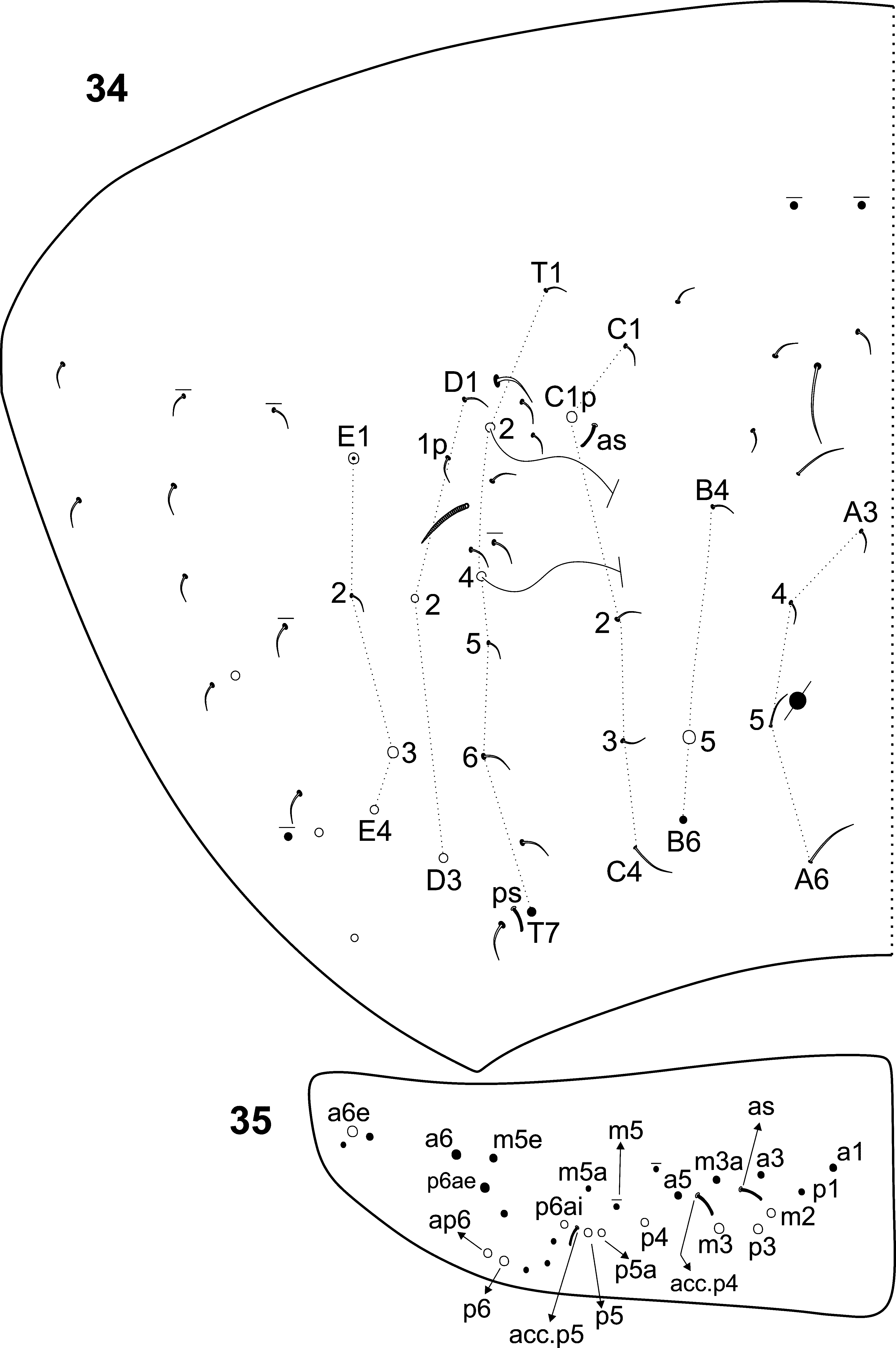
Abdomen chaetotaxy ( Figs 31‒35 View FIGURES 29–33 View FIGURES 34–35 ). Abd. I with 1 ms, 6 anterior (a1–3, a5–6; a1 and a1a absent in some specimens), 5 medial (m2–6) and 2 posterior (p5–6) mic ( Fig. 31 View FIGURES 29–33 ). Abd. II with 1 anterosubmedial sens (as), 4 anterior (a2‒3, a5–6), 7 medial (m2–7), and 5 posterior (p4–p7e?) chaetae. ‘ el ’ present as a smooth mic ( Fig. 32 View FIGURES 29–33 ). Abd. III with 1 ms, 1 anterosubmedial sens (as), 5 anterior (a2–3, a5–7), 7 medial (m2–5, am6, pm6, m7) and 4 posterior (p3, p5–7) chaetae ( Fig. 33 View FIGURES 29–33 ). Abd. IV with 1 posterior (ps) and 1 anterosubmedial sens (as) and several median and lateral smooth mic and mes; mac formula as 1 ‘B’ (B5), 1 ‘C’ (C1p), 2 ‘D’ (D2–3) and 2‒3 ‘E’ (E1, E3–4, E1 as mic in some specimens); central chaetae very similar to long sensilla, difficult to separate ( Fig. 34 View FIGURES 34–35 ). Abd. V with 1 anterosubmedial (as) and 2 accessory sens (acc.p4–5), 5 anterior (a1, a3, a5–6e), 6 medial (m2–3a, m5–5e), 3 posteroanterior (p5a, p6ai–6ae) and 6 posterior (p1, p3–6, ap6) chaetae; mac and mes similar in size on Abd. V ( Fig. 35 View FIGURES 34–35 ).
Legs ( Figs 36‒37 View FIGURES 36–43 ). Subcoxae I, II and III with 2, 2, 1 pseudopores, respectively. Trochanteral organ with about 8 spine-like chaetae ( Fig. 36 View FIGURES 36–43 ). Ungues with 4 inner teeth, two paired basal, 1 unpaired median and 1 minute unpaired distal; outer side with 1 median tooth. Unguiculi acuminate, with smooth lamellae. Tenent hairs smooth and capitate. Tibiotarsus III with a smooth inner distal chaeta, near the unguiculus ( Fig. 37 View FIGURES 36–43 ).
Collophore ( Figs 38‒40 View FIGURES 36–43 ). Anterior side with 2+2 large distal and at least 4+4 thinner ciliated chaetae ( Fig. 38 View FIGURES 36–43 ); posterior face with 1+1 large ciliated chaetae laterally, and 2+2 median smooth chaetae ( Fig. 39 View FIGURES 36–43 ); lateral flap with 2 smooth and 4 weakly ciliated chaetae ( Fig. 40 View FIGURES 36–43 ).
Furcula ( Figs 41‒43 View FIGURES 36–43 ). Manubrium ventral side with 1+1 distal ciliated chaetae; dorsal side with two types of ciliated chaetae: regular and distally weakly ciliated (present on dorso-lateral face of manubrium); manubrial plate with 4 ciliated chaetae and 2 pseudopores ( Fig. 41 View FIGURES 36–43 ). Dens dorsally with rows of ciliated chaetae ( Fig. 42 View FIGURES 36–43 ). Mucro bidentate, with apical tooth slightly longer than subapical; mucronal spine without spinelet, reaching the basal tooth ( Fig. 43 View FIGURES 36–43 ).
Etymology. The name refers to the number of ocelli in each eyepatch of the new species. Pseudosinella triocellata sp. nov. is the first recognized Brazilian species of Pseudosinella with 3+3 eyes.
Distribution and habitat. Same distribution and habitat of Cyphoderus equidenticulati sp. nov. Additionally, Pseudosinella triocellata sp. nov. was also found in spots of more forested habitats, locally known as “Cerradão”, with a dense tree covering which filters out a large part of the sunlight, and a thick layer of leaf litter on the soil.
Remarks. Pseudosinella triocellata sp. nov. resembles mostly P. stewartpecki Katz, Soto-Adames & Taylor, 2016 (in: Katz et al. 2016) and P. intermixta Folsom, 1924 , both from Galápagos Islands, by 3+3 eyes, presence of Ant. IV apical bulb, 1 mac on Th. II (p3) and absence of mac on Th. III. The new species still resembles P. stewartpecki in labial triangle chaetatotaxy with chaetae M1, E, L1, L2 ciliated, r reduced and A1–3 weakly ciliated (described as ‘serrate’ by Katz et al. 2016); 4+4 chaetae and 4+4 scales on head’s ventral groove; Abd. I without mac; Abd. II with 2 mac (m3, m5); Abd. III with 2 mac (pm6, p6). The description of P. intermixta by Folsom (1924) is poorly detailed and don’t allows more precise comparison of dorsal chaetotaxy features or any other structure. Most characters used here were provided by Katz et al. (2016).
The new species is unique by the combination of: 1) dorsal head with 4‒5 mac: A0, A2a–3, S2 (4 in P. stewartpecki— A0, A2a–3, and P. intermixta— A0, A2–3, M2); 2) labial triangle chaetae A4–5 weakly ciliated (smooth in P. stewartpecki ); 3) trochanteral organ with 8 spine-like chaetae (5 in P. stewartpecki ); 4) Collophore anterior side with 6+6 distal ciliated chaetae, 2+2 large (4+4 or 5+ 5 in P. stewartpecki ); and posterior side with 4 distal smooth chaetae (5 in P. stewartpecki ); 5) Ungues with 4 inner teeth: one pair of equally sized basal teeth, 1 median, and 1 minute distal tooth (3 inner teeth in P. stewartpecki : one pair of unequally sized basal teeth and 1 distal tooth; 3 inner teeth in P. intermixta : one pair of minute basal teeth and 1 large distal tooth); 6) Unguiculi acuminate, with smooth lamellae (unguiculi with at least 2 or more minute teeth in P. stewartpecki ). In addition, the dorsal chaetotaxy of Abd. IV of the new species also differs from P. stewartpecki , with C1p as mac (mic in P. stewartpecki ); B6, T6, E2 as mic (mac in P. stewartpecki ); and F1 absent (present as mac in P. stewartpecki ). A summarized comparison among Pseudosinella triocellata sp. nov., P. stewartpecki and P. intermixta is shown in Table 1.
Data based in: 1 Katz et al. (2016). Legends: - = absent,? = unknown, mic = microchaeta(e), mac = macrochaeta(e).


No known copyright restrictions apply. See Agosti, D., Egloff, W., 2009. Taxonomic information exchange and copyright: the Plazi approach. BMC Research Notes 2009, 2:53 for further explanation.