
Pseudopachylus martensi, Kury, Adriano B., 2006
|
publication ID |
https://doi.org/ 10.5281/zenodo.174085 |
|
publication LSID |
lsid:zoobank.org:pub:CA5E2CC2-6382-41A3-9437-65C177DBDDAB |
|
persistent identifier |
https://treatment.plazi.org/id/039E87EA-FF82-FFE1-3437-FEF02D18E66D |
|
treatment provided by |
Plazi |
|
scientific name |
Pseudopachylus martensi |
| status |
sp. nov. |
Pseudopachylus martensi sp. nov.
Figs 1–10 View FIGURES 1 – 3 View FIGURES 4 – 8 View FIGURES 9 – 10
Type material. ♂ holotype ( MNRJ 4435), Brazil, São Paulo, São José do Barreiro, 30.x. 1967, O. A. Roppa leg.
Diagnosis. Eye mound with thick straight spine instead of typical Pseudopachylus hook. All scutal areas and free tergites smooth and unarmed. Ventroretrolateral apophysis of coxa IV simple, straight. Trochanter IV unarmed. Tarsal formula: 4 (3)/ 5 (3)/ 5 / 5. This species is probably most closely related to Pseudopachylus alticola ( H. Soares, 1945) because they share the atypical armature of eye mound, both can be distinguished mainly by the armature of coxa and trochanter IV of male.
Etymology. Species name honors the distinguished German arachnologist Dr Jochen Martens who contributed immensely to the knowledge of the order Opiliones .
Description of ♂ holotype ( MNRJ 4435)
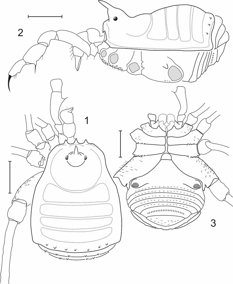
Dorsum ( Figs 1–2 View FIGURES 1 – 3 ): Projections of frontal margin of carapace absent. Anterolateral triangular lobes on carapace absent. Lateral margins of carapace clearly divergent to posterior end. Outline of carapace not projected anterolaterally. Outline of scutum at carapace not pointed. Outline of scutum at groove II not pointed. Lateral margin of opisthosomal scutum smooth and unarmed marginally. Posterior constriction of opisthosomal scutum lost: scutum without any constriction, with sides straight and parallel, giving the scutum the shape of a bell. Posterior border of scutum as wide as the rest with one row of tubercles. Eye mound very high, campaniform, removed from the anterior border of carapace, with unpaired erect pointed spine. Frontal hump low, moderately developed. Mesotergal area I constricted by scutal groove but not divided into two halves and clearly procurved. Mesotergal area I as long as any of the others. Mesotergal area III unarmed. Mesotergal area IV unarmed. Tegument of dorsal scutum fine granular. Free tergites with a transverse row of tubercles each.
Venter ( Fig. 3 View FIGURES 1 – 3 ): Tubercles of scutum and legs I –IV with short bristles. Stigmatic opening placed on a discrete mound. Stigmatic area discrete from coxa IV, separated by a wellmarked groove. Area surrounding stigmata convex, keeping outline sternitescoxa. Sternite III not projected laterally nor anteriorly nor posteriorly. Free sternites with a transverse row of tubercles each.
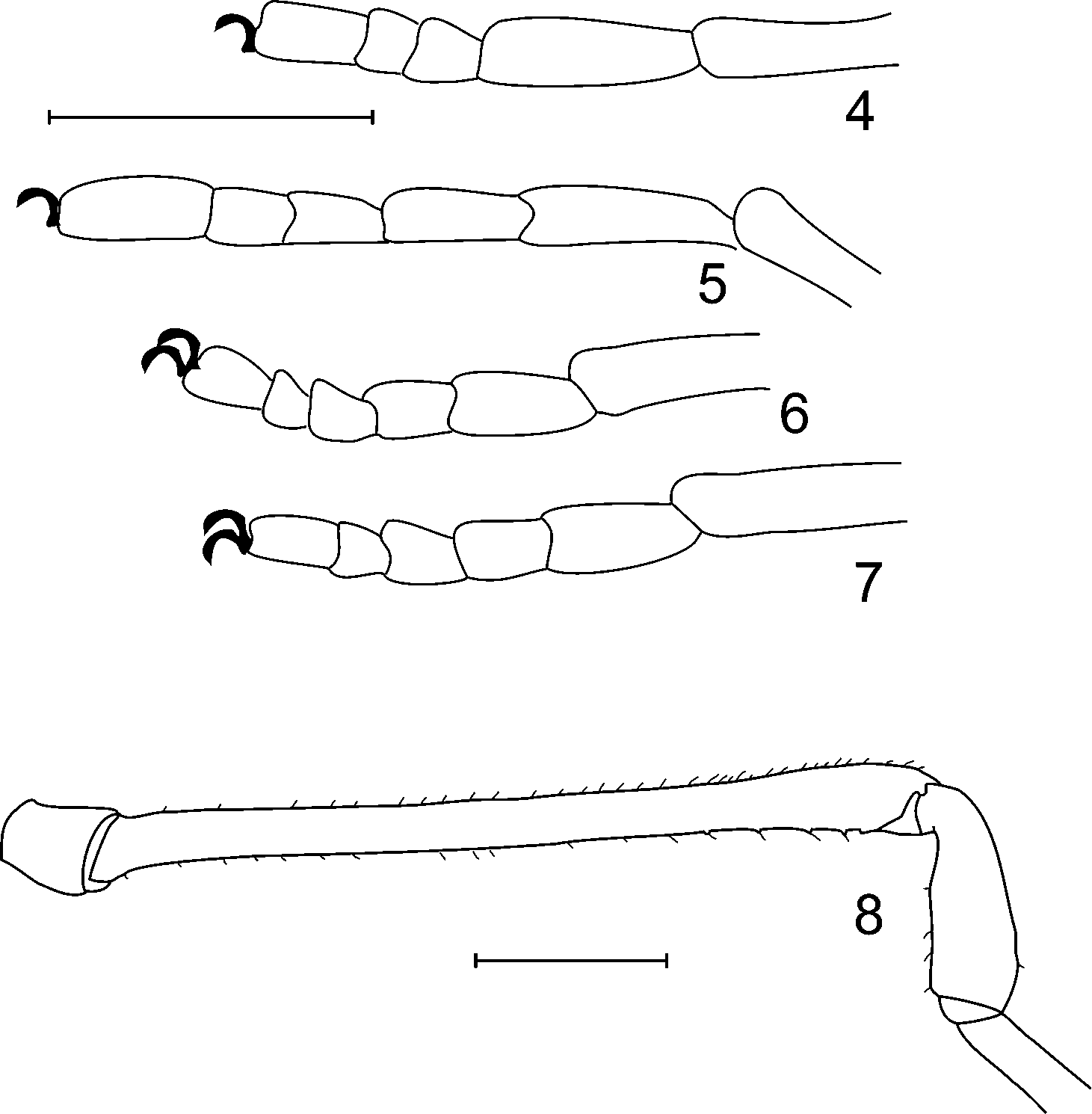
Appendages ( Figs 1–8 View FIGURES 1 – 3 View FIGURES 4 – 8 ): Length of basichelicerite much shorter than carapace. Cheliceral hand unarmed. Cheliceral hand and basichelicerite weak and slender. Pedipalpus well developed, at least as long as dorsal scutum. Pedipalpal femur cylindrical, concave mesobasally, with two ventral setiferous tubercles and subapical mesal setiferous tubercle. Pedipalpal patella unarmed. Distalmost ectal spine of pedipalpal tibia longer than the others ( Fig. 2 View FIGURES 1 – 3 ). Calcaneus of metatarsus I not swollen. Coxa II in situ widely surpassing coxa III in length. Coxa IV very small and short, attaining area II in dorsal view ( Fig. 1 View FIGURES 1 – 3 ). Lateral margin of coxa IV oblique to the main axis of body. Surface of coxa IV finely granular. Coxa IV with dorso apical acuminate oblique apophysis. Coxa IV on ventral surface, retrolateral border with blunt commalike conical apophysis. Coxa IV subapical retrolateral without special features. Trochanter IV ventral retrolateral without any subbasal (alpha) or distal (beta) apophysis. Trochanter IV ventroapically unarmed. Femur IV (probably sexually dimorphic as in all other Pseudopachylus ) straight, unarmed, elongate, not incrassate, without serrulae or pectination, on dorsal surface with fine granulation. Patellae II –IV short. Tibia IV as thick as femur IV, unarmed on retrolateral surface. Tarsal formula: 4 (3)/ 5 (3)/ 5 / 5.
Color: Body and appendages more or less uniform reddish dark yellow with some brown mottling, coxae and trochanters I –IV clearly lighter. Color pattern of femora I –IV uniform, without lighter areas or rings.
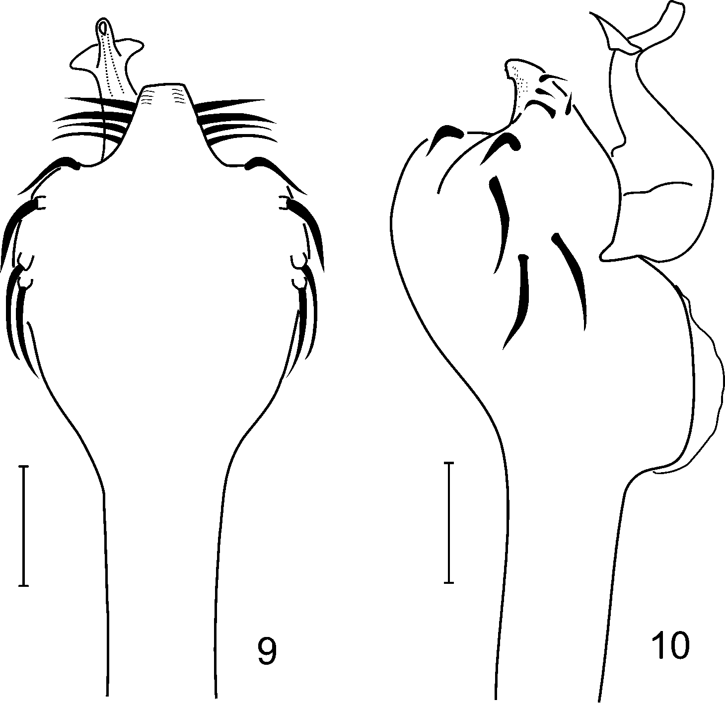
Penis ( Figs 9–10 View FIGURES 9 – 10 , dorsal view not given as it does not bring any relevant supplementary information): Ventral plate of penis with basal portion immensely swollen. Lamina parva with distal patches of rough granules, trapezoid, clearly marked and normal sized. Paired ventral subapical projections of ventral plate absent. Dorso apical hyaline body of truncus present. Four pairs of setae of lamina parva short and straight. Four pairs of setae of basal portion of ventral plate very long and pointing basally roughly similar as in Agoristenidae . Shape of lamina parva clearly trapezoid. Supplementary dorsal shield on glans absent. Position of stylus in relation to the glans apical bent by 90 degrees. Attachment of flabellum to stylus skirtlike.
Distribution. BRAZIL. SÃO PAULO. São José do Barreiro (coordinates 22.6333, 44.5833), 600 m. WWF Ecoregion: NT0703 (Campos Rupestres montane savanna). Biome: Tropical & Subtropical Grasslands, Savannas, & Shrublands. This portion of NT0703 is an “island” surrounded by the Serra do Mar coastal forests (NT0160), therefore it is not certain that the habitat of the species is savanna. It could also be forest patches amidst a savanna biome.
No known copyright restrictions apply. See Agosti, D., Egloff, W., 2009. Taxonomic information exchange and copyright: the Plazi approach. BMC Research Notes 2009, 2:53 for further explanation.