
Pseudamiops springeri Gon & Bogorodsky
|
publication ID |
https://doi.org/ 10.11646/zootaxa.3701.1.8 |
|
publication LSID |
lsid:zoobank.org:pub:59C67BFE-FD32-45F8-A15E-095B6216DC07 |
|
DOI |
https://doi.org/10.5281/zenodo.5678073 |
|
persistent identifier |
https://treatment.plazi.org/id/0FA3AC74-2F2E-47F6-8C77-D57E3B859383 |
|
taxon LSID |
lsid:zoobank.org:act:0FA3AC74-2F2E-47F6-8C77-D57E3B859383 |
|
treatment provided by |
Plazi |
|
scientific name |
Pseudamiops springeri Gon & Bogorodsky |
| status |
sp. nov. |
Pseudamiops springeri Gon & Bogorodsky View in CoL , new species
Figs. 1 View FIGURE 1 a–c, 2a–c; Table 1 View TABLE 1
Holotype. USNM 262240, 24.55 mm, Gulf of Aqaba, Egypt, Al-Hamira, 0–16 m, V.G. Springer et al., 19 July 1969.
Paratypes. KAUMM 8, 24 mm, Saudi Arabia, Al Wajh bank, 25°35'52.86" N, 36°41' 01.80" E, rotenone, 5 m, S.V. Bogorodsky & T. Alpermann, 12 June 2013; SAIAB 190571, 15.15 mm, Saudi Arabia, Rabigh-Masturah, unnamed isolated reef, 23°02.839' N, 38°46.621' E, 18 m, S.V. Bogorodsky, 8 April 2011 (originally KAUMM 6); SMF 34907, 24.8 mm, Saudi Arabia, 60 km south of Al Wajh, 26°03'29.80'' N, 36°35'41.00'' E, fringing reef, rotenone, 11 m, S.V. Bogorodsky & T. Alpermann, 13 June 2013; USNM 262248, 26.85 mm, Egypt, Strait of Jubal, NW edge of Sha’b al Fanadir Reef, rotenone, 0–6 m, H.A. Feldman et al., 7 January 1965.
Comparative material. Pseudamiops pellucidus : SAIAB 787, 21.5 mm, paratype, Baixo Pinda, Mozambique; SAIAB 789, 3: 27.5–33.2 mm, paratypes, Ibo, Mozambique; SAIAB 790, 33.4 mm, paratype, Cape Delgado, Mozambique; SAIAB 26189, 5: 28.5–30.7 mm, non-types, Baixo Pinda, Mozambique. Pseudamiops diaphanes : BPBM 37247, 29.2 mm, holotype, Oahu, Hawai’i. Pseudamiops phasma : BPBM 12651, 33.2 mm, holotype, Nuku Hiva, Marquesas Islands. Pseudamiops sp. ROM 907CS, 16.6 mm, Chagos Archipelago, photograph of cleared and stained specimen.
Diagnosis. Dorsal-fin rays VI + I,8; anal-fin rays II,8; pectoral-fin rays 13; preopercle edge smooth or with 3– 4 minute serrations ventrally; pseudobranch present, with 2–4 filaments; no canine teeth on vomer; genital opening flanked by two slender papillae, one in front and one behind it ( Fig. 1 View FIGURE 1 a); anterior nostril with skin flap, about twice nostril diameter, posteriorly on nostril’s rim, reaching at most half way to posterior nostril; supraneural bones absent.
In alcohol, body cream white to yellowish dark brown; peritoneum with large stellate melanophores usually visible through abdominal body wall; fins pale.
After death, body of smaller paratype was opaque white ( Fig. 1 View FIGURE 1 b), gradually becoming translucent posteriorly and with small dusky spot behind eye on temporal area; gills partially visible through translucent opercle; large peritoneal melanophores clearly visible through abdominal body wall; pupil black; iris silvery, sometimes with irregular blackish markings, denser dorsally; fins clear.
In life, body and head transparent, with red gills, large peritoneal melanophores, series of eggs (opaque white, ovate objects), dark kidney and vertebral column clearly visible; iris grayish brown ( Fig. 1 View FIGURE 1 c).
Description. Proportional measurements of the holotype and paratypes as percentage of the standard length are given in Table 1 View TABLE 1 . Dorsal-fin rays VI + I,8; anal-fin rays II,8; first soft ray in second dorsal and anal fins unbranched; last soft ray in second dorsal and anal fins split to base; pectoral-fin rays 13, first two upper and lower rays un-branched (see Remarks below); principal caudal-fin rays 9+8, mostly damaged; procurrent caudal-fin rays 7 dorsal and 6 ventral (7–8 + 7, respectively); total gill rakers 3 + 10; developed gill rakers 1 + 6; gill rakers on ceratobranchial 7 (6–7); lateral line absent; one median predorsal scale (all lost); body scales lost (cycloid scales in about 24 lateral scale series in largest paratype). Vertebrae 10+14. Supraneural bones absent and first dorsal-fin pterygiophore inserted between 3rd and 4th neural spines.
Pseudamiops springeri P. pellucidus
Body slender, its depth 5.7 (4.9–5.85) in SL and its width 1.6 in its depth; head length 3.1 (3.2–3.4) in SL; snout longer than eye diameter, 4.3 (4.1–4.4); eye moderate, its diameter 5.9 (4.5–5.2); interorbital width 5.0 (4.2– 4.3), all in head length.
Mouth large, oblique, upper lip distinctly thicker anteriorly; upper jaw length 1.8 (1.7–1.8) in head length; maxilla reaching posteriorly well beyond eye, to about middle of cheek; its vertical edge truncate to slightly indented and its ventral edge with short down-pointing spine posteriorly ( Fig. 2 View FIGURE 2 a); maxilla depth 3.4 (3.65–4.2) in upper jaw length; lower jaw sub-terminal, 1.55 (1.6–1.65) in head length, fitting into upper jaw, leaving outer premaxillary teeth exposed; upper jaw with one large symphyseal canine (about half pupil size) on left side ( Fig. 2 View FIGURE 2 c) and two close together on right side; 2–3 irregular series of small teeth starting in front of symphyseal canines and running posteriorly along jaw, those at symphysis slightly enlarged; lower jaw with 3–4 series of small teeth tapering to 2 series posteriorly; teeth of inner row enlarged, starting with large canine at middle of gape ( Fig. 2 View FIGURE 2 c) followed by smaller caniniform teeth posteriorly; vomer and palatines with single, irregular series of small teeth.
Posterior nostril small, oval, next to front edge of eye at its mid-level, about 4–5 times in pupil diameter; anterior nostril smaller, almost at tip of snout (excluding upper lip), with postero-lateral skin flap about twice its diameter ( Fig. 2 View FIGURE 2 b). Head sensory canals with small, simple pores; nasal and orbital parts of supraorbital canal with five pores ( Fig. 2 View FIGURE 2 a); first pore medial to anterior nostril; second pore medial and anterior to rear nostril; last three pores above eye with middle pore removed medially; infraorbital canal with seven pores ( Fig. 2 View FIGURE 2 a), first at midway between posterior nostril and upper jaw, pores 2–5 along ventral edge of suborbital bones with pores 3–4 closer to each other, and pores 6–7 behind eye; mandibular section ( Fig. 2 View FIGURE 2 b) of preoperculo-mandibular canal with five ventrally directed pores, fourth pore paired. Head with network of free neuromasts ( Figs. 2 View FIGURE 2 a, b): top of head with 2 medial longitudinal rows from near tip of snout to above posterior edge of orbit, each with at least 20 neuromasts; rows crossed by 6 transverse series of similar neuromasts spread between tip of snout and interorbital area, more or less at equal distance from each other; each transverse series with 8–9 medial neuromasts (i.e. between longitudinal rows) and 4–7 lateral ones on left and right sides, respectively; anterior half of posterior nostril encircled with free neuromasts; mandibular section of preoperculo-mandibular canal with longitudinal series of free neuromasts and at least five transverse series; some free neuromasts present on mental area, behind tip of lower jaw. No free neuromasts visible on body of holotype due to lost skin and scales (see also Remarks below).
Fin spines slender and feeble; first dorsal spine 5.3 (4.0) in head length; second and third spines damaged, but third dorsal spine of smallest paratype 2.8 in head length; spine of second dorsal fin damaged (2.8–3.9) and longest dorsal-fin ray, second or third, 1.8 (1.5–1.9), both in head length. First and second anal-fin spines damaged in holotype and largest paratype; in other paratypes, first anal-fin spine 2.3–2.9 in second spine and second anal-fin spine 2.7–3.4 in head length; longest anal-fin ray, second or third, 1.9 (1.65–2.0) in head length. Pectoral fin reaching posteriorly over anus, its length 6.0 (5.05), and pelvic fin not reaching anus, its length 6.8 (6.3–6.7), both in SL; pelvic spine damaged (1.7 in pelvic-fin length). Caudal peduncle slender, its depth 3.1 (2.5–2.8) in caudal peduncle length and its length 3.5 (3.6–3.8) in SL. Caudal fin damaged (3.5-3.8) in SL.
Preopercle edge with 3–4 minute serrations ventrally; edges of preopercular ridge, posttemporal and suborbital bones smooth. Distances from snout to first dorsal-fin origin 2.5 (2.7–2.8), to second dorsal- and anal-fin origins 1.8, and to pelvic-fin insertion 3.3 (3.0–3.2), all in SL. Distance from anus to anal-fin origin 3.8 (3.4–4.7) in distance between anal-fin origin and pelvic-fin insertion; latter distance 4.1 (3.7–4.1) in SL. Genital opening with two slender papillae, one in front and one behind it ( Fig. 1 View FIGURE 1 a).
Colour in life and after death as described in Diagnosis above. In alcohol, body brown, head and cheek paler; large, stellate peritoneal melanophores faintly visible through abdominal body wall; single similar melanophore on temporal area of head and two others under skin of occiput; pupil black; fins pale.
Distribution. At present Pseudamiops springeri is known only from the Red Sea where it was collected in the northern part of the Gulf of Aqaba and the Strait of Jubal, both in Egypt, and north of Jeddah, Saudi Arabia.
Etymology. Pseudamiops springeri is named for Dr Victor G. Springer (USNM) who collected the holotype.
Remarks. The caudal fin region in radiographs of these small specimens was not clear enough to observe structural details. The first anal-fin spine is very weak and easily damaged, but its remnant is visible on a radiograph. The branching pattern of the pectoral-fin rays of the holotype is different in each of the fins. Undamaged rays of the left pectoral fin look un-branched, but seem to be regenerated. The middle rays of the right pectoral fin are clearly branched. Judging from both fins, at least the upper and lowermost two, possibly three or four rays are un-branched and this conforms to Smith’s (1954) observation that only the middle rays of the pectoral fin of Pseudamiops are branched. The pectoral and pelvic fins of the smallest paratype are proportionally longer, with the former reaching posteriorly over the base of the second anal-fin ray and the latter reaching close in front of the anus. The symphyseal canines on the upper jaw of the largest paratype (USNM 262248) are broken, but their relatively wide round base is recognizable on the postero-medial extension of the jaw behind symphysis. The lower jaw of this fish has two close-set large lateral canines at middle of gape; the equivalent tooth of the right side is missing and could be the large loose tooth found inside the mouth cavity of this specimen. In the smallest paratype (SAIAB 190571) the inner series teeth of the lower jaw are relatively larger and the lateral canines are only slightly larger than these teeth. The holotype lost all its scales. Some scales are present on the largest paratype, allowing for an estimate of the lateral scale series. The ventral maxillary spine of the smallest paratype is a very small, pointed triangular bony projection.
The largest paratype (USNM 262248) has 13 transverse series of free neuromasts dorsally from tip of snout to first dorsal-fin origin, but the last 5–6 are less obvious. The 6th series is on line with the posterior edge of the eye and the 7th follows the supratemporal head sensory canal. Between these two transverse series there are six short longitudinal ones: one series along the anterior part of the temporal canal on each side of the head and another four closer together medially; a short transverse series connects each of the temporal longitudinal series to the next longitudinal series medially. A series of free neuromasts follows the preopercular section of the preoperculomandibular canal; the mandibular section has six transverse series, the last more or less in line with the ventral maxillary spine; the mandibular longitudinal series is divided into two parts, one from the 2nd mandibular pore to the 4th transverse series and another from the 6th transverse series to ventral preopercular pore. Preopercle with at least four longitudinal series of free neuromasts intersecting two vertical ones; the uppermost longitudinal series immediately below the 7th infraorbital pore and lowermost series behind the lower end of maxilla. Several free neuromasts are present between lower edge of eye and 4th–5th infraorbital pores (skin near other pores is damaged). Although many scales are missing along with the associated skin and neuromasts, short vertical series of free neuromasts were observed in several places on the body of this paratype. These included four series below first dorsal-fin base and three below second dorsal-fin base; 10 series near the ventral edge of the body from next to genital opening to a short distance anterior to lower caudal-fin base; and two series proximally on middle caudalfin rays. In addition, a short longitudinal series was found extending anteriorly from caudal-fin base along middle of body.
Like other species of the genus, P. springeri is very cryptic, living deep inside caves. The smallest paratype was collected about 2–3 m from the entrance of a cave on a seaward reef at a depth of 18m, while other paratypes (KAUMM 8 and SMF 34907) were found in caves at 5 and 11 m depth, respectively.
Comparisons. The number of pectoral-fin rays easily separates Pseudamiops springeri (13 rays) from its Pacific Ocean congeners including P. gracilicauda (15–16), P. diaphanes (16–18 rays) and P. phasma (19 rays). Pseudamiops springeri is closest to Smith’s (1954) P. pellucidus ( Fig. 1 View FIGURE 1 d, Fig. 2 View FIGURE 2 d), so far known from the east coast of Africa, and the unidentified Pseudamiops sp. from Chagos Archipelago (Winterbottom et al. 1989). The photo of a 21 mm fish in Winterbottom et al. (1989: fig. 181) suggests a dark peritoneum and their description makes no mention of a large canine on the vomer of their specimens. In addition, a cleared and stained specimen (ROM 907CS) from the Chagos collection shares with P. springeri the absence of a large canine tooth on vomer, 13 pectoral-fin rays and the first dorsal-fin pterygiophore position behind the third neural spine. However, it seems to differ from other congeners in having a total of eight rays in the anal fin, of which the first two are on the first analfin pterygiophore, an unusual condition in the Apogonidae . The first of these is a short spine, but the nature of the second, relatively long ray is still undecided (R. Winterbottom, pers. comm.). If it is a spine, the Chagos fish is unusual in the genus and family in having anal fin of II,7; a ray will confirm the original observation of Winterbottom et al. (1989) of an anal fin with a single spine.
Pseudamiops pellucidus and P. springeri share the same number of soft dorsal-fin rays, a similar count of developed gill rakers, the placement of the first dorsal pterygiophore behind the third neural spine, and the absence of supraneurals. Smith (1954, 1961), followed by Gon (1986), reported a consistent count of 14 pectoral-fin rays for P. pellucidus , but we counted 13 rays on one pectoral fin of two of Smith’s (1954) paratypes (SAIAB 789 and 790). Pseudamiops pellucidus differs from P. springeri in having usually nine soft anal-fin rays, 33 transverse series of scales, a large canine (occasionally twin teeth) on the vomer ( Fig. 2 View FIGURE 2 d), no skin flap on the anterior nostril, and a reddish pupil. The peritoneum of alcohol-preserved P. pellucidus varies from pale to having scattered dark spots laterally, but in the latter case the spots are not visible through the body wall. By contrast, in alcoholpreserved P. springeri large, dusky stellate melanophores are visible through the body wall. In addition, the caudal peduncle of P. pellucidus is narrower (3.3–4.7 in peduncle depth vs. 2.5–3.1 in P. springeri ) and longer (3.1–3.6 in SL vs. 3.5–3.8 in P. springeri ). Several other morphometric characters (e.g. longest dorsal- and anal-fin rays, see Table 1 View TABLE 1 ) could separate these two species, but considering the small sample size and the size of the specimens all such characters should be confirmed by a larger sample.
The head sensory canal pores of P. springeri ( Figs. 2 View FIGURE 2 a–c) are more conspicuous than those of P. pellucidus ( Fig. 2 View FIGURE 2 d). Pore configuration in P. springeri is similar to that of Pseudamiops pellucidus as described and illustrated by Bergman (2004) based on a single specimen from Mauritius (USNM 349773), except for the mandibular section of the preoperculo-mandibular canal. Bergman (2004) described, but did not illustrate, two pairs of pores at the anterior end of the mandibular canal, and four “nearly equidistant” pores along the lateral margin of the canal which she did illustrate (see her fig. 38C), as well as a single median longitudinal series of free neuromasts. We could not identify the anteriormost pair of pores in our specimens of P. springeri and five P. pellucidus (SAIAB 26189) from the type locality. If they do exist they are probably minute. Pseudamiops springeri has five pores that open ventrally in the mandibular canal on each side of the lower jaw, the anteriormost in the same position as in P. pellucidus of Bergman (2004: fig. 38C) and most likely part of her second, more posterior pair of pores (presumably, one on each side of the lower jaw). Smith (1954) and Randall (2001) found five mandibular canal pores in P. pellucidus and P. phasma , respectively. The distance between the mandibular canal pores increases posteriorly up to the fourth one (= Bergman’s third pore) while the distance from the latter to the last (fifth) pore is about the same as between the third and the fourth pores. This is similar to the pore arrangement described by Smith (1954) for P. pellucidus . A notable difference between Bergman’s fish and P. springeri is the paired fourth pore. Of the five P. pellucidus specimens in SAIAB 26189, at least two had the same number and configuration of mandibular pores as in P. springeri , including a paired fourth pore, and all had transverse series (branches in Smith 1954) of free neuromasts on each side of the median longitudinal series. Paired mandibular pores are not unusual in pseudamine fishes and are found in species of Gymnapogon and Pseudamia (Bergman 2004: figs. 35C and 37C, respectively; Smith 1954: Pseudamia gelatinosa ). The two Pseudamiops species also seem to differ in the size of the ventral pore of the preopercular section of the preoperculo-mandibular canal. In P. springeri this pore is not different from the pores of the mandibular section of the canal, but in P. pellucidus it is apparently much larger, breaking the confluence between the two parts of the canal (Bergman 2004).
The depiction of a rounded caudal fin in P. springeri ( Fig. 1 View FIGURE 1 a, dotted line) is based on the caudal-fin shape of this fin in one of the paratypes ( Fig. 1 View FIGURE 1 b) and in other species of Pseudamiops (Lachner 1953; Smith 1954, 1961; Ida & Moyer 1974; Winterbottom et al. 1989; Randall 1998, 2001). The small depressions on the dorsal and ventral edges of the caudal peduncle of the paratype of P. pellucidus in Fig. 1 View FIGURE 1 d were caused by the string the collectors, JLB and MM Smith, used to tie a label to this fish.
In Fig. 1 View FIGURE 1 c and in Randall’s (1998) underwater photos of P. gracilicauda and P. diaphanes the blood red gills are clearly visible through the thin transparent gill cover. In his photo of P. diaphanes after death the gill cover is whitish masking part of the gills, as is the case in the paratypes of P. springeri ( Fig. 1 View FIGURE 1 b), and in tank photos of Pseudamiops from Chagos Archipelago (Winterbottom et al. 1989) and of P. phasma from the Marquesas Islands (Randall 2001). This suggests that the opercular bones of these fishes become cloudy after death.
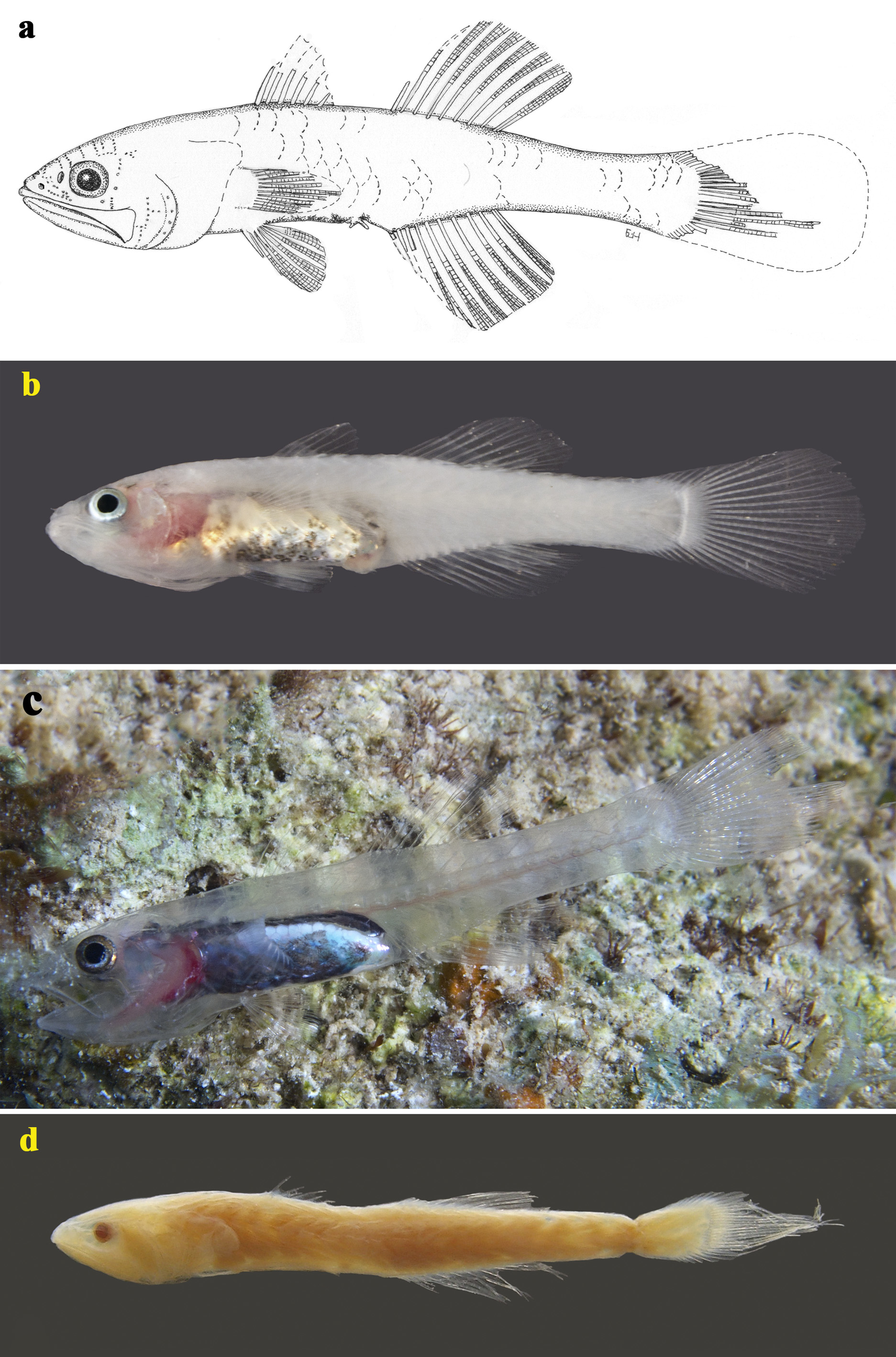

TABLE 1. Proportional measurements (expressed as percentage of the standard length) of Pseudamiops springeri and P. pellucidus.
| Standardlength(mm) Bodydepth Bodywidth Lengthofhead | Holotype USNM 262240 24.55 17.5 10.8 32.6 | Paratype USNM 262248 26.85 19.2 11.7 31.3 | Paratype SAIAB 190571 15.15 20.3 13.2 31.0 | n = 10 21.5–33.4 11.6–18.6 8.5–11.6 27.6–34.2 |
|---|---|---|---|---|
| Lengthofsnout Eyediameter Interorbitalwidth Length of upper jaw Length of lower jaw | 7.5 5.5 6.5 17.7 21.0 | 7.3 6.15 7.45 17.7 19.0 | 7.6 5.9 7.3 17.95 19.3 | 6.0–7.9 4.5–5.7 4.3–6.5 16.3–22.8 18.0–24.9 |
| Maxillawidth Length of first dorsal spine Length of second dorsal spine | 5.2 6.1 Damaged | 4.8 7.8 Damaged | 4.3 Damaged Damaged | 3.0–4.4 6.0–9.7 10.2–11.5 |
| Length of third dorsal spine | Damaged | Damaged | 11.2 | 9.55–9.9 |
No known copyright restrictions apply. See Agosti, D., Egloff, W., 2009. Taxonomic information exchange and copyright: the Plazi approach. BMC Research Notes 2009, 2:53 for further explanation.