
Pseudalataspora kovalevae, Kalavati, Chaganti, Mackenzie, Ken, Collins, Catherine, Hemmingsen, Willy & Brickle, Paul, 2013
|
publication ID |
https://doi.org/ 10.11646/zootaxa.3647.4.4 |
|
publication LSID |
lsid:zoobank.org:pub:D5B8E3C7-36D1-42EE-8785-4C2BB99BC62F |
|
DOI |
https://doi.org/10.5281/zenodo.6150787 |
|
persistent identifier |
https://treatment.plazi.org/id/03E287B9-D556-7361-FF12-A423FA862ED7 |
|
treatment provided by |
Plazi |
|
scientific name |
Pseudalataspora kovalevae |
| status |
sp. nov. |
Pseudalataspora kovalevae n. sp.
Material studied
Type host: Macruronus magellanicus Lönnberg, 1907
Site of infection: gall bladder
Localities, dates and depths: (1) off southern Chile, 55º 30´S, 71º 30´W, October 2007, 350m; (2) off Chiloe Island, Chile, 43º 0 0ˏS, 73º 0 0ˏW, June 2007, 300m; (3) west of Falkland Islands, 51º 00´to 52º 30´S, 62º 00´to 62º 30´W, October 2007, 200– 250m; (4) west of Falkland Islands, 51º 00´to 52º 30´S, 62º 00´to 62º 30´W, October 2009, 200– 250m.
Type locality: (1).
Prevalence: (1) 40% (4 of 9); (2) 64% (16 of 23); (3) 23% (7 of 30); (4) 57% (24 of 42).
Host length range: 25–42 cm.
Collection numbers: NHMUK 2012.3.19.1, 2012.3.19.2, 2012.3.19.3.
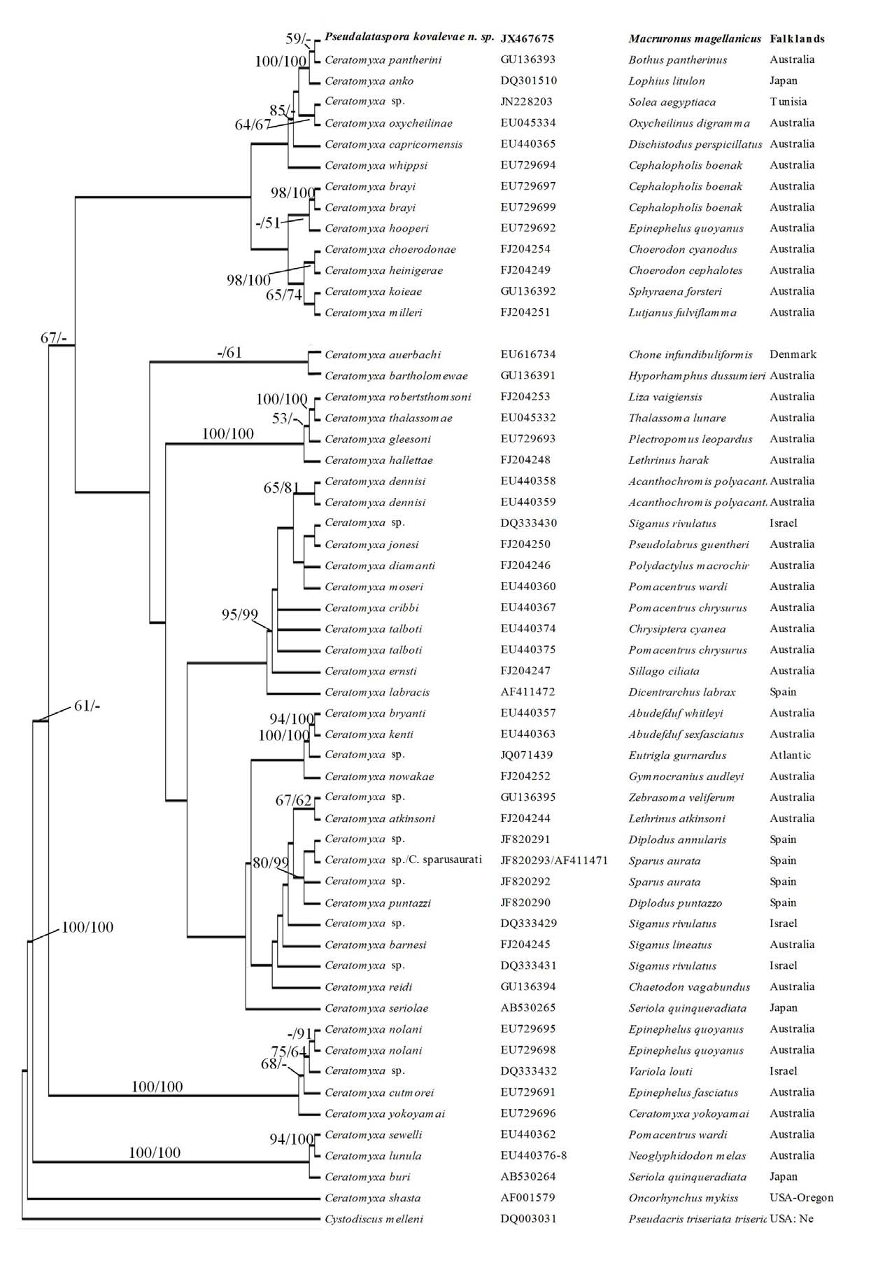
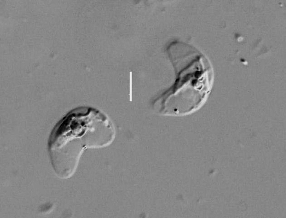
Morphological description. Sporoblast globular, disporic ( Fig. 1 View FIGURES 1 – 5 ). No pseudopodia observed. Ectoplasm dense and clearly demarcated from fine endoplasm. Dimensions, based on 10 fixed specimens: 28.4–38.4 x 28.0– 36.0.
Spore ( Figs. 2–6 View FIGURES 1 – 5 View FIGURE 6 ) triangular with sharply pointed tips in side view, flat anteriorly and curved in apical view. Sutural line prominent and raised. Sporoplasm deeply staining and binucleate. Valves drawn out into two delicate broad alate processes joined together at their proximal extremities to form a parachute-like structure over the valves. Polar capsules spherical, close together. Polar filament thick, number of coils not visible. Dimensions, based on 30 fixed spores, as ranges with means ± SD in parentheses: spore length 8.0–10.5 (9.1 ± 0.68); spore thickness, including alate processes 30.0–35.5 (31.8 ± 1.52), excluding alate processes 8.8–11.6 (9.0 ± 1.2); spore width 14.0–20.0 (15.7 ± 1.57); polar capsule diameter 2.5–3.0 (2.7 ± 0.16); polar capsule length: spore length = 1: 2.8–3.8; spore length: spore width = 1: 3.0–4.9.
Molecular results: Pseudalataspora kovalevae . PCR amplification of the myxosporean 18S rRNA gene from four infected gall bladders from sample (4) above generated a product of approximately 1650 nucleotides in both of the samples processed from each fish. Forward and reverse 18S rRNA gene sequences were obtained for PCR products from both samples from each individual host, and internal sequences from a single sample from each host. All sequences were identical. Two polymorphic positions were found. A final sequence of 1594 nucleotides was submitted to Genebank under Accession No. JX467675 View Materials .
The sequence was novel with respect to previously generated data from myxosporean species. Pseudalataspora kovalevae 18S rRNA gene sequence showed closest sequence identity to Ceratomyxa anko Freeman et al., 2008 ( DQ301510 View Materials ) and Ceratomyxa pantherini Gunter et al., 2010 ( GU136393 View Materials ) based on BLAST analysis with 92% and 97% identity over 92% and 85% of its sequence respectively. Following removal of gaps and ambiguous bases, 1034 nucleotides remained for phylogenetic analyses. Cystodiscus melleni (Jirku et al., 2006) was chosen as the outgroup based on its position in the distance tree generated following initial BLAST analyses of the P. kovalevae sequence.
MP and ML trees showed similar topologies, P. kovalevae grouping with C. pantherini and C. anko in both cases, with strong bootstrap support (100) ( Fig. 7 View FIGURE 7 ).
Discussion. Twelve species of the genus Pseudalataspora have been described from the gall bladders of marine fishes but none has been sequenced to date. In addition, we were made aware of an unpublished manuscript discovered in the late Professor Kovaleva’s laboratory in Kaliningrad, which includes the morphological description of Pseudalataspora pacifica Kovaleva and Grudnev , a new species found in the gall bladder of M. magellanicus caught off the coast of Chile in 1983. The authors described the new species as being similar to P. umbraculiformis , originally described by Gaevskaya and Kovaleva (1984) from the gadid fish Gaidropsarus mediterraneus (L., 1758) in the Northeast Atlantic. However, they considered the two species to differ sufficiently in certain features to be considered different species. The main differences they identified were in the shape and size of the spores, the diameter of the polar capsules, and the number of coils in the polar filament. Because the description of P. pacifica remains unpublished, it cannot be considered to be a valid species, but in recognition of Professor Kovaleva’s discovery we decided to name our new species after her. Table 1 compares P. kovalevae with both P. umbraculiformis and P. pacifica . Pseudalataspora kovalevae is from the same host and locality as P. pacifica and these two species are clearly more similar in morphology than either is to P. umbraculiformis .
Gunter et al. (2009) suggested that the Ceratomyxa clade currently contains a number of as yet undifferentiated genera and that additional sampling and further morphological and genetic data are needed to resolve the divisions within the group. As previously mentioned, there are currently no sequence data deposited in GenBank for any species of Pseudalataspora , and the new P. kovalevae 18S rRNA gene sequence grouped most closely with some Ceratomyxa species. Morphologically the spores of these two genera are very similar, with the presence or absence of delicate alate processes being the main distinguishing feature between them. The morphological and molecular similarities between these genera therefore suggest that they are closely related, but they are currently classified in different families. The alate processes are fragile and not always easily observed, which raises the possibility that some species currently assigned to the genus Ceratomyxa may turn out on closer inspection to belong in Pseudalataspora .
Feature P. umbraculiformis P. “ pacifica ”(*) P. kovalevae
Plasmodium shape Club-shaped or spherical, Spherical, disporous Spherical, disporous disporous
Plasmodium dimensions 18.0–21.0 29.28–32.28 28.4–38.4 x 28.0–36.0
Spore shape Wedge-shaped, expanded Rounded tops, sharply Triangular with pointed
anteriorly, narrowed curved anteriorly extremities in side view; curved posteriorly and flat anteriorly in apical view
Spore length 8.0–9.3 7.98–12.63 8.0–10.5
Spore thickness with alate 14.6–17.3 27.93–35.91 30.0–35.5
processes
Spore thickness without alate 6.7–8.0 Not given 8.8–11.6
processes
Spore width 14.6–17.3 Not given 14.0–20.0
Sutural line Clear and straight Clear and straight Prominent, raised
Polar capsule diameter 2.7 3.0-3.32 2.5–3.0
No. of polar filament coils 5 6 Number not observed
No known copyright restrictions apply. See Agosti, D., Egloff, W., 2009. Taxonomic information exchange and copyright: the Plazi approach. BMC Research Notes 2009, 2:53 for further explanation.