
Promephitis majori Pilgrim, 1933
|
publication ID |
https://doi.org/ 10.5252/g2016n4a5 |
|
publication LSID |
urn:lsid:zoobank.org:pub:528EED12-2BF4-4BA6-A3DC-7706ED8C5072 |
|
persistent identifier |
https://treatment.plazi.org/id/03D587C0-1E05-6F64-CEA5-FA56FCBEF80D |
|
treatment provided by |
Felipe |
|
scientific name |
Promephitis majori Pilgrim, 1933 |
| status |
|
Promephitis majori Pilgrim, 1933
HOLOTYPE. — Well-preserved cranium AMNH 20585 View Materials , from Quarry 1 in Samos, Upper Miocene, probably close to 7 Ma ( Koufos et al. 2009).
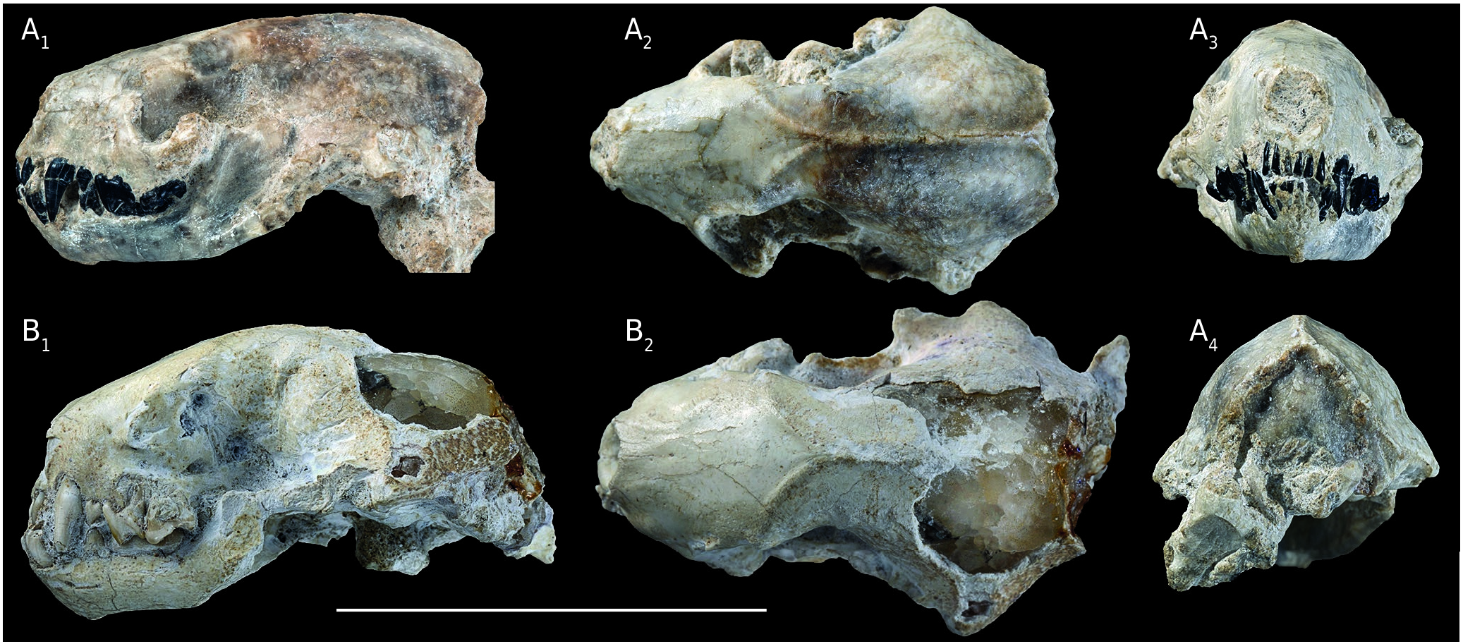
REFERRED MATERIAL FROM BULGARIA. — HD- 9505, cranium with mandible.
DIAGNOSIS. — A species slightly smaller than the type species, P. lartetii . Snout less inflated, occipital relatively higher.
DESCRIPTION
HD-9505 is an almost complete, well-preserved specimen that includes the cranium and attached mandible ( Fig. 1A View FIG ). Being virtually undistorted, it was chosen for CT-scanning, but poor contrast between bone and sediment and imperfect internal preservation made observation of fine structures very limited. The description below includes both its externally visible features and those only visible in the 3D reconstruction.
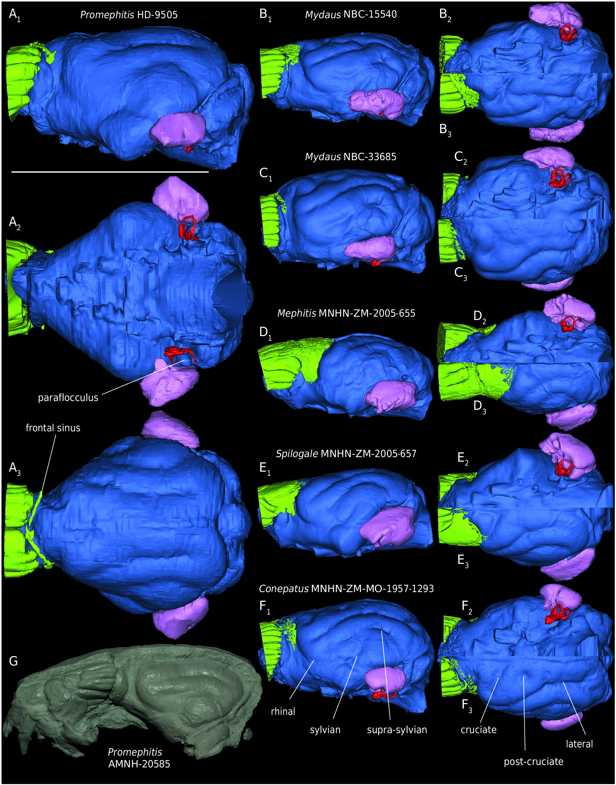
The skull is small (measurements: Table 1), about as large as that of Spilogale putorius (L.), and much more similar, in its short, broad face to American mephitids than to the south-Asian Mydaus . The facial sutures are not discernible. The nasal opening is slightly higher than broad; the anterior border of the orbit is located above the metacone of P4 but the face is short because the teeth are crowded rostrally. The snout narrows rostrally and is inflated, as in American mephitids, with no concave part, except shallow depressions between the canine and nasal aperture, and at the infra-orbital foramen, which is single and small. On the palate, the incisive foramina are large, and the choanae reach the level of the middle of M1; they are separated by a bony septum. The orbit is small, and limited posteriorly by a blunt but conspicuous post-orbital process of the frontal; the zygomatic arch is broken off. The temporal lines converge slightly behind the post-orbital constriction, where the skull is narrower than across the orbits, to form a long, low sagittal crest that diverges, at the top of the occipital, into almost straight lambdoid crests that bor- der a high, triangular occipital. The paroccipital processes are of moderate size, and antero-posteriorly compressed. A condyloid canal is present; the condylar (hypoglossal) foramen is located close to the occipital condyle. The jugular foramen is large. The large post-glenoid foramen is located above the anterior part of the long external auditory duct. There is no supra-meatal fossa. The bulla is poorly inflated. In ventral view, postero-laterally to it, the ventral floor of the mastoid sinus forms a smooth, concave area between the paroccipital and mastoid processes. The mastoid processes do not protrude significantly, but the skull is much broader at this level than across the paroccipital processes. Above them, the lateral walls of the braincase are laterally displaced by the expansion of the underlying mastoid sinus. In the endocranium, a distinctive feature is the very deep subarcuate fossa, housing a long paraflocculus ( Fig. 2A View FIG ).
Because of cracks in the walls of the braincase, the reconstructed parts of the endocranium lack precision, but the main features are discernible. The mastoid sinus is mostly lateral with respect to the brain and labyrinth and extends far laterally; it is roughly pyramidal, with an apex above the mastoid. The frontal sinus is restricted to minute canals.
The brain shape, as reflected by the endocranial reconstruction ( Fig. 2A View FIG ), is remarkably primitive. The olfactory bulbs are not very large and not quite distinct from the rest of the brain. All sulci are shallow; the rhinal sulcus is located high; the sylvian sulcus is not discernible; the supra-sylvian sulcus is moderately arched, and short; the lateral sulcus is barely arched; there is no cruciate sulcus.
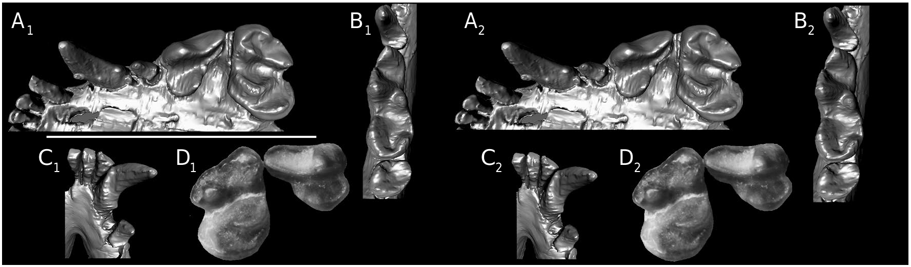
The CT-scan allowed examination of the well-preserved teeth ( Fig. 3 View FIG A-C). The upper incisors are small, with I3 slightly larger than the central ones. The canine bears small but distinct cuspids at the base of the mesiolingual and distal keels. On the left side only, there is a minute P2, displaced lingually by the closeness of C and P3. P3 is a simple tooth, broadest distally, with two fused roots. P4 is a short, triangular tooth. There is a minute parastyle; the paracone is much longer and taller than the metacone, and the blade is not notched. A crest descends from the distobuccal angle of the tooth, along its distal margin, towards the main lingual cusp, but is interrupted before reaching it. This cusp, which assumes the shape of a mesiobuccally to distolingually compressed cone along the distolingual margin of the tooth, is a hypocone, because it interlocks with the trigonid of m1. The protocone is indistinct as a cusp, but forms a small shelf instead. M1 is slightly longer than P4, but much broader than long. The paracone and metacone are subequal in size; an extremely strong buccal cingulum greatly increases the width of the tooth; its mesial half forms a high, strong ridge around the paracone, and almost forms a cusp near the middle of the tooth. The protocone is crescentic, and separated from the buccal blade by a narrow valley, but not too much weight should be given to this morphology, because there is much variation in the shape of the postprotocrista in modern forms; the hypocone consists of a long crescent along the distolingual edge of the tooth. Dental measurements are given in Table 2.
The mandibular symphysis has a moderately inclined ventral border. The masseteric fossa is limited ventrally by a sharp crest that descends antero-ventrally from the condyle; ventrally to this ridge, the angle of the mandible is reduced, so that the ventral border of the mandible clearly ascends from below m1 to the angular process, which is located close to the condyle.
The lower incisors are vertically inserted, and mesiodistally narrow, with i3 slightly larger than i1 and i2. The canine has a strong ridge around its lingual half.There are only two premolars, but on neither side is the area between p3 and the canine well preserved, so that the presence of a p2 cannot definitely be ruled out. The p3 and p4 have two roots and are tall and slender, with a steep mesial edge, a distinct cingulum mesially and a broad distal part, bordered by a robust cingulum on p4.
On m1, the trigonid is slightly longer than the talonid. The protoconid is by far the tallest cuspid; it is unconnected to the metaconid, but a cristid descends towards the hypoconid, from which it is separated by a notch. In connection with the long, oblique hypocone of P4, the metaconid is distinctly more posterior than the protoconid, and the paraconid is long and longitudinally oriented, so that the three cuspids form an angle distinctly larger than 90°. The talonid is deeply basined, the central depression occupying most of the area, and the ridge-like cuspids being marginally located. The hypoconid is taller than the entoconid; in front of the latter is a second lingual cuspid, separated from the metaconid by a notch, deeper in the right m1. The CT-scan shows that the left m1 has a single accessory buccal root while the right one also has an additional lingual one; we observed the same variability in CT-scanned Mydaus skulls, with NBC-33685 having a single, accessory lingual root and NBC-14540 having both lingual and buccal accessory roots.The m2 is shorter than broad, and also consists of a central basin circled by ridge-like elements.
No known copyright restrictions apply. See Agosti, D., Egloff, W., 2009. Taxonomic information exchange and copyright: the Plazi approach. BMC Research Notes 2009, 2:53 for further explanation.
|
Kingdom |
|
|
Phylum |
|
|
Class |
|
|
Order |
|
|
Family |
|
|
Genus |