
Praunus flexuosus (Muller, 1776)
|
publication ID |
https://doi.org/ 10.1093/zoolinnean/zlac083 |
|
DOI |
https://doi.org/10.5281/zenodo.7814237 |
|
persistent identifier |
https://treatment.plazi.org/id/D96287D1-4B76-3214-D1E0-15CEFBE6114D |
|
treatment provided by |
Plazi |
|
scientific name |
Praunus flexuosus |
| status |
|
PRAUNUS FLEXUOSUS View in CoL View at ENA
The first and second thoracopods (Thp1 and Thp2) are modified in comparison to the third to eighth pairs (Thp3– 8) of pediform thoracopods. The following descriptions relate only to Thp1, based on a detailed examination of the right Thp1 of five male individuals of P. flexuosus .
Cuticle and skeletal structures of Thp 1 in P. flexuosus
The more or less rectangular protopod (more than twice as wide as long) consists of coxa and basis. The thorax cuticle proximal to the coxa is robust medially, anteriorly and posteriorly, but membranous laterally.
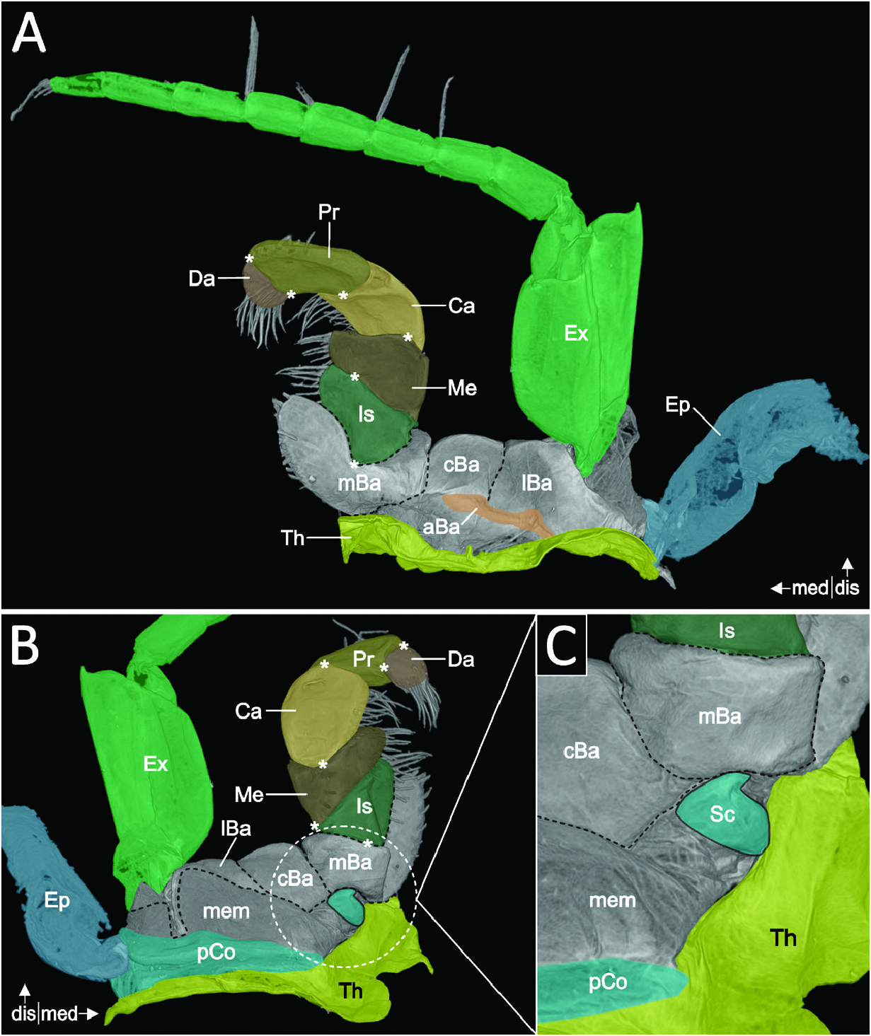
The coxa is formed as a narrow, incomplete proximal ring of moderate robustness, open (i.e. membranous) anteromedially. The ends of the open ring are in contact with the thorax posteromedially and anteriorly, potentially establishing two articulation points, which would form a bicondylar articulation, mostly for adduction/abduction. Anteriorly, the narrow coxa is almost entirely hidden by the thorax. Posteriorly, the coxa forms a transversal invagination from its most medial to its most lateral extent (pCo in Fig. 13B, C View Figure 13 ), where it forms an articulation with the epipod. Proximally to that articulation, the coxa continues by curving around the lateral side into the membranous area of the thorax and towards anterior. From its most lateral point, a sclerite reaches dorsad and forms another articulation with the epipod. Distal to the posterior invagination of the coxa, a wide membrane (mem in Fig. 13B View Figure 13 ) connects the coxa to the basis. Posteromedially within this membrane sits a sclerite (presumably a fragment of the coxa), apparently articulating the basis with the thorax (Sc in Fig. 13C View Figure 13 ).
Laterally attached to the coxa, a lamellar epipod is present (Ep in Fig. 13A, B View Figure 13 ). Dorsally, it forms an articulation via a sclerite with the lateral coxa, while posteriorly, it articulates with the lateral extent of the coxa invagination. An anterior contact point with the lateral basis (lBa in Fig. 13A View Figure 13 ) might constitute a third articulation point.
The basis appears subdivided by two longitudinal constrictions (from anterior over ventral to posterior) into three distinguishable portions: a medial, a central and a lateral portion. The medial portion (mBa in Fig. 13A–C View Figure 13 ) is fitted closely to the thoracic sternum ( Fig. 13B, C View Figure 13 ) and carries an endite anteromedially that extends in a distomedial direction ( Fig. 13A, B View Figure 13 ). Distally, the medial portion articulates via an anterior and a posterior articulation point (+ in Fig. 13A, B View Figure 13 ) with the endopod (i.e. the ischium). The constriction at the transition of the medial to the central portion forms a medial–central apodeme, serving as attachment site for one extrinsic exopod muscle (BEx1). The central and the lateral portions together form the distal (or ventral) wall of the basis (cBa and lBa in Fig. 13A, B View Figure 13 ). The proximal anterior margin of these portions forms a transversal invagination (aBa in Fig. 13A View Figure 13 ), serving as attachment site for two muscles (TB3 and BEx3). This invagination is most pronounced at its most medial and most lateral extent, respectively. The posterolateral margin of the lateral portion of the basis extends proximally towards the direction of the coxa, as a narrow cuticular beam; the basis and coxa are, in this area, separated by a wide interpodomeral membrane ( Fig. 13B View Figure 13 ). Anterolaterally, the lateral portion of the basis curves proximolaterally, establishing a contact point (and possibly an articulation) with the epipod (Ep in Fig. 13A View Figure 13 ).
Distolaterally within the membrane of the lateral portion of the basis, the exopod (Ex in Fig. 13A, B View Figure 13 ) articulates via an anterior and a posterior hinge point with the basis. The peduncle of the exopod comprises a short stem and a much longer distal portion, followed by a flagellum. The peduncle is broad and anteroposteriorly flattened, with a ventral bulge and a prominent distodorsal pointed protrusion. The peduncle articulates with the flagellum via an anteroventral articulation point. The cuticle of the first annulus is slightly more robust around this joint. All individuals examined exhibited a total of eight annuli ( Fig. 13A View Figure 13 ).
The crescent-shaped endopod consists of the ischium, merus, carpus, propodus and dactylus. Anteriorly, the endopod describes a strong concave curvature ( Fig. 13A View Figure 13 ). The ischium (Is in Fig. 13A, B View Figure 13 ) is about as long as the proximodistal extent of the basis and carries medially a more or less distinct endite. A bicondylar articulation with the merus is realized by an anteromedial and a lateral articulation point, respectively (+ in Fig. 13A, B View Figure 13 ).
The merus (Me in Fig. 13A, B View Figure 13 ) is about as long as the ischium and carries medially a more or less distinct endite. A bicondylar articulation with the carpus is formed by an inwardly folded posterior edge and an anterior articulation point (+ in Fig. 13A, B View Figure 13 ).
The carpus (Ca in Fig. 13A, B View Figure 13 ) is about twice as long as the merus. A bicondylar articulation with the propodus is formed by an anterior and a posterior articulation point, respectively (+ in Fig. 13A, B View Figure 13 ).
The propodus (Pr in Fig. 13A, B View Figure 13 ) is about half as long as the carpus. Its distal opening is not oriented distally but is ~90° displaced in a medial direction. A bicondylar articulation with the dactylus is formed by a medial and a lateral articulation point, respectively (+ in Fig. 13A, B View Figure 13 ).
The dactylus is relatively short and rounded (Da in Fig. 13A, B View Figure 13 ), with its distal setose tip facing a proximomedial direction.
No known copyright restrictions apply. See Agosti, D., Egloff, W., 2009. Taxonomic information exchange and copyright: the Plazi approach. BMC Research Notes 2009, 2:53 for further explanation.
|
Kingdom |
|
|
Phylum |
|
|
Class |
|
|
Order |
|
|
Family |
|
|
Genus |