
Polysyncraton catillum, Kott, 2010
|
publication ID |
https://doi.org/ 10.1080/00222930701359218 |
|
persistent identifier |
https://treatment.plazi.org/id/0A49A339-DF4C-600F-FE52-C3E5DA78FD9D |
|
treatment provided by |
Felipe |
|
scientific name |
Polysyncraton catillum |
| status |
sp. nov. |
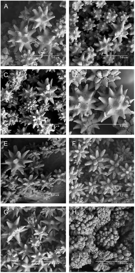
Polysyncraton catillum View in CoL sp. nov.
( figure 16F View FIG )
Distribution. Type locality: Western Australia (northern side of Ashmore Reef, 6–16 m, coll. L. Marsh, 16 September 1986, holotype WAM 1046.88).
Description. Colonies are almost circular, thin, brittle, plates with conspicuous stellate branchial apertures along each side of the deep primary common cloacal cavities that surround solid stands of test in which the ventrum of each thorax is embedded. Conspicuous stellate branchial apertures are outlined in spicules. Spicules are in a single layer in the paper-thin surface test, are crowded in the solid stands of test in which the ventrum of each thorax is embedded and also are crowded in a layer on the base of the colony. Spicules are not present in a layer of test in the floor of the common cloacal canals and are sparse around the abdomina, which are embedded in the basal test. Spicules are small, to 0.04 mm diameter. They have 9–11 long, thick and almost rod-like rays in optical transverse section.
Zooids have relatively large but delicate thoraces. The oesophageal neck is relatively short and a short, thin retractor muscle projects from it. Stigmata are long and fusiform, nine are in each of the first two rows, eight in the third and seven in the last row. A huge atrial aperture exposes much of the branchial sac directly to the common cloacal cavity and has a bifid atrial tongue of varying size. The long gut loop is typical of Polysyncraton with a long rectum. Four coils of the vas deferens surround about eight male follicles.
Remarks. Apart from the small, thin, plate-like colonies, this species is distinguished from others by its spicules, which are relatively small and have long, almost cylindrical rays. The spicules of P. cuculliferum are larger and although they have the same number of rays, they are shorter and more pointed than those of the present species. The conspicuous circular common cloacal cavities around the solid stands of test also distinguish the species from P. cuculliferum . Polysyncraton scobinum Kott, 2001 has similar but larger spicules with more rays than the present species and although P. pseudorugosum has similar common cloacal systems, its spicules are larger than the present species with more conspicuously conical pointed rays.
Polysyncraton cuculliferum ( Sluiter, 1909) View in CoL
( figure 16G View FIG )
Diplosomoides cuculliferum Sluiter, 1909: 90 View in CoL .
Polysyncraton cuculliferum: Kott, 2002c: 30 View in CoL and synonymy.
Not Didemnum cuculliferum: Kott View in CoL : 2001: 167 (v Didemnum nekozita Tokioka, 1967 View in CoL ).
Distribution. Previously recorded (see Kott, 2002): Northern Territory (Darwin), Queensland (Heron I. to Lizard I.), Indonesia. New records: Northern Territory (South Shell I., QM G308604; Angler Reef, QM G308616).
Description. The in situ photographs of newly recorded colonies from Darwin Harbour resemble those from Queensland (see Kott, 2001: pl. 5B, C). They are large, encrusting and surrounding debris, with the surface raised into lobes. The common cloacal canals are thoracic but roomy, the surface layer of test in the roof of the canal being very thin and brittle. Spicules are in a single layer in the surface test and are in a crowded layer on the base of the colony. They are not in the test in the floor of the common cloacal canals and are sparse in the basal test where the abdomina are embedded. They are to 0.057 mm diameter with 9–11 strong, sharply pointed conical rays in optical transverse section. Some of the spicules have rays of variable size. Branchial apertures are conspicuous on the surface of the colony with six petal-like lobes crowded with spicules. Internally the test is translucent and cloudy, with the zooids in clumps surrounded by deep primary common cloacal canals that extend the full length of the zooids and occasionally become posterior abdominal.
Larvae are in one of the colonies (QM G308608) collected in May. They have a conspicuous horizontal ampulla on the right side of the trunk which projects posteriorly.
Remarks. The newly recorded specimens lack the large papilla often associated with each branchial aperture in this species. However, all other characters conform with previous descriptions of the species, especially the large thorax, roomy common cloacal canals and relatively small spicules with sharply pointed rays. Although Kott (2002c) reported that the external larval ampulla on the right side of the trunk was vertical, it projects horizontally in the newly recorded specimens. Polysyncraton pseudorugosum has similar spicules with the same number of pointed conical rays, but they are larger than the spicules of the present species and are present throughout the colony. The spicules of P. catillum and P. peristroma have longer almost cylindrical or rod-shaped rays that distinguish them from the present species.
Polysyncraton dromide Kott, 2001
( figure 16H View FIG )
Polysyncraton dromide Kott, 2001: 99 View in CoL ; 2002c: 32.
Distribution. Previously recorded (see Kott, 2001, 2002c): Northern Australia (Darwin, Torres Strait), Western Australia (Cockburn Sound). New records: Northern Territory (Darwin, QM G308596, G308670).
Description. In preservative the colonies are firm and gelatinous, but thin, beige and translucent lamellae or sheets, with the surface divided into a mosaic of elevated blister-like oval zooid-free areas surrounded by circular depressions over the narrow circular primary common cloacal canals that are lined on each side by the zooids. One (QM G308670) of the newly recorded colonies is aspiculate. Parts of the other are aspiculate, but other parts have a superficial aspiculate layer containing brown pigment cells over a thin layer of evenly spaced spicules. Spicules are absent from the remainder of the colony. The spicules (to 0.04 mm diameter) are almost burr-like, with 15–17 rays in optical section. The ray tips are sometimes rounded and the spicules almost globular but other spicules have short pointed rays.
Thoraces are large with a long, tulip-shaped branchial siphon, a bifid atrial lip, a long retractor muscle from the top of the oesophagus, four coils of the vas deferens and eight or nine testis follicles. Larvae, present in the basal test of specimens collected in August have 12 lateral ampullae on each side of the three antero-median adhesive organs and larval blastozooids.
Remarks. The large thoraces of the newly recorded colonies with their bifid atrial tongues are exactly like those of the type material as are the colonies, the spicules and the larvae with bladder cells packed in their test. Although Kott (2001) reported only three coils of the vas deferens for this species, four have been found on re-examination of the type material (see also Kott, 2002c). The newly recorded colonies differ from the type material only in their fewer testis follicles, 20 having been reported previously (see Kott, 2001). In life this species has a dramatic appearance, the blister-like elevations of the surface test being opaque, and sometimes quite metallic in appearance.
No known copyright restrictions apply. See Agosti, D., Egloff, W., 2009. Taxonomic information exchange and copyright: the Plazi approach. BMC Research Notes 2009, 2:53 for further explanation.
|
Kingdom |
|
|
Phylum |
|
|
Class |
|
|
Order |
|
|
Family |
|
|
Genus |
Polysyncraton catillum
| Kott, Patricia 2010 |
Polysyncraton cuculliferum: Kott, 2002c: 30
| KOTT, P. 2002: 30 |
Diplosomoides cuculliferum
| SLUITER, C. P. 1909: 90 |