
POLYCYSTIDINAE Schockaert and Karling, 1970
|
publication ID |
https://doi.org/ 10.1080/00222933.2018.1444212 |
|
publication LSID |
lsid:zoobank.org:pub:098ADF23-763B-41F6-A2F5-54F6AA8620E8 |
|
DOI |
https://doi.org/10.5281/zenodo.5187222 |
|
persistent identifier |
https://treatment.plazi.org/id/6640516D-3335-FFD3-CF04-034E81E7FC59 |
|
treatment provided by |
Felipe |
|
scientific name |
POLYCYSTIDINAE Schockaert and Karling, 1970 |
| status |
|
Subfamily POLYCYSTIDINAE Schockaert and Karling, 1970 View in CoL
Genus Austrorhynchus Karling, 1952 View in CoL
Austrorhynchus wennersgaardi Volonterio and Ponce de León , sp. nov.
( Figures 1–4 View Figure 1 View Figure 2 View Figure 3 View Figure 4 )
Diagnosis
Austrorhynchus species with eyes and white to greyish parenchyma. Type II prostate stylet, double-walled; funnel about 20–25% of total stylet length, with the proximal rim rolled upon itself to a varying degree; tube of the outer stylet slightly bent, with the inner stylet extending throughout its length; a strongly bent hook arises at the level of the junction between the funnel and the tube; hook and tube of about the same size. Type III stylet with a foot and a shorter style connected by a long bridge, defining a broad window; style expands to a thin comb-bearing plate with large teeth, decreasing in size and thickness towards the base of the flagellum; in general, the third to seventh teeth from the free end of the plate are the larger ones, and their bases are frequently placed at a higher level than those of the rest; foot continues into a sinuous to straight, narrow flagellum, bearing a distal expansion; a row of progressively shorter, fine teeth extends from the base of the flagellum to the proximal part of the expansion. Oviparous.
Description
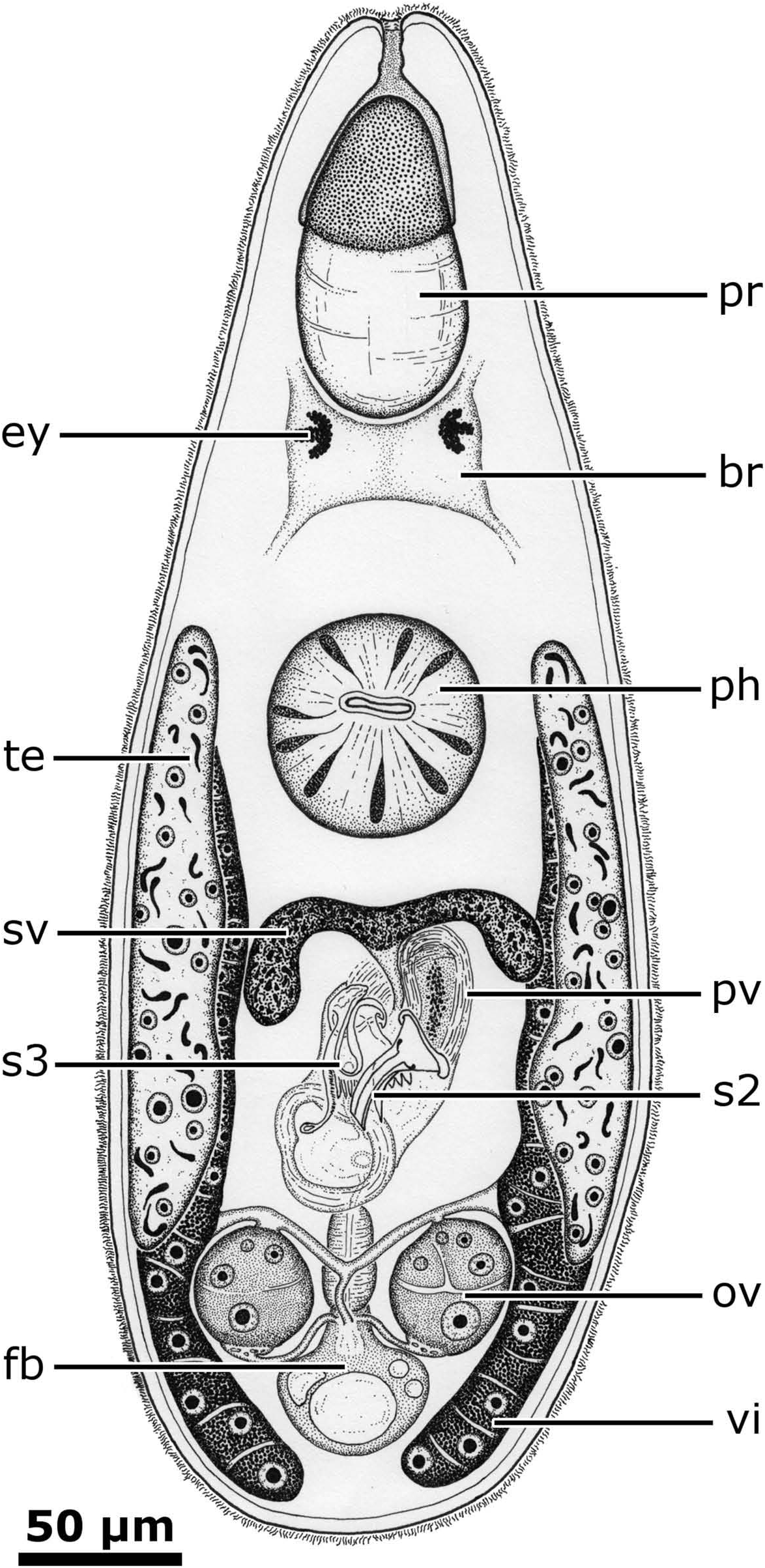
General organization. Preserved specimens are 431–1044 µm (mean 711.0, SD 191.5) long, white to greyish, rather opaque and bear two eyes ( Figure 1 View Figure 1 ). Proboscis about 10–20% of body length. Pharynx 68–158 µm (mean 123.7, SD 30.9) in diameter.
Testes paired. Seminal vesicle tapers into an ejaculatory duct opening into proximal end of male genital canal, between bases of type II and III prostate stylets. Prostate vesicle of type II associated with type II stylet. Compact muscle sheet connects wall of prostate vesicle to base of type III stylet.
Paired ovaries and vitellaria. Ovo-vitelloducts of each side join into a common ovovitelloduct. Female bursa connected to a female duct type I, which opens into posterior wall of genital atrium. Common ovo-vitelloduct and insemination ducts open into bursal stalk. Uterus opens into anterior wall of distalmost portion of atrium.
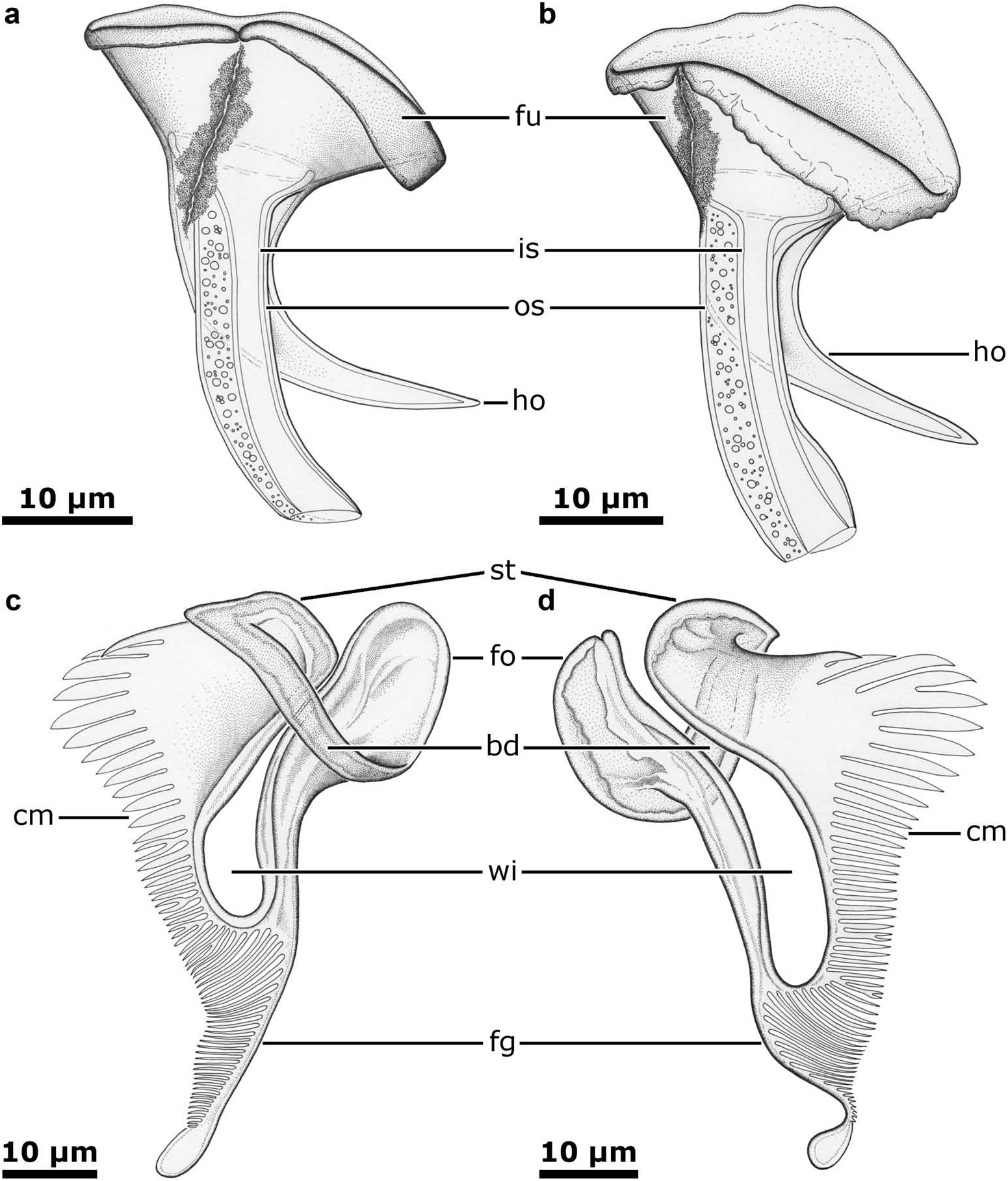
Sclerotized parts of copulatory organ. The double-walled type II prostate stylet ( Figure 2 View Figure 2 (a,b)) is 40–49 µm (mean 44.5, SD 2.2) long and consists of a funnel, a distal tube and a hook. The funnel is 8–14 µm (mean 10.4, SD 1.6) long (about 20–25% of the total stylet length) and 22–33 µm (mean 28.9, SD 2.8) wide proximally; its proximal rim is broad and rolled upon itself to a varying degree. Tube of outer stylet slightly bent, 27–34 µm (mean 31.1, SD 2.0) long, 11–17 µm (mean 13.8, SD 1.6) wide proximally and 5–8 µm (mean 6.4, SD 1.1) wide distally; inner stylet extends throughout its length. In a few specimens, distal portion of tube of outer stylet is dilated ( Figure 2 View Figure 2 (b)). A large, 28–34 µm (mean 31.0, SD 2.1) long, strongly bent hook arises at the level of the junction between the funnel and the tube.
Type III prostate stylet ( Figure 2 View Figure 2 (c,d)) has a foot and style connected by a long bridge, defining a broad window. Style expands to a thin comb-bearing plate carrying 22–27 µm (mean 24, SD 1.9) large teeth. In general, third to seventh teeth from free end of plate are the larger ones, measuring 10–15 µm (mean 12.1, SD 1.6) in length; their bases are frequently placed at a higher level than those of the rest ( Figure 2 View Figure 2 (d)). Remaining teeth on comb decrease in size and thickness towards base of flagellum. Flagellum, 29–37 µm (mean 32, SD 2.6) in length, is sinuous to straight and has a distal expansion; a row of progressively shorter, fine teeth extends up to proximal part of latter. Distance from proximal end of foot to tip of flagellum is 59–77 µm (mean 71.5, SD 4.7).
Histology. At the histological level, the overall characteristics of the new species are in agreement with the previous histological descriptions of Austrorhynchus ( Karling 1952; Brunet 1965); the relevant differences are described below.
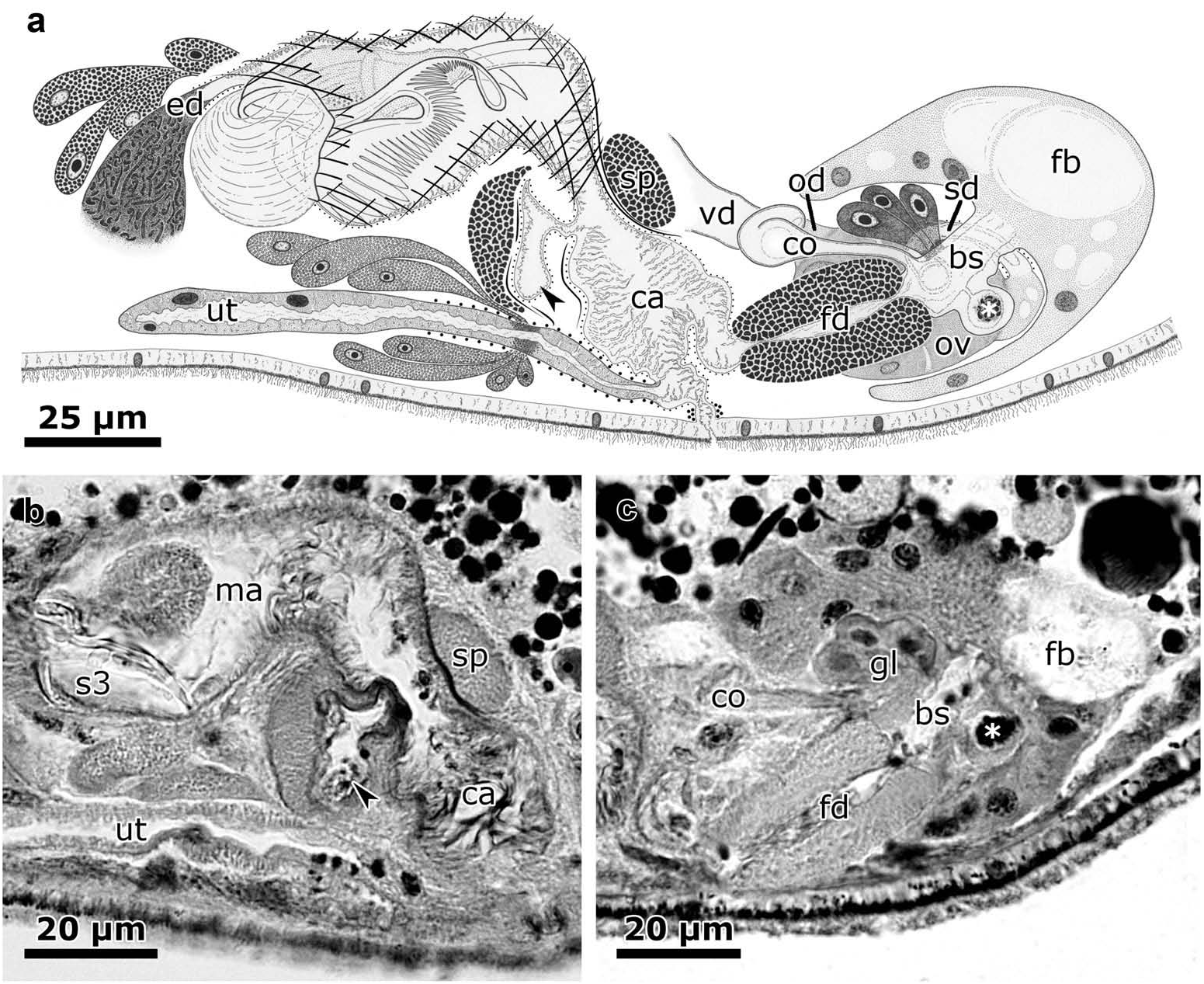
The genital canal can be divided into three sections based on the histology of its wall ( Figure 3 View Figure 3 (a)): (i) the proximal portion of the male genital canal, with an epithelium bearing pseudo-cilia surrounded by an internal layer of thin circular muscle fibres and two external layers composed of thick, oblique fibres; (ii) the distal portion of the male genital canal, where the internal oblique fibres adopt a longitudinal direction and the external ones are replaced by a sphincter; (iii) the atrium inferius sensu Karling 1952, with an epithelium bearing long pseudo-cilia and a mono-layer of thin circular muscle fibres surrounded by scarce longitudinal fibres. Most of the longitudinal fibres present in the distal portion of the male genital canal leave the wall of the canal immediately below the sphincter and attach to the ventral body wall close to the gonopore.
In a few specimens (for example, NHMUK 2017.11.3.11), the anterior wall of the genital canal is dilated at the level of the sphincter, forming a pouch ( Figure 3 View Figure 3 (a,b), indicated with an arrowhead). Sperm cells seen in the male genital canal were always located in the vicinity or at the level of the sphincter, either in the main cavity of the canal or inside the pouch.
Gland cells open into the dorsal wall of the bursal stalk, just posteriorly to the opening of the common ovo-vitelloduct. In a few specimens, the ventral wall of the bursal stalk presents an expansion filled with sperm cells ( Figure 3 View Figure 3 (a,c), indicated with an asterisk).
Material examined
Holotype. One whole mount of a specimen from Balvino Point , King George Island , South Shetland Islands (62.188889ºS, 58.903889ºW), Antarctica, collected by Odile Volonterio and Rodrigo Ponce de León on 22 January 2009, deposited in the Natural History Museum (NHM), London (accession number: NHMUK 2017.11.3.1). GoogleMaps
Paratypes. Eight slides with whole mounts of specimens from Balvino Point , Maxwell Bay , King George Island , South Shetland Islands (62.188889ºS, 58.903889ºW), Antarctica, collected by Odile Volonterio and Rodrigo Ponce de León on 19 December 2006 (two slides), 5 January 2008 (one slide), 6 January 2009 (one slide), 24 January 2009 (two slides) and 6 February 2009 (two slides with gravid specimens); one slide with a whole mount of a specimen from Pata de Perro Point , Maxwell Bay , King George Island , South Shetland Islands (62.185278°S, 58.878889°W), Antarctica, collected by Odile Volonterio and Rodrigo Ponce de León on 29 January 2010 GoogleMaps . Deposited in the Natural History Museum (NHM), London (accession numbers: NHMUK 2017.11 View Materials .3.2–9 and 2017.11.3.10 respectively) .
Additional material. Two slides with sagittal sections of two specimens from Balvino Point , King George Island , South Shetland Islands (62.188889ºS, 58.903889ºW), Antarctica, collected by Odile Volonterio and Rodrigo Ponce de León on 22 January 2009, deposited in the Natural History Museum (NHM), London (accession number: NHMUK 2017.11 View Materials .3.11–12) GoogleMaps .
Biology
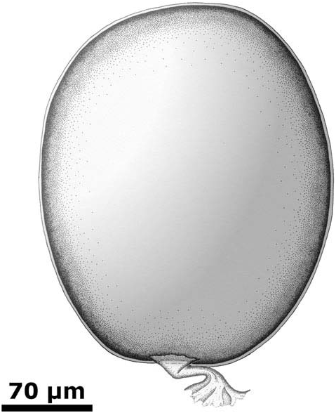
The species is oviparous. In February 2009 two gravid specimens, each carrying a single egg, were found; eggs are oval, 282–299 µm (mean 290.5, SD 12.0, n = 2) long, 231–243 µm (mean 237.0, SD 8.5, n = 2) wide and present a short adhesive peduncle ( Figure 4 View Figure 4 ).
Distribution
King George Island , South Shetland Islands, Maritime Antarctic .
Etymology
Specific name in honour of the Norwegian sailor Ole Wennersgaard, a crew member of the Swedish Antarctic Expedition who died during the extreme Antarctic winter of 1903, when part of the expedition was forced to remain in Poulet Island until rescued by the vessel Uruguay ( Ekelof 1904).
| NHMUK |
Natural History Museum, London |
No known copyright restrictions apply. See Agosti, D., Egloff, W., 2009. Taxonomic information exchange and copyright: the Plazi approach. BMC Research Notes 2009, 2:53 for further explanation.
|
Kingdom |
|
|
Phylum |
|
|
Order |
|
|
Family |
POLYCYSTIDINAE Schockaert and Karling, 1970
| Volonterio, Odile & Ponce de León, Rodrigo 2018 |
Austrorhynchus wennersgaardi Volonterio and Ponce de León
| Volonterio and Ponce de Leon 2018 |
Austrorhynchus
| Karling 1952 |