
Platorchestia paludosus, Cheng, Yu-Ting, Nakazono, Kenji, Lin, Kirk & Chan, Benny K. K., 2011
|
publication ID |
https://doi.org/ 10.5281/zenodo.207935 |
|
DOI |
https://doi.org/10.5281/zenodo.6185339 |
|
persistent identifier |
https://treatment.plazi.org/id/03EF87A0-FFED-CC1F-49FF-FC6BBC58F931 |
|
treatment provided by |
Plazi |
|
scientific name |
Platorchestia paludosus |
| status |
sp. nov. |
Platorchestia paludosus View in CoL sp. nov.
(Figures 2–9)
Platorchestia japonica View in CoL . ― Miyamoto & Morino 2004: 83 –88 (non P. japonica ).
Materials examined. Holotype (NMNS—6519-001) 1 male, Guan-du, Taipei County, Taiwan, Aug 2008, Coll. Y- T. Cheng. Paratypes (NMNS—6519-002–06519-009) 4 males and 4 females, data same as holotype. Paratypes (AM—P.84807) 2 males and 2 females, data same as holotype. Paratypes (ASIZCR—000230) 2 males and 2 females, data same as holotype.
Comparative material. Platorchestia japonica ( Tattersall, 1922) . Lake Biwa, Japan. 2 males 2 females.
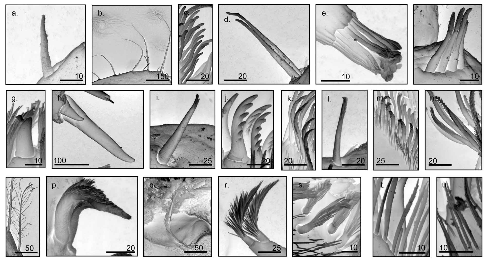
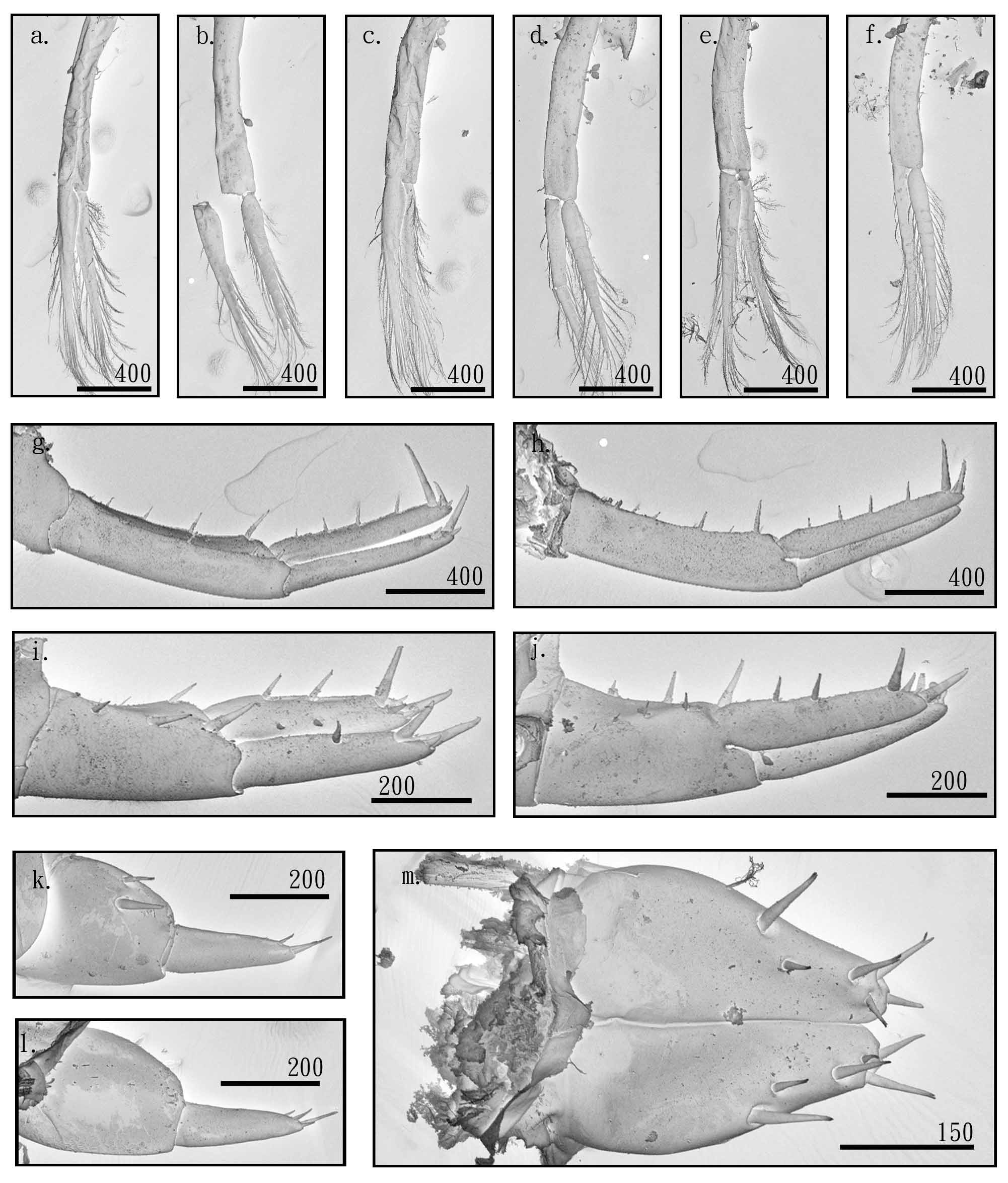
Description. Male. Body smooth, without protuberance. Eyes sub-rounded, larger than 1/3 of head height. Antenna 1 ( Figs 3 View FIGURE 3 –a, b, c and d) short with 6–7 articles (3 peduncle articles, 3–4 flagellated articles), slightly longer than article 4 of antenna 2, peduncle articles 1–3 equal in length, dorsal side convex, ventral side flattened, each peduncle article with simple setae, article 4 slightly curved, each flagellated article with 2 groups of simple setae type IV ( Fig. 1 View FIGURE 1 –d) on the inner and outer side of the dorsal surface.
Antenna 2 ( Figs 3 View FIGURE 3 –e, f, and g) up to 1/3 of the body length, outer margin of ventral side of peduncle article 3 with 3–5 simple setae type IV ( Fig. 1 View FIGURE 1 –d), a medial furrow extend from distal 1/3 of peduncle article 4 to entire peduncle article 5, length of article 5 equals to combined length of article 3 and 4, 11–14 flagellated articles each with medial furrow, flagellum subequal to peduncle 3–5 combined in length; tip of antenna 2 with simple setae type V ( Fig. 1 View FIGURE 1 –e).
Upper lip entire, apical margin with fine setule.
Incisor of mandible ( Figs 3 View FIGURE 3 –i, j) 5 to 6 dentate, left lacinia mobilis 5–dentate, right lacinia mobilis bi-fid, with serrated cutting edges; molar finely serrated, right molar convex, left molar concave; 3–4 pappose setae type I ( Fig. 1 View FIGURE 1 –p) located in the region between the lacinia mobilis and molar, 1 pappose seta type II ( Fig. 1 View FIGURE 1 –q) on the dorsal side of molar.
Lower lip bi-lobed, lobes wide, with fine setule on the inner margin.
Inner plate of maxilla 1 ( Figs 3 View FIGURE 3 –l, m) narrow, with 2 apical papposerrate setae type I ( Fig. 1 View FIGURE 1 –r); outer plate with 9 serrate setae type I ( Fig. 1 View FIGURE 1 –j), with a row of 13–19 setule type I ( Fig. 1 View FIGURE 1 –t) on ventral base of inner 4 serrate setae; palp very small, 2 articulate.
Inner plate of maxilla 2 ( Fig. 3 View FIGURE 3 –n, o, p, q, r and s) smaller than outer plate, distal margin with 17–19 simple setae type III ( Fig. 1 View FIGURE 1 –c), a row of 8–10 serrate setae type II ( Fig. 1 View FIGURE 1 –k) on ventral and a row of 6–11 serrate setae type IV ( Fig. 1 View FIGURE 1 –m) on dorsal, inner margin with 2 papposerrate setae type I ( Fig. 1 View FIGURE 1 –r), inner one longer; distal margin of outer plate with 15–20 simple setae type III ( Fig. 1 View FIGURE 1 –c), a row of 9–12 serrate setae type II ( Fig. 1 View FIGURE 1 –k) on ventral surface, a row of 3 serrate setae type IV ( Fig. 1 View FIGURE 1 –m) and 2 serrulate setae type I ( Fig. 1 View FIGURE 1 –n) on dorsal surface.
FIGURE. 2. Platorchestia paludosus sp. nov. a. external view, male. b. female gnathopod 1. c. female gnathopod 2.
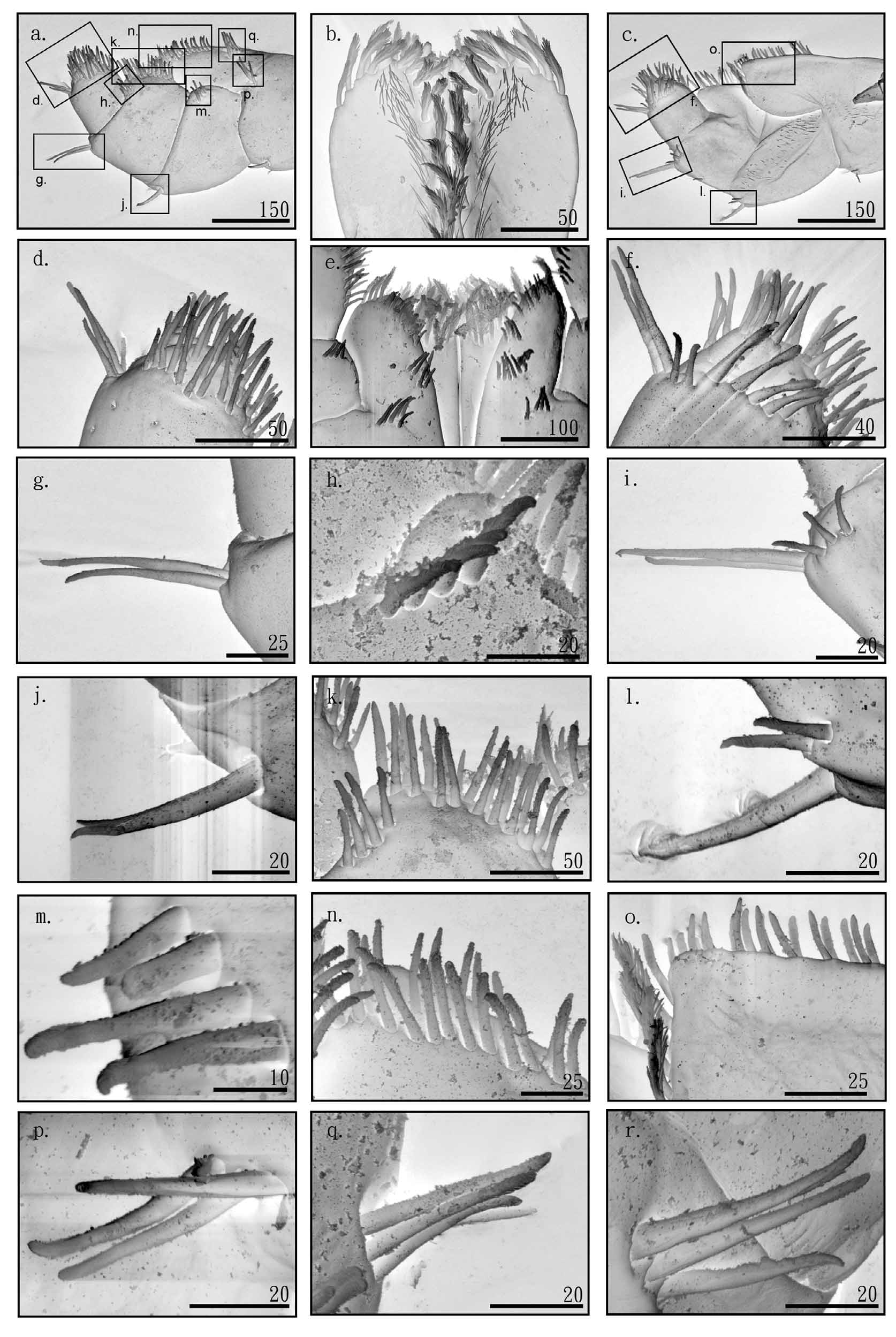
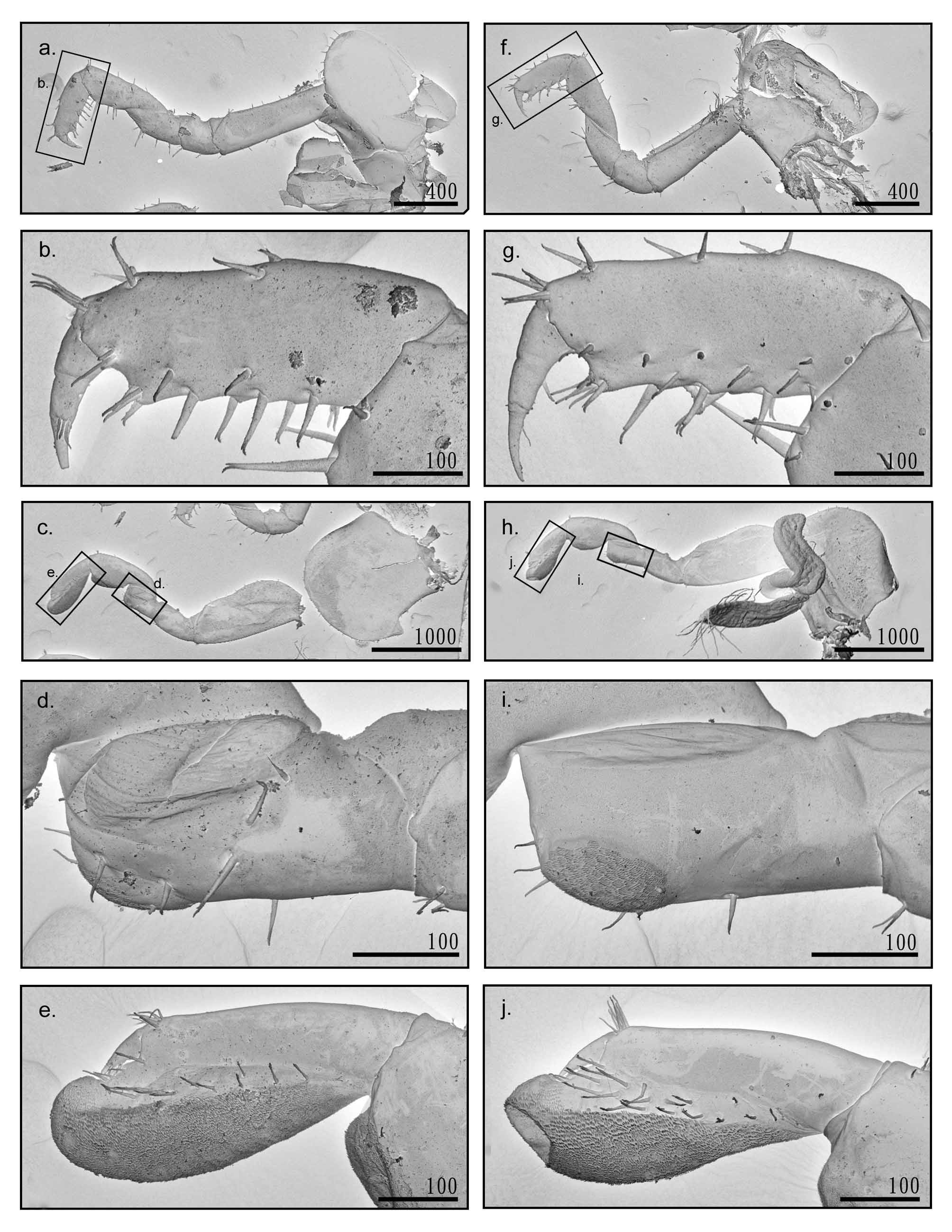
Distal end of maxilliped inner plate ( Figs 4 View FIGURE 4 –b, e) with 3 cuspidate setae type I ( Fig. 1 View FIGURE 1 –g), a row of 3–4 papposerrate setae type II ( Fig. 1 View FIGURE 1 –s) behind the cuspidate setae, dorsal surface ( Fig. 4 View FIGURE 4 –b) with a row of papposerrate setae type II ( Fig. 1 View FIGURE 1 –s) along inner margin, with fine setae situating behind the papposerrate setae; ventral surface ( Fig. 4 View FIGURE 4 –e) with several papposerrate setae type II ( Fig. 1 View FIGURE 1 –s); distal end of outer plate ( Figs 4 View FIGURE 4 –n, o) with a row of 4–5 papposerrate setae type II ( Fig. 1 View FIGURE 1 –s) along margin, along inner margin to ventral surface of distal corner with 2 rows of simple setae type VI ( Fig. 1 View FIGURE 1 –f), ventral surface of medial inner margin ( Fig. 4 View FIGURE 4 –q) with a row of 3–5 simple setae type VI ( Fig. 1 View FIGURE 1 –f); palp article 1 wider than high, with a row of 3–4 simple setae type VI ( Fig. 1 View FIGURE 1 –f) on ventral surface of distal inner corner ( Fig. 4 View FIGURE 4 –a, m), distal outer margin ( Figs 4 View FIGURE 4 –a, c, j and l) with 1–2 cuspidate setae type III ( Fig. 1 View FIGURE 1 –i) and 2–3 simple setae type VI ( Fig. 1 View FIGURE 1 –f); distal inner corner of ventral surface of palp article 2 ( Figs 4 View FIGURE 4 –a, and h) with a row of 3–4 simple setae type VI ( Fig. 1 View FIGURE 1 –f), along the inner margin ( Figs 4 View FIGURE 4 –a, and k) with 2 rows of simple setae VI ( Fig. 1 View FIGURE 1 –f), distal outer margin ( Figs 4 View FIGURE 4 –a, c, g and i) with 1–2 cuspidate setae type III ( Figs 1 View FIGURE 1 –i) and 2–3 simple setae type VI ( Fig. 1 View FIGURE 1 –f), article 3 rounded apically, dorsal surface ( Figs 4 View FIGURE 4 –c, and f) with a row of 4–5 serrate setae type III ( Fig. 1 View FIGURE 1 –l) and 2–3 simple setae type VI ( Fig. 1 View FIGURE 1 –f) along the base of palp article 4, ventral surface with ( Figs 4 View FIGURE 4 –a, d) several simple setae type VI ( Fig. 1 View FIGURE 1 –f), outer distal corner ( Fig. 4 View FIGURE 4 –a, c, d and f) with a group of 2–3 simple setae type IV ( Fig. 1 View FIGURE 1 –d); article 4 small with several simple setae type III ( Fig. 1 View FIGURE 1 –c) and simple setae type VI ( Fig. 1 View FIGURE 1 –f) apically.
Gnathopod 1 ( Figs 5 View FIGURE 5 –a, and b) coxa with straight or convex anterior margin, ventral margin convex with a few setae; basis slightly expanded posterodistally, subequal with merus and carpus when combined in length; ischium shortest, anteroproximal margin with a notch; merus sub-triangular, rounded posteriorly; carpus ( Figs 5 View FIGURE 5 –c, and d) with scabrous tumescent protuberance posterodistally, outer lateral surface ( Figs 5 View FIGURE 5 –c) with a row of 2–3 serrate setae type III ( Fig. 1 View FIGURE 1 –l) and 2–3 cuspidate setae type III ( Fig. 1 View FIGURE 1 –o) at base of tumescent protuberance, inner lateral surface ( Fig. 5 View FIGURE 5 –d) with 7–10 serrate setae type III ( Fig. 1 View FIGURE 1 –l) without cuspidate setae; propodus ( Figs 5 View FIGURE 5 –e, f) with scabrous tumescent protuberance posterodistally, outer lateral surface with a row of 4–6 serrate setae type III ( Fig. 1 View FIGURE 1 –l) along posteroproximal margin to medial base of tumescent protuberance, outer lateral surface with a group of 4–5 simple setae type IV ( Fig. 1 View FIGURE 1 –d) on protuberance base distal end, dactylus base with a group of 2–3 simple setae type IV ( Fig. 1 View FIGURE 1 –d), anterodistal corner with a group of 2–3 simple setae type IV ( Fig. 1 View FIGURE 1 –d); length of dactylus subequal to palm, and not reach posterodistal angle of propodus.
Gnathopod 2 ( Figs 5 View FIGURE 5 –g, h) coxa as wide as deep with posterior process, ventral margin convex, basis anterior margin straight, posterior margin convex and proximal end necked; ischium larger than merus, with a notch on proximal end of anterior margin; length of merus subequal to carpus, with convex posterior margin; carpus with convex anterior margin; propodus oval, palm margin curved, without notch, both lateral surface with a row of cuspidate setae type III ( Fig. 1 View FIGURE 1 –i); dactylus curved, as long as palm.
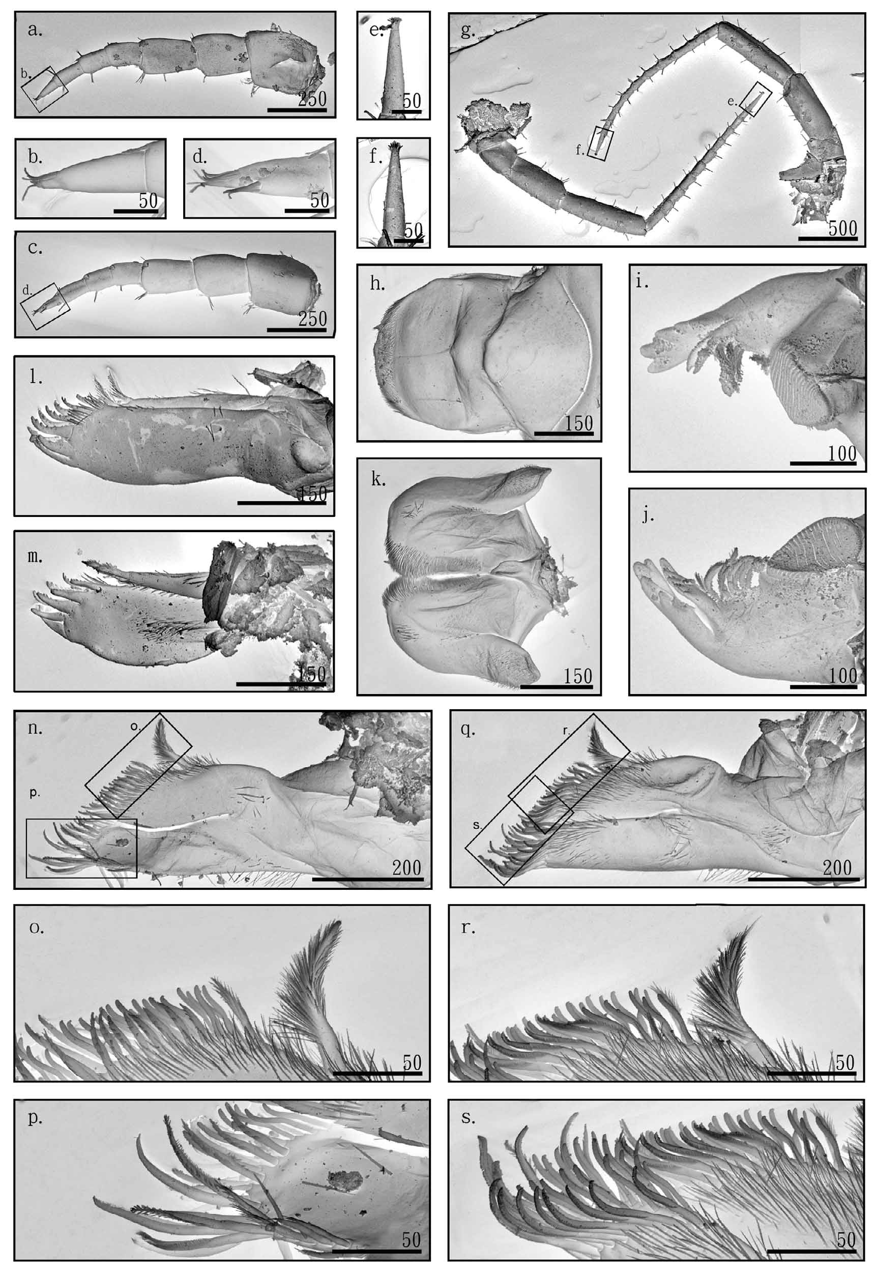
Pereopods 3–7 cuspidactylate. Pereopod 3 coxa ( Fig. 6 View FIGURE 6 –a) subquadrate with posterior process, ventral margin slightly convex; basis longest, subequal to combined length of merus and carpus; ischium shortest, anteroproximal margin notched; merus longer than carpus, anterior margin slightly convex, posterior margin straight; carpus anterior and posterior margins parallel; propodus slender, longer than carpus, slightly shorter than merus; dactylus ( Fig. 6 View FIGURE 6 –c) subequal to ischium in length, posterior nail base with 1 simple setae type I ( Fig. 1 View FIGURE 1 –a).
Pereopod 4 ( Figs 6 View FIGURE 6 –d, and e) similar to pereopod 3, shorter, coxa ( Fig. 6 View FIGURE 6 –d) subquadrate, with posterior process, ventral margin straight; basis longest, subequal to combined length of merus and carpus, slightly convex; ischium shortest, anteroproximal margin notched; merus longer than carpus, anterior margin slightly convex, posterior margin straight; carpus shorter than that of pereopod 3, both margins parallel; propodus slender, subequal to merus, dactylus ( Fig. 6 View FIGURE 6 –f) pinched, posterior nail base with 1 simple seta type I ( Fig. 1 View FIGURE 1 –a).
Pereopod 5 coxa ( Fig. 6 View FIGURE 6 –g) bilobed, anterior lobe larger than posterior lobe; basis oval; ischium shortest, posteroproximal margin notched; merus subequal to carpus in length, anterior margin straight, posterior margin slightly convex; carpus both margins parallel; propodus slender; dactylus ( Fig. 6 View FIGURE 6 –i) with 1 simple seta type I ( Fig. 1 View FIGURE 1 –a) on anterior nail base.
Pereopod 6 coxa ( Fig. 6 View FIGURE 6 –j) bilobed, anterior lobe very small, anterior margin of posterior lobe nearly vertical, anterodistal corner strongly curved; basis oval; ischium shortest, posteroproximal margin notched; merus subequal to carpus in length, anterior margin straight, posterior margin slightly convex; carpus both margins parallel; propodus slender, longest; dactylus ( Fig. 6 View FIGURE 6 –l) with 1 simple seta type I ( Fig. 1 View FIGURE 1 –a) on anterior nail base.
Pereopod 7 coxa ( Fig. 6 View FIGURE 6 –m) rounded posterodistally; basis oval, posterodistal lobe broader than pereopod 5–6; ischium shortest, posteroproximal margin notched; merus subequal to carpus, anterior margin straight, posterior margin slightly convex; carpus anterior and posterior margins parallel; propodus slender, longest; dactylus ( Fig. 6 View FIGURE 6 – o) with 1 simple seta type I ( Fig. 1 View FIGURE 1 –a) on anterior nail base.
Pleopods ( Figs 7 View FIGURE 7 –a, b, c, d, e and f) biramous, peduncle and ramus subequal in length, pleopod 3 peduncle shortest, ramus with 7–10 articulated segments.
Peduncle of uropod 1 ( Figs 7 View FIGURE 7 –g, h) slightly longer than ramus, with 1 row of 4–5 cuspidate setae type III ( Fig. 1 View FIGURE 1 –i) along each dorsal lateral margin; outer ramus with 3–5 cuspidate setae type II ( Fig. 1 View FIGURE 1 –h) on distal end, without seta on medial region; inner ramus with 3–5 cuspidate setae type II ( Fig. 1 View FIGURE 1 –h) on distal end, a row of 3–4 cuspidate setae type III ( Fig. 1 View FIGURE 1 –i) along dorsal margin, without 1–2 cuspidate setae type III ( Fig. 1 View FIGURE 1 –i) along outer lateral region.
Peduncle of uropod 2 ( Figs 7 View FIGURE 7 –i, j) subequal to ramus in length, with 3 cuspidate setae type III ( Fig. 1 View FIGURE 1 –i) on dorsal region; outer ramus with 3 cuspidate setae type II ( Fig. 1 View FIGURE 1 –h) on distal end, with 1 cuspidate seta type III ( Fig. 1 View FIGURE 1 – i) on medial region; inner ramus with 5 cuspidate setae type II ( Fig. 1 View FIGURE 1 –h) on distal end, 2–3 cuspidate setae type III ( Fig. 1 View FIGURE 1 –i) on dorsal margin, with 1–2 cuspidate setae type III ( Fig. 1 View FIGURE 1 –i) on outer lateral region.
Uropod 3 ( Figs 7 View FIGURE 7 –k, l), 1– 2 cuspidate setae type III ( Fig. 1 View FIGURE 1 –i) on peduncle medial region, ramus with a group of 3 simple setae type I ( Fig. 1 View FIGURE 1 –a) on distal end, 0–1 simple setae type I ( Fig. 1 View FIGURE 1 –a) on medial region.
Telson ( Figs 7 View FIGURE 7 –m) bilobed, longer than wide, with a group of 3–4 cuspidate setae type III ( Fig. 1 View FIGURE 1 –i) on distal end, 1–2 cuspidate setae type III ( Fig. 1 View FIGURE 1 –i) as a group on dorsal lateral margin.
Female. Antenna 1 short, slightly beyond article 4 of antenna 2, with 6–7 articles (3 peduncle articles, 3–4 flagellated articles), peduncle articles 1–3 equal in length, article 4 slightly curved, each flagellated article with two groups of simple setae on the inner and outer side of dorsal surface.
Antenna 2 up to 1/3 of body length. Outer margin of ventral side of article 3 with 3–5 simple setae, a medial furrow extending from distal 1/3 of article 4 to the entire article 5, length of article 5 equals to total of article 3 and 4, 12–14 flagellated articles, each with medial furrow. Tip of antenna 2 with a group of simple setae.
Coxa of gnathopod 1 ( Figs 8 View FIGURE 8 –a, f) anterior margin straight or convex, ventral margin convex; basis straight; carpus without tumescent protuberance, posterior margin with 2 long and 4–6 short cuspidate setae type III ( Fig. 1 View FIGURE 1 – i); propodus ( Figs 8 View FIGURE 8 –b, g) without tumescent protuberance, inner lateral surface medial region with 4–5 serrate setae type III ( Fig. 1 View FIGURE 1 –l), outer lateral surface medial region with a row of 2–3 cuspidate setae type III ( Fig. 1 View FIGURE 1 –i) along margin, posterior margin with 2 rows of cuspidate setae type III ( Fig. 1 View FIGURE 1 –i), outer lateral surface with a group of 3–4 simple setae type IV ( Fig. 1 View FIGURE 1 –d) on dactylus base, 3–4 simple setae type IV ( Fig. 1 View FIGURE 1 –d) on posterodistal corner, anterodistal corner with 3–6 simple setae type IV ( Fig. 1 View FIGURE 1 –d); dactylus longer than width of propodus.
Gnathopod 2 ( Figs 8 View FIGURE 8 –c, h) coxa as wide as deep with posterior process, convex ventral margin, basis expanded anteroproximally, posterior margin straight; ischium subequal to merus in length; merus ( Figs 8 View FIGURE 8 –d, i) with small scabrous region posterodistally; carpus with scabrous tumescent protuberance posterodistally; propodus ( Figs 8 View FIGURE 8 –e, j) with posterodistal tumescent protuberance elongated distally, scabrous region from posteroproximal end to posterodistal end, outer lateral surface with 10–14 serrate setae type III ( Fig. 1 View FIGURE 1 –l) along scabrous region, inner lateral surface with 16–19 serrate setae type III ( Fig. 1 View FIGURE 1 –l) along scabrous region, palm margin short, and outer lateral palm margin with a row of 4–5 simple setae type I ( Fig. 1 View FIGURE 1 –a), anterodistal corner with 6–8 simple setae type I ( Fig. 1 View FIGURE 1 –a); dactylus curved as long as palm.
Habitat. This salt marsh in Guan-du, Taiwan is dominated by Phragmites communis (L.) Trin. and Brachiaria mutica (Forsk) Stapf. , with irregular flooding after raining. Platorchestia paludosus sp. nov. was found under leaflitter of Brachiaria mutica (Forsk) on the water front of estuarine marshes.
Etymology. The word ‘paludosus’ indicates that the species is common in marshy habitats.
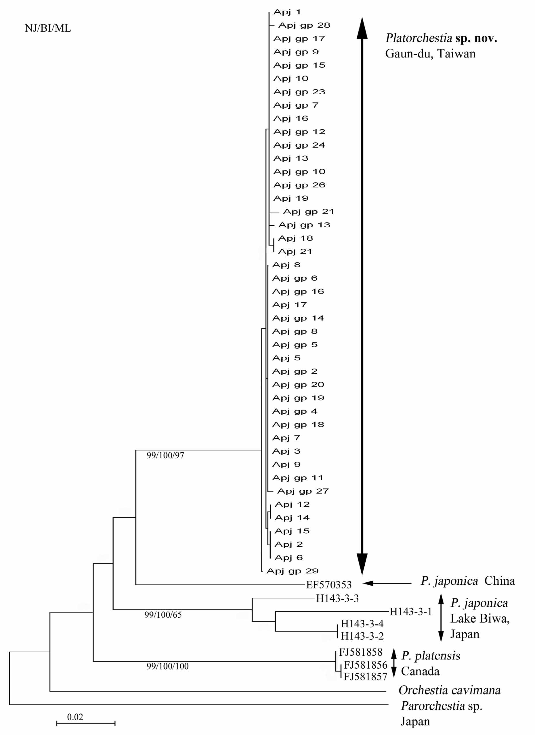
Molecular analysis. We got 618 b.p. aligned sequence of COI (GC ratio: 0.359), from 43 samples of P. paludosus sp. nov. and four samples of P. japonica . The number of total polymorphic sites was 102 within which 75 were parsimony informative sites (PIS), defining 13 haplotypes. There were 9 (3 PIS) and 50 (11 PIS) polymorphic sites for P. paludosus and P. japonica with 10 and 3 haplotypes respectively. The haplotype diversity (h) and nucleotide diversity (π) of P. paludosus samples were 0.743 ± 0.047 and 0.00183 ±0.00024. Those of P. japonica samples were 0.833 ±0.222 and 0.04396 ± 0.01253 (total: 0.784 ± 0.043 and 0.02088 ± 0.00826). Neighbor joining tree of P. paludosus , P. japonica and P. platensis ( Radulovici et al. 2009, NCBI, FJ581856 View Materials – FJ581858 View Materials ) formed three distinct clades, with bootstrap value> 99 suggesting the clades were well separated ( Fig. 9 View FIGURE 9 ), and topology of BI and ML trees were essentially consistent with NJ. Average sequence divergence ( Tab. 2) of P. paludosus from P. japonica was 13.3% (11.9–15.3%) from P. japonica , which was comparable to the interspecific difference of other Platorchestia species including P. platensis ( Radulovici et al. 2009, NCBI, FJ581856 View Materials – FJ581858 View Materials ; 14.5 and 17.2%).
Platorchestia P. japonica P. japonica P. platensis Orchestia cav- Parorchestia sp. nov. Japan China imana sp.
Platorchestia sp. nov. 0.2
P. japonica ( Japan) 13.3 4.8
P. japonica ( China) 10.4 14.3 _
P. p l a t e n s i s 14.5 17.2 16.1 0.01
Orchestia cavimana 19.1 21.1 19.9 23.2 _
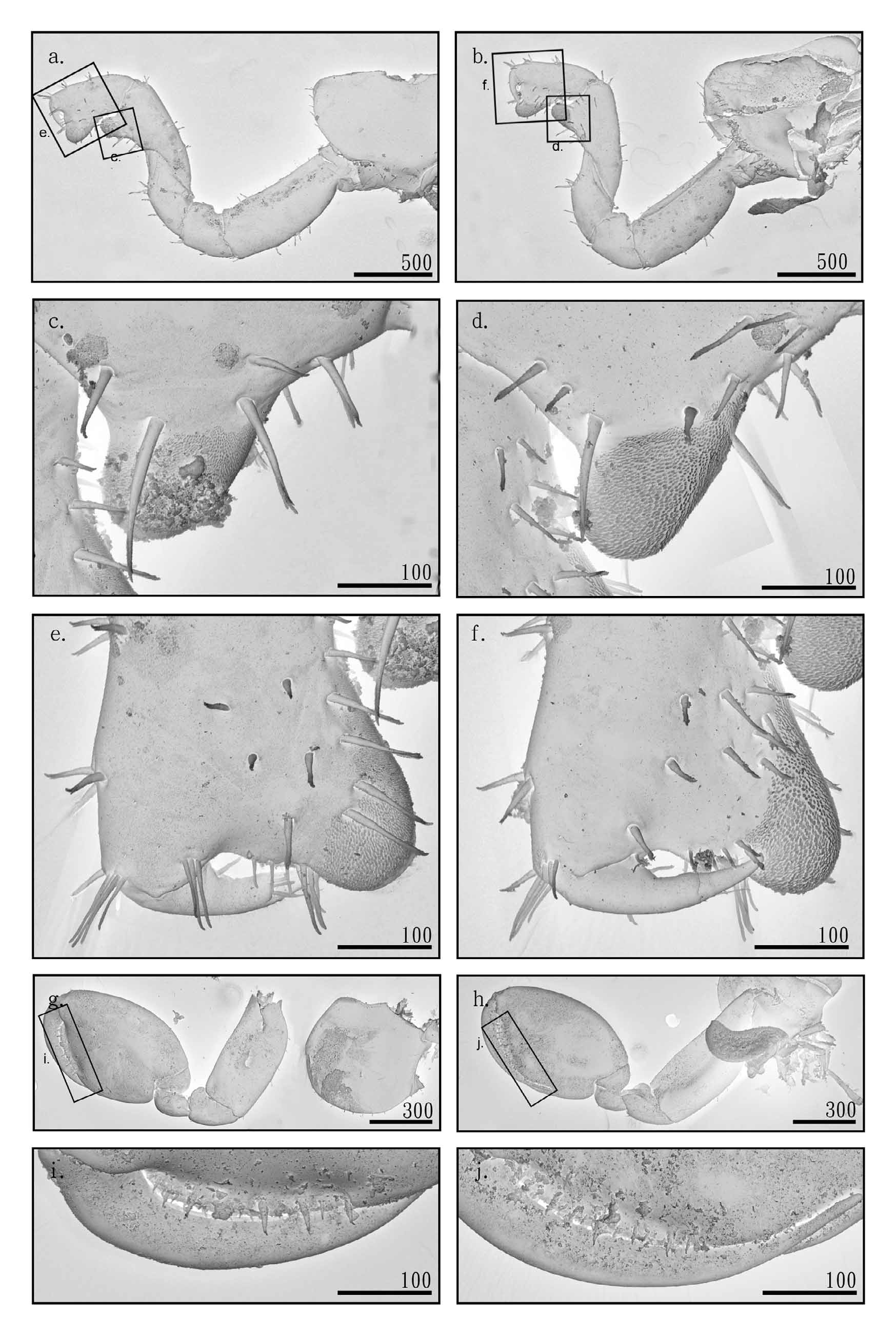
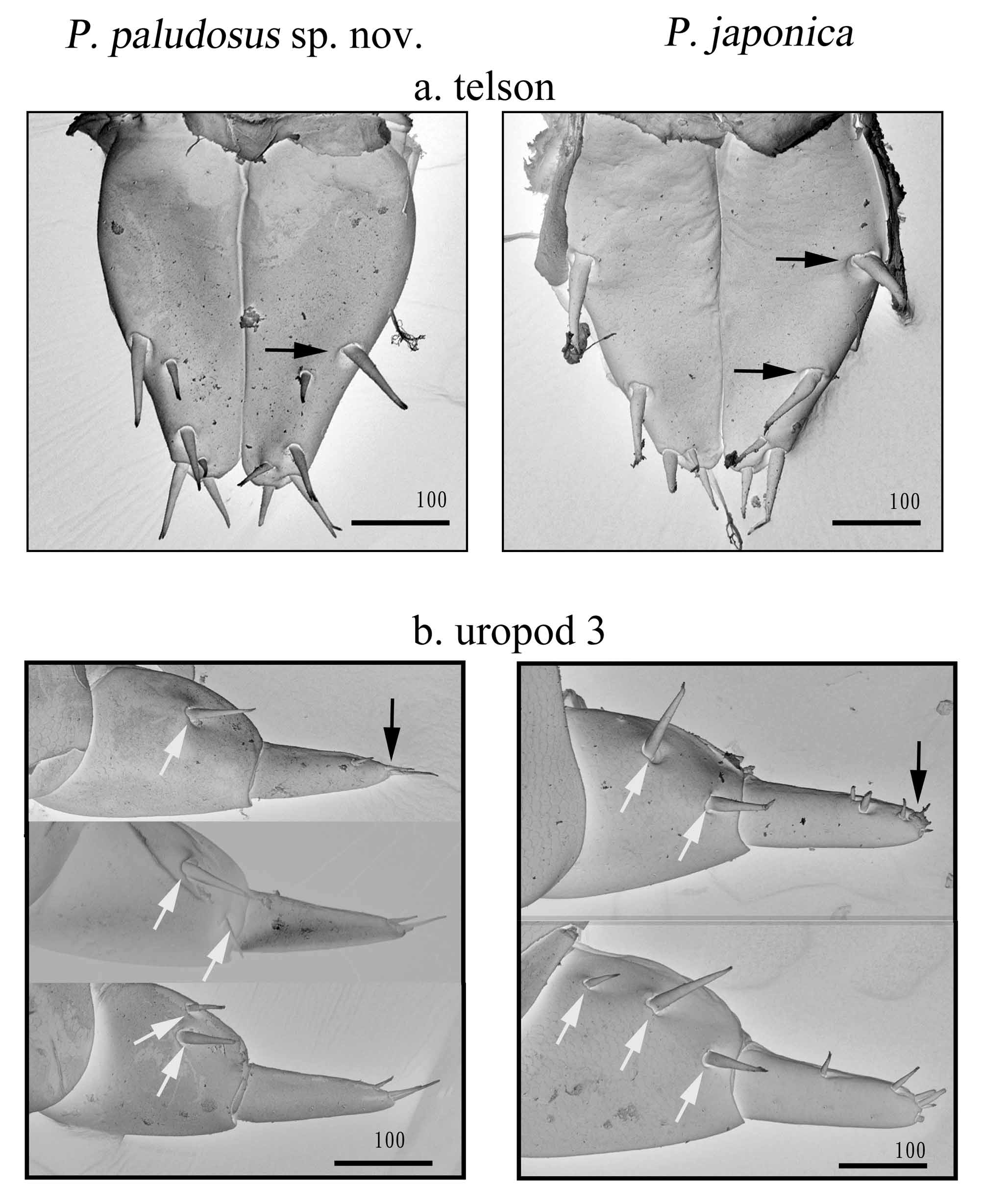
Parorchestia sp. 22.0 24.0 23.4 22.6 26.0 _ Discussion. In the present study, Platorchestia paludosus View in CoL sp. nov. is considered as a new species based on morphological and molecular evidence. The morphology is very close to P. japonica sensu stricto ( Tattersall 1922). Miyamoto & Morino (2004) considered their material collected from North Taiwan as P. japonica View in CoL and Miyamoto & Morino (2004) noted their Taiwanese specimens differed from P. japonica View in CoL collected from the Lake Biwa, Japan in the degree of sexual dimorphisms in peduncle articles 4 and 5 of antenna 2 and carpus of pereopod 7, shape of coxal plate of pereopod 6 and anterodistal corner of the posterior, morphology of marginal spines in peduncles of pleopods 2 and 3 and the spine patterns in pereopod 6 and 7. In the present study, we found that P. japonica View in CoL identified by Miyamoto & Morino (2004) in Taiwan was P. paludosus View in CoL identified in the present study. We used scanning electron microscopy and additionally noted consistent morphological differences between P. paludosus View in CoL (10 individuals) and P. japonica View in CoL (4 individuals) collected from the Lake Biwa, in the distribution pattern of setae on the lateral margin of telson, and in the shape of the ramus tip of uropod 3. Platorchestia paludosus View in CoL has one group of cuspidate setae type III on the lateral margin of telson, whilst P. japonica View in CoL contains two groups of cuspidate setae type III on the lateral margin of telson ( Fig. 10 View FIGURE 10 a). P. paludosus View in CoL has a sharper ramus of uropod 3, whilst the tip of ramus in uropod 3 of P. japonica View in CoL is blunt ( Fig. 10 View FIGURE 10 b). However, the number of setae on peduncle of uropod 3 varied from 1–3 in both P. paludosus View in CoL and P. japonica View in CoL , and thus, this cannot be considered as a taxonomic important character ( Fig. 10 View FIGURE 10 b).
Miyamoto & Morino (2004) divided the genus Platorchestia View in CoL into three groups based on the different gradient of sexual dimorphism character of antenna 2 and pereopod 6 and 7. The “group 3” defined by Miyamoto & Morino (2004) included species “displaying no sexual dimorphism” and having “antenna 2 and pereopod 6 and 7 barely (or slightly) sexual dimorphic”. Currently six species are placed within this group, including P. japonica ( Tattersall 1922) View in CoL , P. humicola View in CoL , P. k a a l e n s i s, P. lanipo View in CoL and P. pickeringi View in CoL , and the P. paludosus View in CoL in the present study also belong to this group. Platorchestia paludosus View in CoL can be differentiated from P. pickeringi View in CoL , P. kaalensis View in CoL and P. lanipo View in CoL by having normal maxilliped palps (vs. slender maxilliped palps) and differs from P. humicola View in CoL by possessing longer pleopod ramus: 0.8–0.9 times as long as peduncle (v.s. 0.5–0.6 times in P. humicola View in CoL ). Results from DNA barcoding further supports P. paludosus View in CoL as different from P. japonica View in CoL and is therefore a new species. Average sequence divergence of P. paludosus View in CoL from P. japonica View in CoL was 13.3% (11.9–15.3%), which is comparable to the interspecific difference P. platensis View in CoL (NCBI, FJ581856 View Materials – FJ581858 View Materials ; 14.5 and 17.2%). Platorchestia platensis View in CoL differs from P. japonica View in CoL in the morphology of palm of mature male gnathopod 2 and degree of sexual dimorphism in antenna 2 and pereopod 6 and 7 ( Miyamoto & Morino 2004). Our phylogenetic analysis also included a COI sequence (from GenBank) of P. japonica View in CoL collected from Hebei, China (NCBI, EF570353 View Materials ; Hou, Fu et al. 2007). Sequence divergence of P. japonica View in CoL from China and specimens from Lake Biwa, Japan reached about 14.3% and P. japonica View in CoL from China is closer to P. paludosus View in CoL , in having a sequence divergence of 10.4 %. Miyamoto & Morino (2004) claimed their P. japonica View in CoL collected from Taiwan had slight morphological differences (in the sharpness of coxal plate of pereopod 6 and number of margin spine in uropods 1–3) from P. japonica View in CoL from China in Morino & Dai (1990). Morphological and molecular data suggests that P. japonica View in CoL from the mainland China could be another cryptic species and awaits further morphological and molecular analysis.
Iwasa (1965) recorded Orchestia platensis Krøyer View in CoL [= Platorchestia platensis (Krøyer) View in CoL , Bousfield 1982] from Tanshui, N. Taiwan but there were no detailed text or figure illustrations for this species in Iwasa (1965). It is therefore difficult to check the validity of the record from Iwasa (1965). However, Miyamoto & Morino (2004) described their collections in Taiwan from 1976–1981 but they have not reported any P. platensis (Krøyer) View in CoL , and their records in N. Taiwan were ‘ P. japonica View in CoL ’.
Species name Gender Id. no. GenBank no. P. paludosus View in CoL sp. nov. m Apj 1 HQ 010294 View Materials
(Taipei, Taiwan.) f Apj 2 HQ 010295 View Materials
f Apj 3 HQ010296 View Materials
m Apj 5 HQ010297 View Materials
m Apj 6 HQ010298 View Materials
m Apj 7 HQ010299 View Materials
m Apj 8 HQ 010300 View Materials
f Apj 9 HQ010301 View Materials
m Apj 10 HQ 010302 View Materials
* Apj 12 HQ 010303 View Materials
* Apj 13 HQ 010304 View Materials
* Apj 14 HQ 010305 View Materials
continued next page Species name Gender Id. no. GenBank no. f Apj 15 HQ 010306 View Materials * Apj 16 HQ 010307 View Materials * Apj 17 HQ 010308 View Materials * Apj 18 HQ 010309 View Materials * Apj 19 HQ 010310 View Materials * Apj 21 HQ010311 View Materials m Apj gp 2 HQ010312 View Materials m Apj gp 4 HQ010313 View Materials m Apj gp 5 HQ010314 View Materials m Apj gp 6 HQ010315 View Materials m Apj gp 7 HQ010316 View Materials m Apj gp 8 HQ010317 View Materials m Apj gp 9 HQ010318 View Materials m Apj gp 10 HQ010319 View Materials m Apj gp 11 HQ010320 View Materials m Apj gp 12 HQ010321 View Materials m Apj gp 13 HQ 010322 View Materials f Apj gp 14 HQ010323 View Materials m Apj gp 15 HQ010324 View Materials m Apj gp 16 HQ010325 View Materials m Apj gp 17 HQ010326 View Materials m Apj gp 18 HQ010327 View Materials m Apj gp 19 HQ010328 View Materials m Apj gp 20 HQ010329 View Materials m Apj gp 21 HQ010330 View Materials m Apj gp 23 HQ010331 View Materials m Apj gp 24 HQ010332 View Materials m Apj gp 26 HQ010333 View Materials m Apj gp 27 HQ010334 View Materials m Apj gp 28 HQ010335 View Materials m Apj gp 29 HQ 010336 View Materials P. japonica View in CoL m H143-3-1 HQ 010293 View Materials (Lake Biwa, Japan.) f H143-3-2 HQ 010337 View Materials f H143-3-3 HQ010338 View Materials m H143-3-4 HQ 010339 View Materials Parorchestia sp. f H100-4-1 HQ 010292 View Materials (Lake Biwa, Japan.)

No known copyright restrictions apply. See Agosti, D., Egloff, W., 2009. Taxonomic information exchange and copyright: the Plazi approach. BMC Research Notes 2009, 2:53 for further explanation.
|
Kingdom |
|
|
Phylum |
|
|
Class |
|
|
Order |
|
|
Family |
|
|
Genus |
Platorchestia paludosus
| Cheng, Yu-Ting, Nakazono, Kenji, Lin, Kirk & Chan, Benny K. K. 2011 |
Platorchestia japonica
| Miyamoto 2004: 83 |