
Plakina muricyae, Cruz-Barraza & Vega & Carballo, 2014
|
publication ID |
https://doi.org/ 10.1111/zoj.12137 |
|
persistent identifier |
https://treatment.plazi.org/id/4617186B-FFA7-FF9C-FEF2-EEAFFAD83DA2 |
|
treatment provided by |
Marcus |
|
scientific name |
Plakina muricyae |
| status |
sp. nov. |
PLAKINA MURICYAE View in CoL SP. NOV.
( FIGS 2A–C View Figure 2 , 3 View Figure 3 )
Material examined
Holotype: MNCN-1.01/694, Antiguo Corral del Risco (Nayarit), 20°46′20″N, 105°32′49″W, 3 m depth, 29.iii.2012 GoogleMaps . Paratypes: LEB-ICML-UNAM-1108, Punta Mita (Nayarit), 20°46′20″N, 105°32′49″W, 3 m depth, 19.ii.2005 GoogleMaps . LEB-ICML-UNAM-1213, La Entrega (Oaxaca) 15°42′50″N, 96°05′20″W, 4 m depth, 4.v.2005 GoogleMaps . LEB- ICML-UNAM-1523, Bahía Tiburones, Isabel Island (Nayarit), 21°50′38″N, 105°52′14″W, 2 m depth, 21.vii.2005 GoogleMaps . LEB-ICML-UNAM-1651, Playa Blanca, Socorro Island (Revillagigedo), 18°48′59″N, 111°02′42″W, 0.5 m depth, 6.v.2008 GoogleMaps . LEB-ICML-UNAM-2011, Caleta de Bines, Socorro Island (Revillagigedo), 18°44′10″N, 110°57′37″W, 6 m depth, 7.xi.2009 GoogleMaps . LEB-ICML-UNAM- 2068, Antiguo Corral del Risco (Nayarit), 20°46′20″N, 105°32′49″W, 3 m depth, 29.iii.2012 GoogleMaps , LEB-ICML-UNAM- 2069, Punta Mita (Nayarit), 20°46′20″N, 105°32′49″W, 3 m depth, 29.iii.2012 GoogleMaps .
Specimens examined for comparison: Plakina monolopha from the collection of Laboratorio de Biología Marina (LBM) of Sevilla University ( Spain): LBM- 379 bahía de Algeciras, Spain, 9.vi.1992 ; LBM-511 Bahía de Algeciras , Spain, 28.ix.1992 .
ZooBank LSID: urn:lsid:zoobank.org:act:5BE60630-02A9-4A54-91E5-FE93D60795AB .
Etymology
The species is named after Dr Guilherme Muricy for his contribution to the study of the homosclerophorid taxonomy.
Diagnosis
Whitish to light green yellowish, thinly encrusting sponge, with smooth surface and rounded borders, characteristically with abundant subectosomal spaces regularly distributed. Spicules are diods, triods, and simple calthrops, which constitute a dense and sometimes confuse choanosomal alveolar structure, whereas the monolophose calthrops are arranged in a thin ectosomal layer.
Description
Thinly encrusting sponge from 1 to 4 mm thick, under coral rubbles and fragments of death corals, where it covers areas from 1 to 5 cm in diameter ( Fig. 2A, B View Figure 2 ). The surface varies from smooth to irregular, with lightly elevated, rounded borders, which are visible to the naked eye. Some small lobules, from 140–170 μm in diameter and 120–200 μm high, are observed under the microscope. Subectosomal spaces are abundant and regularly distributed on the surface, giving it a punctate appearance; they measure from 100 to 315 μm in diameter. Oscula are scarce, circular in shape, of about 1 mm in diameter, slightly elevated, and distributed irregularly over surface. Consistency is fleshy and firm. Ectosome is a translucent membrane not easily detachable. Choanosome with circular or oval canals, about 100 μm in diameter. Colour is whitish to light green yellowish in life; preserved is ochre.
Spicules: diods, triods, simple calthrops and monolophose calthrops ( Table 2). Diods are large, irregularly curved, some of them with a swelling slightly marked in the middle shaft ( Fig. 3A View Figure 3 ). Diods always present, but they are less abundant than triods. They measure 35–92.5 μm long and 0.5–5 μm wide. Triods are the most abundant spicules. They possess equidistant rays, although can be irregular with ‘T’ or ‘Y’ shape ( Fig. 3B View Figure 3 ). Rays measure from 6.25 to 37.5 μm long (spicules’ total length 12.5–72.5 μm). Simple calthrops are scarce; they may possess rays with sharp point or with small spines and commonly one of the rays is slightly larger than the others ( Fig. 3C View Figure 3 ). Calthrop rays measure from 6.25 to 30 μm long (total length 12.5–50 μm). Monolophose calthrops are common but not abundant; lophose rays have a simple ramification pattern: they ramify once at a medial position into two to four smaller rays with spined tips, sometimes smaller rays have a second bifurcation. Simple rays can end in a sharp or spiny point ( Fig. 3D View Figure 3 ). Rays measure 7.5–25 μm long (total length 15–50 μm).
Skeleton: ectosomal structure is armed by a thin layer of monolophose calthrops with the ramified ray directed toward the surface. Choanosomal skeleton is formed by diods, triods, and calthrops, forming an alveolar, dense, and sometimes confuse reticulation, with meshes of 7.5–15 μm in diameter ( Fig. 3E View Figure 3 ).
DNA barcode sequence data
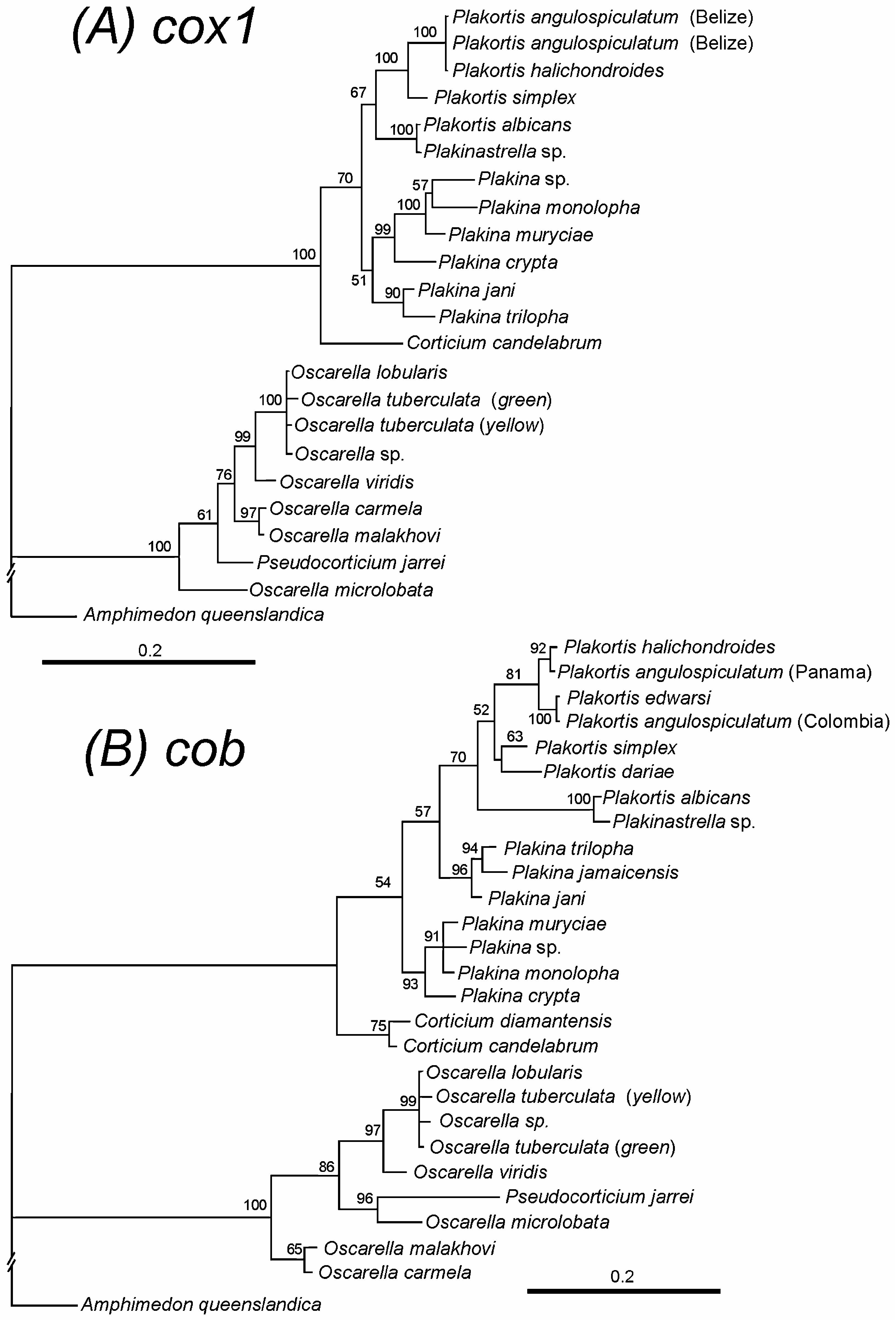
We obtained sequences of three individuals from Isabel Island, (Nayarit, Mexico), which were designat- ed here as the holotype and two paratypes. The three have an identical sequence for cox1 and cob. Therefore, these results, together with the morphological similarity between the specimens, confirm that they belong to the same species. Comparison of our cox1 ‘barcode fragment’ with similar sequences available in GenBank showed that Plakina muricyae sp. nov. differs by 4% from the nearest se- quence, which corresponds to Plakina monolopha ( HQ269351 View Materials ) from Sète, France. Cob comparison shows a similar result but differing by only 2% from Plakina monolopha .
Sequences of Plakina muricyae sp. nov. have been deposited in GenBank with accession number: KJ162928 View Materials (cob) and KJ162930 View Materials (cox1).
Ecology and distribution
The specimens were collected in coral reefs from the Mexican Pacific coast; Isabel Island (Nayarit), Oaxaca, and Socorro Island (Revillagigedo) ( Fig. 1 View Figure 1 ), from 0.5 to 6 m depth. All specimens were found in cryptic habitats, mainly under coral bases of the genus Pocillopora . Specimens from Punta Mita show a colour variation from white to light green yellowish, whereas at Revillagigedo only light green yellowish specimens were found.
Remarks
Plakina muricyae View in CoL sp. nov. displays similar characteristics to the Plakina monolopha View in CoL species complex, shares monolophose calthrops as only lophate spicules. Plakina monolopha View in CoL was described from the Mediterranean Sea ( Schulze, 1880), and it was then considered as a cosmopolitan species (see Muricy et al., 1998; Table 2). However, some authors mentioned that the non- Mediterranean specimens could be misidentified (e.g. Muricy et al., 1998). Here, we compared specimens of Plakina muricyae View in CoL sp. nov. with specimens of Plakina monolopha View in CoL from the Mediterranean Sea ( Carballo, 1994; Plakina monolopha View in CoL : LBM-379 Algeciras bay 9.vi.1992; LBM-511 Algeciras bay, 28.ix.1995). One of the main differences found between the two species is the presence of non-lophose calthrops with rays ending in three or four tiny spines in our specimens, which were not found in Plakina monolopha View in CoL . Additionally, there are differences in spicule size between the species; Plakina monolopha View in CoL possesses diods 50–67 μm long and 2–3 μm wide; triods with rays 44–55 μm long and 2–2.5 μm wide; calthrops with rays 20–30 μm long. Colour also varies between the species; Plakina monolopha View in CoL is white or rose, whereas Plakina muricyae View in CoL sp. nov. is whitish to light green yellowish. DNA sequence comparison of cox1 and cob fragments between Plakina muricyae View in CoL sp. nov.
M, monolophose; S, simple.
and Plakina monolopha (from France) showed that the species are closely related but with a significant genetic distance ( Fig. 8 View Figure 8 ). We considered that these morphological and molecular differences, together with the ample geographical distance, are sufficient to consid- er Plakina muricyae sp. nov. and Plakina monolopha to be different species. Morphological differences from other specimens cited as Plakina monolopha around the world can be seen in Table 2.
The only species from the eastern Pacific region with monolophose calthrops as seen in Plakina muricyae sp. nov. is Plakina pacifica Desqueyroux-Faúndez & van Soest, 1997 . Both species possess triods, but they are relatively scarce in Plakina pacifica , and very abundant in Plakina muricyae sp. nov., in which they constitute the main skeleton. In addition, our specimens of Plakina muricyae sp. nov. possess monolophose calthrops in a single size category with rays measuring from 7.5 to 25 μm, whereas P. pacifica has monolophose calthrops in two size categories: rays measuring 13–15 μm and 19–45 μm long ( Table 1).


No known copyright restrictions apply. See Agosti, D., Egloff, W., 2009. Taxonomic information exchange and copyright: the Plazi approach. BMC Research Notes 2009, 2:53 for further explanation.
|
Kingdom |
|
|
Phylum |
|
|
Class |
|
|
Order |
|
|
Family |
|
|
Genus |
Plakina muricyae
| Cruz-Barraza, José Antonio, Vega, Cristina & Carballo, José Luis 2014 |
Plakina muricyae
| Cruz-Barraza & Vega & Carballo 2014 |
Plakina muricyae
| Cruz-Barraza & Vega & Carballo 2014 |
Plakina muricyae
| Cruz-Barraza & Vega & Carballo 2014 |
Plakina muricyae
| Cruz-Barraza & Vega & Carballo 2014 |
Plakina muricyae
| Cruz-Barraza & Vega & Carballo 2014 |
P. muricyae
| Cruz-Barraza & Vega & Carballo 2014 |
P. muricyae
| Cruz-Barraza & Vega & Carballo 2014 |
P. muricyae
| Cruz-Barraza & Vega & Carballo 2014 |
P. muricyae
| Cruz-Barraza & Vega & Carballo 2014 |
P. muricyae
| Cruz-Barraza & Vega & Carballo 2014 |
P. muricyae
| Cruz-Barraza & Vega & Carballo 2014 |
P. muricyae
| Cruz-Barraza & Vega & Carballo 2014 |
P. muricyae
| Cruz-Barraza & Vega & Carballo 2014 |
Plakina pacifica
| Desqueyroux-Faundez & van Soest 1997 |
P. monolopha sensu
| Thomas 1970 |
P. monolopha sensu
| Koltun 1964 |
P. monolopha sensu
| Arndt 1927 |
P. monolopha sensu
| Thiele 1898 |
Plakina monolopha
| Schulze 1880 |
Plakina monolopha
| Schulze 1880 |
Plakina monolopha
| Schulze 1880 |
Plakina monolopha
| Schulze 1880 |
Plakina monolopha
| Schulze 1880 |
Plakina monolopha
| Schulze 1880 |
Plakina monolopha
| Schulze 1880 |
Plakina monolopha
| Schulze 1880 |