
Plagiodontes patagonicus (d’Orbigny, 1835)
|
publication ID |
https://doi.org/ 10.1080/00222930902903244 |
|
DOI |
https://doi.org/10.5281/zenodo.5216713 |
|
persistent identifier |
https://treatment.plazi.org/id/E25D87A9-2275-3516-FE67-AB2CFE5FFF88 |
|
treatment provided by |
Felipe |
|
scientific name |
Plagiodontes patagonicus (d’Orbigny, 1835) |
| status |
|
Plagiodontes patagonicus (d’Orbigny, 1835) View in CoL
The pallial complex ( Figure 19A,B View Figure 19 ) was briefly described by Hylton-Scott (1952), so we add only some additional details.
It is about 30 mm long. The kidney is triangular and occupies less than 20% of the lung length. Although Hylton-Scott (1952) described the secondary ureter as closed, we determined that it is open, being the ureteric pore located at the level of the lower third of the kidney length. The conspicuous pulmonary vein branches in the afferent marginal vein that occupies 60–70% of its length. The vascularization in the ad-rectal area and in the area between the veins is conspicuous. The marginal vein, branching from the last portion of the pulmonary vein and bordering the pallial border, is branched in minor veins. The pallial border has a brown spongy area, the pallial gland.
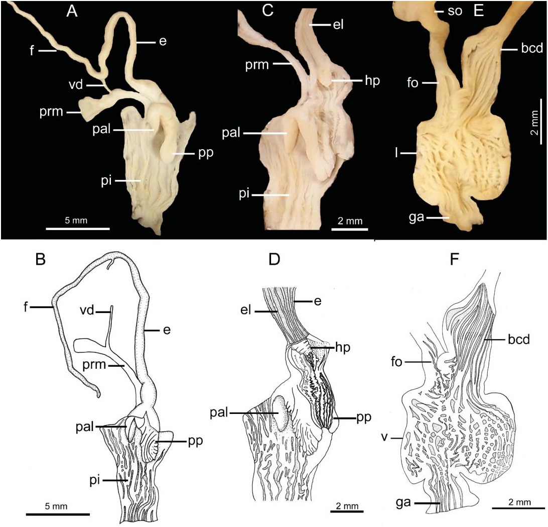
Anatomy of the genital system and its morphometric variations were already described by Hylton-Scott (1952), Cazzaniga and Fernández-Canigia (1985) and Cazzaniga et al. (2005), but the inner anatomy of the penis and vagina has not been analysed until now. Figure 20A,B View Figure 20 shows the general aspect of the genital system.
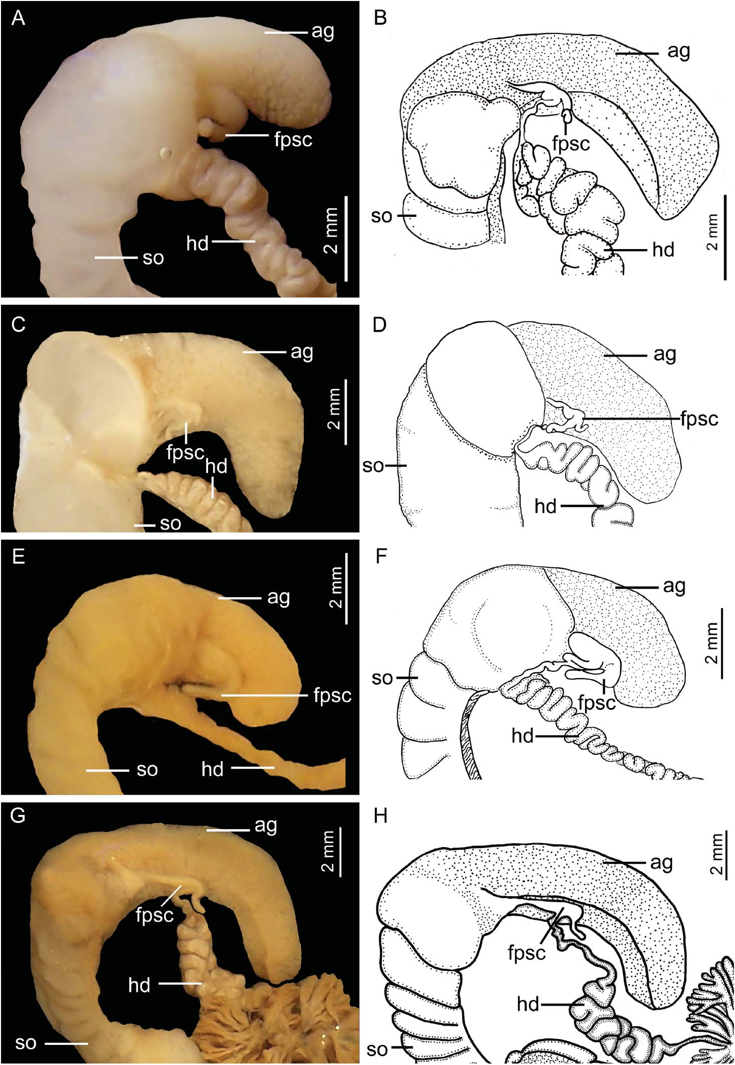
Internally, the penis bears a simple penial papilla or verge ( Figure 18A,B View Figure 18 ), proximally rounded and swollen, with transverse slits bordering its opening; it is distally triangular, elongated and smooth. The penis inner wall has voluminous and undulated pilasters with only a few anastomoses. While the area surrounded by the penial sheath and the genital atrium has multiple straight, thin folds, the area below the penial papilla is smooth.
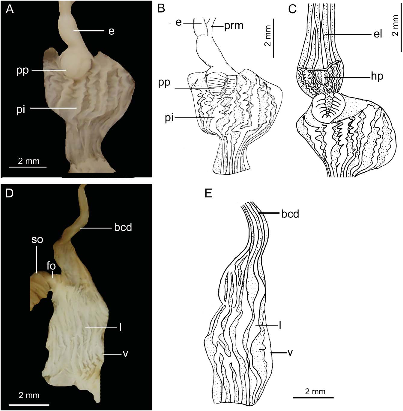
The transition between penis and epiphallus is constricted, the epiphallus being proximally swollen and distally cylindrical. This cylindrical portion has five to seven straight thin folds ( Figure 21C View Figure 21 ). It is separated from the swollen part by the same structure found in the remaining Plagiodontes species. The flagellum, cylindrical and shorter than the epiphallus, has a straight fold in its inner wall. There is a noticeable change of diameter between the epiphallus and the flagellum. The vagina, shorter than the penis and twice as long as it is wide, has its inner surface longitudinally folded, with minor anastomoses that give it a reticulate aspect ( Figure 21D,E View Figure 21 ). The bursa copulatrix duct is distally swollen and has internal longitudinal straight folds ( Figure 21D,E View Figure 21 ).

No known copyright restrictions apply. See Agosti, D., Egloff, W., 2009. Taxonomic information exchange and copyright: the Plazi approach. BMC Research Notes 2009, 2:53 for further explanation.
|
Kingdom |
|
|
Phylum |
|
|
Class |
|
|
Order |
|
|
Family |
|
|
Genus |