
Plagiodontes multiplicatus parvus ( Hylton-Scott, 1952 )
|
publication ID |
https://doi.org/ 10.1080/00222930902903244 |
|
DOI |
https://doi.org/10.5281/zenodo.5216711 |
|
persistent identifier |
https://treatment.plazi.org/id/E25D87A9-2278-3510-FEE6-AD53FE18F8E7 |
|
treatment provided by |
Felipe |
|
scientific name |
Plagiodontes multiplicatus parvus ( Hylton-Scott, 1952 ) |
| status |
|
Plagiodontes multiplicatus parvus ( Hylton-Scott, 1952) View in CoL
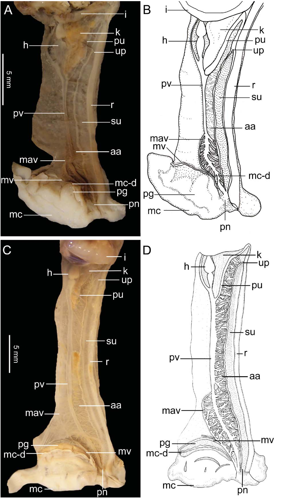
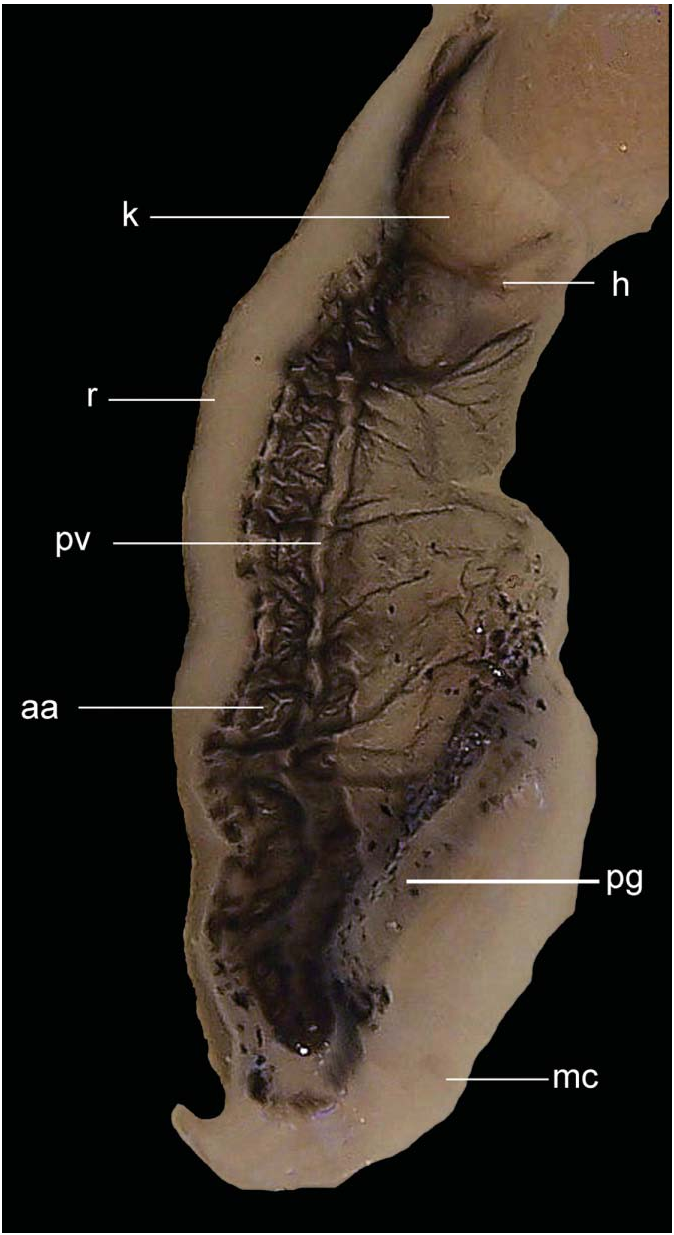
The pallial complex is about 26–31 mm long ( Figure 14C,D View Figure 14 ). A triangular kidney occupies 20% of the lung length. The secondary ureter opens above the level corresponding to the midpoint of the kidney, and is delimited by two ridges of which the ad-rectal one is more developed. Within the pericardium the auricle is much wider than the ventricle.
The pulmonary vein is prominent. The afferent marginal vein branches out at its midpoint. There is a conspicuous vascularization in the ad-rectal area and between the two main veins. The marginal vein, with no branching, runs along the mantle collar, which has a spongy pallial gland and several indentations that correspond to the position of the apertural teeth.
The genital system is relatively large ( Figure 15C,D View Figure 15 ). The orange-brownish ovotestis is formed by six groups of digitiform acini. The hermaphroditic duct, cream to light-brown in colour, is convoluted and centrally swollen. The fertilization pouch– spermathecal complex, located in the inferior portion of the albumen gland, is white and proximally swollen; it is distally formed by a free L-shaped blind sac. The albumen gland is light brown to pale orange, elongated, of rectangular to triangular shape, and variable in size ( Figure 16C,D View Figure 16 ).
The spermoviduct is a tubular and lobed organ formed by the hyaline white uterus and the opaque white prostatic portion, of glandular aspect. The vas deferens emerging just above the bifurcation of the vagina has the same characteristics as in the remaining species.
The vagina is subcylindrical, short (the penis is from 1.5 times to twice as long), as wide as, or slightly wider than, the penis. Its internal surface is composed of thin anastomosed lamellae forming a reticulum ( Figure 18E,F View Figure 18 ).
The bursa copulatrix is subspherical and its duct, of about 20–26 mm long, is internally provided with thin, straight lamellae that are intermingled with short undulated ones in the first portion of the duct.
The club-shaped penis is from 3.3 to 4.3 times as long as wide and about the same length as the epiphallus. The penial sheath is of medium length. The internal penis structure shows a verge with an accessory lobe. The verge, prolongation of the epiphallus, has an internal hollow tubule. The accessory lobe, proximally united to the papilla, is compact, of triangular shape and one-third the size of the papilla. The inner penis wall has a variable sculpture of protruding longitudinal and undulated pilasters with several branches and anastomoses. The pilasters become thin and straight in the area that is externally surrounded by the penial sheath; they are absent under the papilla ( Figure 18A,B View Figure 18 ).
The transition from penis to epiphallus is marked by a constriction. The epiphallus is proximally swollen but otherwise cylindrical. Internally, the cylindrical portion bears five to seven straight thin folds or lamellae; it is separated from the swollen part by a small structure formed by the merging folds. This structure, of probable glandular function, was sometimes covered with a hard translucent cover. The swollen part has some internal longitudinal folds with lateral branches, and continues in a cylindrical tubule inside the verge. This tubule also bears internal longitudinal folds, which are more voluminous and have more branches and anastomoses ( Figure 18E,F View Figure 18 ) than those in the swollen part. The flagellum, longer than the epiphallus, is cylindrical and has only one internal longitudinal fold. The epiphallus–flagellum transition is marked by the insertion of the vas deferens and a slight reduction of the diameter towards the flagellum.

No known copyright restrictions apply. See Agosti, D., Egloff, W., 2009. Taxonomic information exchange and copyright: the Plazi approach. BMC Research Notes 2009, 2:53 for further explanation.
|
Kingdom |
|
|
Phylum |
|
|
Class |
|
|
Order |
|
|
Family |
|
|
Genus |