
Phallomedusa solida (Martens, 1878)
|
publication ID |
https://doi.org/ 10.5281/zenodo.176773 |
|
DOI |
https://doi.org/10.5281/zenodo.6240292 |
|
persistent identifier |
https://treatment.plazi.org/id/03F887BA-9912-5421-118F-C0D4FC29FDE0 |
|
treatment provided by |
Plazi |
|
scientific name |
Phallomedusa solida (Martens, 1878) |
| status |
|
Phallomedusa solida (Martens, 1878) View in CoL
Ampullacera fragilis ; Quoy & Gaimard, 1832 (in part): 201–203, pl. XV, figs. 10–12 (not of Lamarck 1822). Ampullarina quoyana ; Angas, 1867: 232 (not of Potiez & Michaud 1838).
Amphibola solida Martens View in CoL , in Schacko, 1878: 1 –9, pl. I, fig. 1.
Amphibola fragilis ; Bouvier 1892: 146 –153 (not of Lamarck 1822).
Salinator quoyana ; Cotton & Godfrey 1932: 150 (not of Potiez & Michaud 1838). Salinator solida View in CoL ; Woolacott 1945: 2, pl. III, figs. 1, 2, 7, 9.
Type material: Holotype ( Fig. 2 View FIGURE 2 H) from ‘Australia’, Robillard ( ZMB, 22207).
Other material examined: Australia, Queensland: Port Douglas, Dickson’s Inlet, in mangroves, 3 Jun 1977, I. Loch, P.H. Colman and R. Creese (AMS C.400788); Proserpine River, opposite Flying Fox Island, in mangroves, 1 May 1975, W.F. Ponder (AMS C.446518). Australia, New South Wales: Brisbane Waters, Fagan's Bay, on mud in saltmarsh, 10 Mar 2003, R.E. Golding, W.F. Ponder and H. Fukuda (AMS C.442245); South side of Narrabeen Lakes, on sandy mud amongst reeds and mangroves, Mar–Sep 2004, R.E. Golding and M. Hill (AMS C.442241, C.442277, C.442286); Lane Cove River, Boronia Park, on mud in open mangrove forest at low tide, Dec 2003 – Sep 2004, R.E. Golding, M. Hill and P.M. Golding (AMS C.442276, C.442284, C.442266); Parramatta River, Homebush Bay, on mud amongst dense mangroves, Dec 2003 – Feb 2004, R.E. Golding (AMS C.442247, C.442259, C.442262); Lake Illawarra, Kully Bay, creek near lake with sparse saltmarsh vegetation, 3Mar 2004, R.E. Golding and P.M. Golding (AMS C.442239). Australia, Tasmania: Stanley, Sawyer Bay, East Inlet, damp mudflats at low tide, 20 Apr 2004, W.F. Ponder (C.446517).
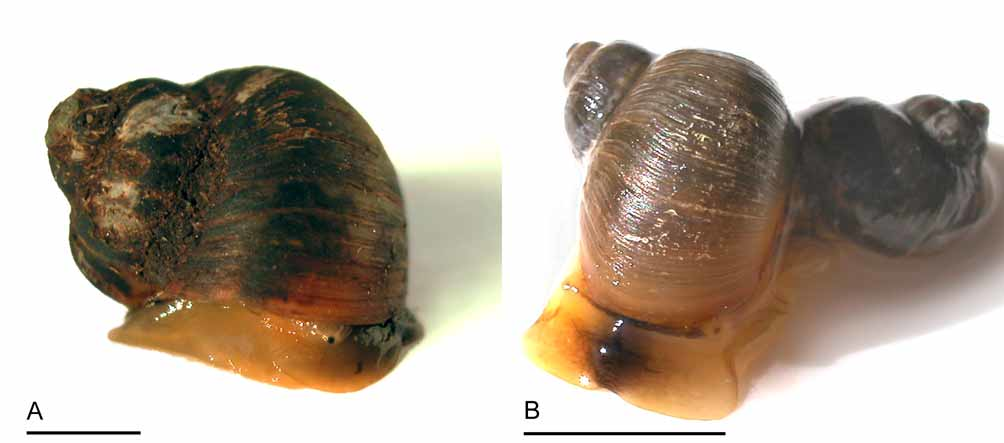
Redescription: Shell ( Figs. 2 View FIGURE 2 H, I): Solid, opaque, spire relatively tall, conical, shell diameter to 20 mm, axial striae indistinct. Whorls rounded, lacking shoulder. Outer lip of aperture slightly thickened at base. Exterior white with brown, narrow zigzag lines on all whorls. Interior of aperture white.
Operculum ( Figs. 3 View FIGURE 3 D, 4A): Thick, dark brown, corneous, elliptical, columellar edge straight, outer edge curved forming rounded point in upper left corner (viewed from exterior). Nucleus paucispiral, eccentric. Prominent hollow keel curving around nucleus on interior of operculum, containing muscular projection from foot.
External morphology ( Figs. 5 View FIGURE 5 A, 6C): Head-foot dark grey in preserved specimens, protruding only a short way beyond shell when animal is crawling; diffuse black pigment across snout. Eyes at central base of small, triangular tentacles.
Mantle organs ( Fig. 7 View FIGURE 7 E): Narrow, opposed ciliary tracts restricted to exhalant canal in mantle cavity; roof of mantle cavity with numerous blood vessels. Hypobranchial gland at right anterior mantle cavity roof pyriform, flat, light brown with dark brown flecks in fresh and formalin-preserved specimens, outer surface smooth.
Digestive system: As for A. crenata .
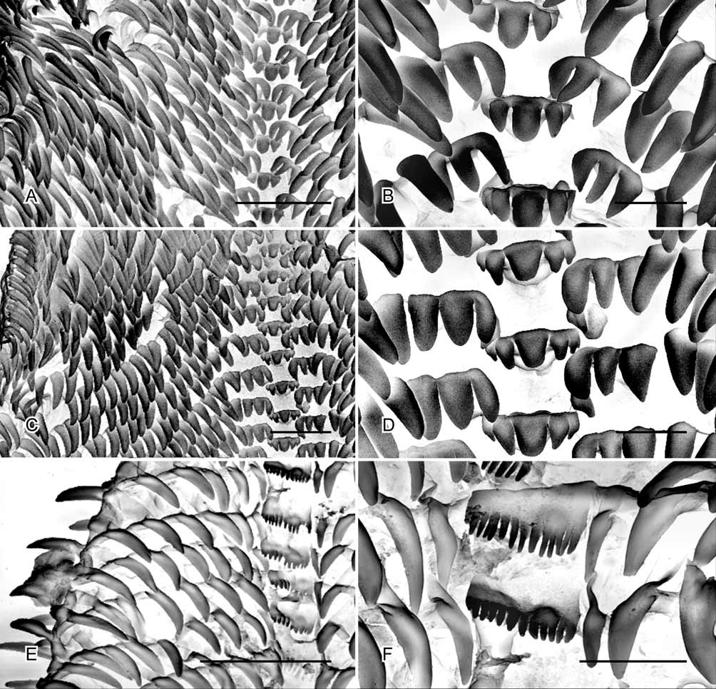
Radula ( Figs. 11 View FIGURE 11 A, B): Each row with central tooth, inner and outer lateral teeth and, on each side, approximately 25 marginal teeth. Central tooth with five wide cusps, mesocone enlarged, rectangular; inner lateral teeth unicuspid, narrow; outer lateral teeth equal in size to central tooth, with three wide cusps increasing in size towards centre of radula; marginal teeth elongate, unicuspid, wide, increasing in length towards outer edge of radula.
Central nervous system ( Fig. 12 View FIGURE 12 F): Pleural ganglia separated from adjacent cerebral, pedal and parietal ganglia by short connectives. Procerebrum, parapedal and subcerebral commissures present. Right parietal ganglion slightly larger than left, right parietal-visceral commissure shorter than left parietal-visceral commissure.
Reproductive system ( Figs. 13 View FIGURE 13 C, 15A, B, 17A, B): Ovotestis composed of numerous lobes uniting to form single acinule duct. Seminal vesicle diverticulum from upper hermaphrodite duct, posterior to junction with acinule duct. Hermaphrodite duct coiling anteriorly, swollen with pale pink, iridescent sperm. Numerous (>10) seminal receptacles present as yellow, spherical projections from hermaphrodite duct. Spermoviduct embedded in right body wall distally, emerging to divide into muscular oviduct/vagina and long vas deferens. Muscular vagina long, with thick, folded walls and two lateral bulges proximally, narrowing distally. Small flagellum of vas deferens extending beyond junction with oviduct; vas deferens very long, coiled, yellow to orange in fresh and formalin-preserved specimens, muscular exterior lined internally with tall narrow prostatic cells containing spherical secretory vesicles. Complex penis with more spiral flange coiling more than three times around penis; spiral base with unciliated external sperm groove between spirally coiled lateral flanges. Penis attached near genital aperture, retractor muscle inserting into base of penis. Flange forming large shield distally and dividing into numerous moderately long tentacle-like appendages. Penial appendages and inner surface of spiral flange with long, forked, chitinous papilla-like microsculpture. Egg mass narrow, cylindrical string deposited in coil on surface of substratum.
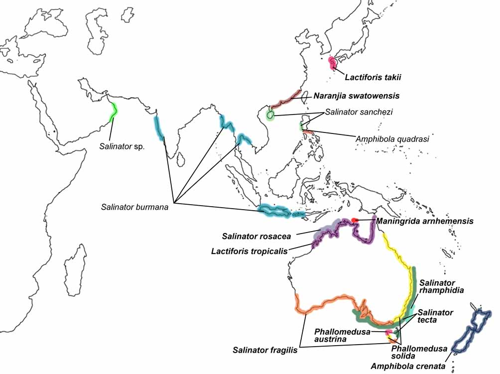
Distribution and habitat ( Fig. 1 View FIGURE 1 ): East and south-east coast of Australia, from north Queensland to South Australia and the north-west of Tasmania (based on AMS collections). Found in upper littoral mangrove, saltmarsh and mudflat habitat, often very abundant.
Remarks: The shell of P. s o l i d a was first figured by Quoy & Gaimard (1832) as a variety of S. fragilis ; although it is not clear where their figured specimen was collected, it appears to be similar to material from south-east Australia. The species has also been incorrectly attributed to S. quoyana by Angas (1867) and Cotton & Godfrey (1932) from Adelaide (South Australia). Phallomedusa solida is dominant in saltmarshes and at the back edge of mangrove habitat along the east coast of Australia.


| ZMB |
Museum für Naturkunde Berlin (Zoological Collections) |
No known copyright restrictions apply. See Agosti, D., Egloff, W., 2009. Taxonomic information exchange and copyright: the Plazi approach. BMC Research Notes 2009, 2:53 for further explanation.
|
Kingdom |
|
|
Phylum |
|
|
Class |
|
|
Family |
|
|
Genus |
Phallomedusa solida (Martens, 1878)
| Golding, Rosemary E., Ponder, Winston F. & Byrne, Maria 2007 |
Salinator quoyana
| Woolacott 1945: 2 |
| Cotton 1932: 150 |
Amphibola fragilis
| Bouvier 1892: 146 |
Amphibola solida
| Schacko 1878: 1 |