
Paratachardina ternata (Chamberlin)
|
publication ID |
https://doi.org/ 10.5281/zenodo.179122 |
|
DOI |
https://doi.org/10.5281/zenodo.6247490 |
|
persistent identifier |
https://treatment.plazi.org/id/0397AD19-FF8B-FF9B-C6CD-FB60453A6F78 |
|
treatment provided by |
Plazi |
|
scientific name |
Paratachardina ternata (Chamberlin) |
| status |
|
Paratachardina ternata (Chamberlin) View in CoL
( Figs 2 View FIGURE 2 G, 11)
Tachardina (Tachardina) ternata Chamberlin, 1923: 208 View in CoL .
Tachardina ternata ( Chamberlin) View in CoL ; Chamberlin, 1925: 41.
Paratachardina ternata (Chamberlin) ; Varshney, 1968: 489.
Type material studied. Lectotype, hereby designated. Adult female. on slide labelled as " Holotype ", INDIA: Kerala, Travancore, ex Acacia sundra , coll. Mahdihassan, from Green 1922 (BME). Paralectotypes. Same label data as lectotype, 2(5) (BME); 2 boxes of dry material (each with a few dry tests of adult females), same label data as lectotype except that both boxes have a red label " TYPE MATERIAL" and one box has an additional label: " ternata , Tachardina / 2 lots / TYPE India " (BME); adult females slide-mounted by PJG 2006 from the BME dry material labelled as: " Tachardia ternata (Green, ms) / on Acacia sundra / Travancore, India / coll. S. Mahdihassan", 6(21: 3 adult females + 29 embryonic first-instar nymphs) (BME).
Adult female
Unmounted material ( Fig. 2 View FIGURE 2 G). Test more or less massed on trees; subglobular and subtrilobate, with distinct flutings running from base to apex; dirty reddish brown in colour ( Chamberlin, 1923).
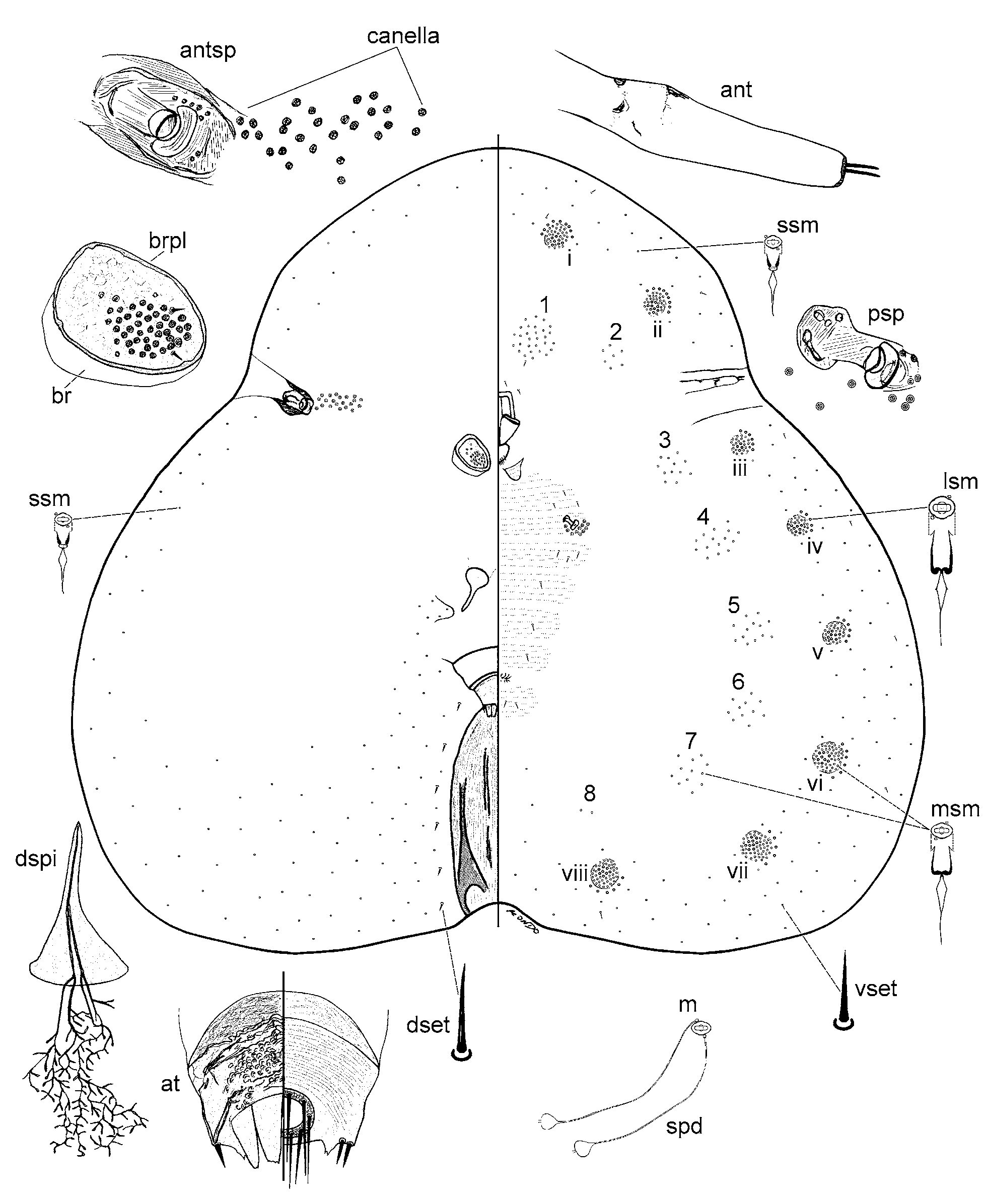
Mounted material ( Fig. 11 View FIGURE 11 ). Body outline broadly pyriform, slightly trilobate to transversely oval, often with a constriction at anterior stigmatic areas. Body 1.8–2.3 mm long, 1.8–2.5 mm wide (n = 9).
Dorsum. Brachia membranous, becoming slightly sclerotized at maturity, short, less than about half length of a brachial plate. Brachial plates subcircular to oblong, often with an irregular outline, each plate 105–123 µm long, 70–80 µm wide; brachial crater very shallow, with a subcircular group of 35–60 pseudospines on narrower side of plate; with 1–5 brachial pores just anterior to group of pseudospines, each pore with 5-loculi; with about one seta on each side of pseudospine group. Anterior spiracles each 58–72 µm long, 28–35 µm wide, surrounded by a sclerotized area; with a group of 6–12 pores within spiracular sclerotization; canella represented by a group of 9–30 spiracular pores on area just outside spiracular sclerotization; canellar and spiracular pores similar, each 5.0–6.0 µm wide with 5 loculi. Dorsal spine well developed, with an opening at apex, length: 88–103 µm, 80–83 µm wide at base; membranous pedicel no longer than length of dorsal spine, slightly wider than base of dorsal spine. Anal tubercle well developed, tapering, highly sclerotized; preanal plate plus supra-anal plate, 188–205 µm long, 150–190 µm wide at widest point, pre-anal plate with a fibrous texture, supra-anal plate with a granular texture. Pygidial apodemes well developed, fairly sclerotized, extending from base of each anal tubercle towards body apex. Anal fringe probably incomplete, each anal fringe plate serrated or at least with some teeth. Anal ring entire, 38–40 µm wide, tip of setae surpassing anal fringe. Microducts scarce, each ca. 3 µm wide, present submarginally, with several ducts present on each antero-anal lobe. Spermatoid ducts: 1–3 associated with each microduct. Dorsal setae each 4–6 µm long marginally or submarginally, with longer setae, each 7–10 µm long, lateral to pygidial apodeme.
Venter. Antennae 128–158 µm long, 2 segmented, segmentation poorly defined, with a sclerotized area delineating both segments, with 2 longer setae and 2 or 3 shorter setae on sclerotized area at apex of terminal segment. Clypeolabral shield 126–139 µm long, 90–95 µm wide. Labium apparently 1 segmented, 43–73 µm long, 65–73 µm wide. Pre-oral lobes elongate, poorly developed, present along margins of clypeolabral shield on each side; post-oral lobes present, dome shaped, with microtrichia. Legs completely absent. Posterior spiracles each 43–50 long, peritremes each 20–25 µm wide; with 12–16 spiracular pores present around each spiracle within spiracular pocket, each pore 5.0–6.0 µm wide. Marginal duct clusters distinct, subcircular, 8 pairs in total, each composed of two types of microducts: medium-sized microducts, elongate oval, each ca. 4 µm wide, most abundant, making bulk of each marginal duct cluster, and large-sized microducts, subcircular each ca. 5 µm wide, present on outer rim of cluster closest to outer margin, and on inner areas of each cluster. Ventral duct clusters in loose subcircular or irregular groups, all composed of medium-sized microducts, 7 or 8 pairs in total, pair just anterior to mouthparts (vdc-1) largest, a small cluster (vdc-2) with 2–6 microducts, close to mdc-ii, often hard to detect, and 5 or 6 pairs of clusters posterior to antennae, if 8th pair (vdc-8) present, with 1–4 microducts. Microducts outside ventral and marginal duct clusters smallest, each ca. 3.0 µm wide, present marginally and submarginally, abundant particularly around marginal duct clusters. Spermatoid ducts similar to those on dorsum, detected around body margin, appearing most numerous within each marginal duct cluster. Ventral setae usually 4–6 µm long, longer setae, each 14–18 µm long, present on abdomen between vulva and body apex.
Diagnosis. Paratachardina ternata is characterized by following features: (i) body broadly pyriform or trilobed; (ii) 7 or 8 pairs of ventral duct clusters, with vdc-2 close to mdc-ii but often difficult to detect; (iii) 35–60 pseudospines on each brachial plate; and (iv) sclerotized pygidial apodemes. Paratachardina ternata appears closest to P. mithila and P. t h e a e as all three have a trilobed body as mounted on the slide. However, P. ternata can be separated from the other two species by the following features (character states of P. mithila and P. t h e a e in parentheses): (i) vdc-2 close to mdc-ii (vdc-2 close to mdc-iii); (ii) absence of a pair of round outpockets of membranous cuticle with a reticulate surface in the area surrounded by the anal tubercle, dorsal spine and pre-anal lobes (present in P. t h e a e at least); and (iii) well-developed pygidial apodemes present (pygidial apodemes absent).
Notes. The type locality is misspelt as "Travencore" in Ben-Dov (2006).

No known copyright restrictions apply. See Agosti, D., Egloff, W., 2009. Taxonomic information exchange and copyright: the Plazi approach. BMC Research Notes 2009, 2:53 for further explanation.
|
Kingdom |
|
|
Phylum |
|
|
Class |
|
|
Order |
|
|
Family |
|
|
Genus |
Paratachardina ternata (Chamberlin)
| Kondo, Takumasa & Gullan, Penny J. 2007 |
Tachardina ternata (
| Chamberlin 1925: 41 |
Tachardina (Tachardina) ternata
| Chamberlin 1923: 208 |