
Paralamyctes wellingtonensis, Edgecombe, Gregory D., 2003
|
publication ID |
https://doi.org/ 10.5281/zenodo.156319 |
|
DOI |
https://doi.org/10.5281/zenodo.6277022 |
|
persistent identifier |
https://treatment.plazi.org/id/03D5C216-FFD8-FF9F-FEDD-07E1B2EEFCC7 |
|
treatment provided by |
Plazi |
|
scientific name |
Paralamyctes wellingtonensis |
| status |
sp. nov. |
Paralamyctes wellingtonensis View in CoL n. sp.
Figs. 131 View FIGURES 1 3 View FIGURES 4 7
Paralamyctes cf. P. chilensis (Gervais in Walckenaer & Gervais, 1847): Edgecombe et al. 2003, figs. 3AC.
Diagnosis: Paralamyctes with 1720 (usually 19) antennal articles. Female gonopod with long, slender process on first article, bearing two relatively stout, blunt spurs. Females with 2,2,2,2/2,2,2,2 to 2,3,3,3/2,3,3,3 coxal pores, inner pore tiny when three present; males mostly with 2,2,2,2/2,2,2,2 coxal pores, maximum 2,3,3,2/2,3,3,2.
Holotype: Male MCZ 52636 ( Fig. 1 View FIGURES 1 3 ), from Puerto Edén, Isla Wellington, Chile, ca 49ºS [49°08'S 74°27'W], leg. P.J. Darlington, 1013 December 1962.
Paratypes: All from type locality, leg. P.J. Darlington, 715 December 1962. MCZ 27838, 1 female, 3 males, from type collection; MCZ 27836, 2 males; MCZ 47468, 1 female; MCZ 47470, 4 females.
Other material: MCZ DNA 100408, 2 males, near Refugio Chileno, Parque Nacional Torres del Paine, Provincia Esperanza, 50º56'45"S 72º55'00"E, leg. J. Miller & I. Agnarsson, 8 December 2000.
Etymology: After Isla Wellington.
Description: Length (anterior margin of head shield to end of telson) up to 16.6 mm; length of head shield up to 1.6 mm. Colour (based on specimens in absolute ethanol): head shield and tergites pale yellow to pale orange with purple mottling; dark purple patch around ocellus and on cephalic pleurite beneath ocellus; antenna yellowbrown to moderate orange; maxillipedes yellow to pale orange, deeper orange on distal part of telopodite; tergites with purple longitudinal median band; anterior sternites pale yellowbrown, posterior sternites variably orange; legs yellow with purple mottling on femur and tibia, also on prefemur of legs 14 and 15; tarsus pale orange, deeper on posterior legs; genital sternite and gonopods orange.
Head shield smooth, 90% width of widest tergites (TT8 and 10). Anterior margin with deep median notch; longitudinal median furrow deeply impressed to transverse suture ( Fig. 8), variably impressed behind suture, in some specimens extending back to midlength of head shield ( Fig. 1 View FIGURES 1 3 ). Posterior margin of head shield gently concave. Border slightly wider posterolaterally than posteromedially. Antenna up to 3.9 times length of head shield; 19 articles on both antennae in most specimens, single specimens with 19/17, 18/19 and 19/20 articles (15 articles on one antenna in holotype possibly teratological: Fig. 1 View FIGURES 1 3 ); antenna usually extends back to anterior part of T5; proximal two articles slightly larger than third and fourth; most articles substantially longer than wide, with many trichoid sensilla ( Fig. 12); terminal article nearly twice length of penultimate. Ocellus relatively large, domed. Tömösváry organ small, elliptical, its outer margin just inside lateral edge of cephalic pleurite ( Fig. 11).
Clypeus bearing transverse band of four setae posteromedially, just in front of labrum. Labrum with transverse seta on sidepiece pit that projects towards midpiece ( Fig. 9); labral margin with weak break in curvature where fringe of branching bristles projects; bristles with many slender, spinelike marginal branches along their lengths ( Fig. 10).
Maxillipede: Dental margin with 4+5, 5+4, 5+5, 5+6 or 6+5 pointed teeth; inner tooth smaller when more than four teeth present; each half of dental margin gently ( Fig. 15) to moderately convex with rounded shoulder laterally; median notch shallow. Setation on coxosternite slightly concentrated anteriorly, with numerous mostly short setae, though setae are scattered over posterior part of coxosternite as well ( Fig. 14). Pretarsal part of tarsungulum slender, up to 1.4 times length of tarsal part ( Fig. 13); setation on inner margin of tarsal part of tarsungulum, tibia and femur of similar density to that on outer margin.
Mandible: Four paired teeth ( Fig. 16). 913 aciculae; up to 27 blunt barbs along both margins of all aciculae ( Fig. 17). Fringe of branching bristles skirts aciculae ( Fig. 19), bristles in ventral part of mandible narrowbased, evenly branching to their bases, splaying into two or more thicker, elongate spines distally; bristles gradually shortening dorsally, grading into multiramous scales that form a dense fringe of two largely intermixed scale rows; fringe extending to furry pad ( Fig. 18). Accessory denticle field with strong grooved ridge on three dorsal teeth, ridge bearing numerous angular denticles ( Fig. 20); most accessory denticles triangular, with pointed apices, grading into rodshaped denticles then small elongate scales near fringe of branching bristles. Furry pad strongly differentiated from accessory denticle field, composed of abundant mostly simple bristles, mostly curled back towards dorsalmost tooth ( Fig. 18).
First maxilla: Bellshaped sternite of typical size for genus, anterolateral margins set in arthrodial membrane ( Fig. 21). Coxal process with blunt apex bearing about 13 simple setae ( Fig. 22), this cluster separated from a few setae on medial edge of coxal process. Cluster of six sensilla microtrichoidea between coxal process and telopodite. Distal article of telopodite with two rows of long, plumose setae along inner margin; slender branches developed along distal half of these setae ( Fig. 23); short, simple setae near bases of plumose setae on their ventral side; main, more sclerotised field of distal article of telopodite with a few scattered setae.
Second maxilla: Sternite small, triangular, fused with coxae, sutures distinct. Band of about six short setae along anterior part of coxa. Trochanterprefemur joint defined by incision on inner margin; tarsus bearing numerous simple setae on outer surface, about 11 plumose setae on inner surface; branches on plumose setae confined to distal half ( Fig. 24). Claw composed of five digits, the middle the thickest and longest ( Fig. 25), pair of slender digits between it and stout outer pair.
Tergites gently wrinkled and turned up laterally. T1 about 86% width of head shield, weakly trapezoidal, with transverse or faintly concave posterior margin; TT3 and 5 with subparallel lateral margins, posterior angles rounded, posterior margins gently concave with most curvature medially ( Fig. 1 View FIGURES 1 3 ); TT614 bordered laterally only; posterior margin of T7 moderately, evenly concave, posterior angle slightly projected; posterior margins of TT8, 10 and 12 concave, increasingly so more posteriorly, posterior angles form obtuse, blunt corners; posterior margins of TT9, 11 and 13 with largely transverse median extent or T13 weakly convex; posterior angle of T9 with blunt projection, T11 and, more so, T13, with strong projections but not toothed; posterior margin of T14 gently concave, with sharp posterior angles lacking projection; posterior margin of tergite of intermediate segment concave in both sexes, with rounded posterior angle. Tergites of first genital segment and telson well sclerotised in both sexes.
Numerous short, fine setae along lateral borders of tergites; several fine setae along posterior margins of all tergites, numerous from T4; surface of tergites scattered with short setae. Sternites with several setae in two illdefined transverse bands across anterior third; two or three longer setae at anterolateral margin.
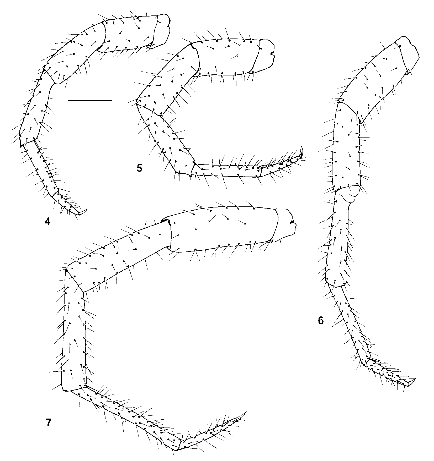
Articulations between two tarsomeres strong, continuous on dorsal side even on anterior legs, with condyle on legs 1315. Setae with similar densities and thicknesses on all legs; setae along inner edge of distitarsus sloping distally on legs 114. Sharp distal spinose projection on tibiae of legs 113 ( Figs. 4, 5 View FIGURES 4 7 ), spinose projection usually present on leg 14, exceptionally blunt ( Fig. 6 View FIGURES 4 7 ). Distitarsus 5254% length of basitarsus on leg 14, 5157% on leg 15; leg 15 basitarsus about 11 times longer than wide ( Fig. 7 View FIGURES 4 7 ). Posterior pretarsal accessory claw slightly longer than anterior accessory claw, 3545% length of main claw ( Fig. 26); scutes weakly defined on accessory claws ( Fig. 27); elongate scutes defined by sutures along length of main pretarsal claw, including its dorsoproximal part ( Fig. 26), without pores; posteroventral spine lacking.
Coxal pores on legs 1215, round, numbers as in Diagnosis; pore rows not set in grooves.
Female ( Figs. 2 View FIGURES 1 3 , 2931): Sternite of segment 15 transverse or faintly concave posteromedially, numerous setae on posterolateral margin, a few on posterior margin and scattered over surface. Sternite of first genital segment with up to 48 short to moderately long setae scattered over most of surface, with slight to distinct concentration near posterolateral margin. Basal article of gonopod bearing 1320 setae, with two blunt spurs of about equal size at distal end of slender, elongate process ( Fig. 31); dorsal surface of spurs concave, with several low tubercles ( Fig. 30); second article of gonopod with 59 setae; third article with two or three setae. Claw undivided, bearing many strong pores on its dorsodistal surface, each pore housing a sensillum coeloconicum ( Fig. 31).
Male ( Figs. 3 View FIGURES 1 3 , 28): Sternite of segment 15 transverse posteromedially; setae sparsely scattered on its anterior half, about 12 setae fringing posterolateral and posteromedian margins. Sternite of first genital segment undivided, posterior margin bulging backwards between gonopods; up to 26 setae scattered over sternite, a few long setae variably present on posterior margin ( Fig. 28). Basal two articles of gonopod each bearing a single row of three to seven setae, usually four or five on first article; third article with four to seven scattered setae; terminal process tapering, spinelike.
Discussion: Paralamyctes wellingtonensis n. sp. is most obviously distinguished from P. chilensis (Gervais in Walckenaer & Gervais, 1847) by its more elongated spurbearing process on the female gonopod (compare Figs. 2 View FIGURES 1 3 , 29 herein with Fig. 24 B of Edgecombe 2001). Analysis of molecular sequence data and combined molecular and morphological data indicates that the two species are each others' closest relative ( Edgecombe & Giribet 2003). Morphologically, they share the usual presence of 19 mostly elongate antennal articles, relatively large, bulging eyes ( Fig. 8), a small Tömösváry organ ( Fig. 11 versus Edgecombe 2001, fig. 25M), four or five pointed teeth on each dental margin of the maxillipede in most specimens ( Fig. 15), and strong articulations between tarsomeres. The mandibles are very similar, sharing many small, barblike pinnules along both margins of the aciculae ( Fig. 17 versus Edgecombe 2001, fig. 25K) and abundant bristles in the furry pad that curl back towards the dorsalmost tooth ( Fig. 18 versus Edgecombe et al. 2002, fig. 7D).
Paralamyctes chilensis View in CoL was assigned by Edgecombe (2001) to the otherwise Australian subgenus P. ( Nothofagobius ). Morphological support for this assignment is also shared by P. wellingtonensis View in CoL , including the extended first article of the female gonopod and bulging posterior margin of the male first genital sternite ( Fig. 3 View FIGURES 1 3 ). However, molecular data and their combination with morphological data ( Edgecombe & Giribet 2003) instead favour a closer relationship between the two Chilean species and the Australasian subgenera P. (Thingathinga) Edgecombe, 2001, and P. ( Haasiella View in CoL ) Pocock, 1901. The most congruent parameter set for combined data resolves the Chilean species with P. ( Haasiella View in CoL ) in particular. That relationship accounts for some shared morphological characters, notably bifid aciculae on the mandible, the only cases of this character state within Paralamyctes View in CoL . In P. ( Haasiella View in CoL ) and in P. wellingtonensis View in CoL the median furrow on the head shield is variably incised behind the transverse suture ( Fig. 1 View FIGURES 1 3 ). However, most parameter sets resolve P. ( Haasiella View in CoL ) closer to P. (Thingathinga) than to the Chilean species ( Edgecombe & Giribet 2003). Because the relationships of the Chilean clade are unstable, a subgeneric assignment within Paralamyctes View in CoL is not made.

No known copyright restrictions apply. See Agosti, D., Egloff, W., 2009. Taxonomic information exchange and copyright: the Plazi approach. BMC Research Notes 2009, 2:53 for further explanation.